Unit 1 QCAA Biology
 Call Kai
 Learn
Learn Practice Test
 Spaced Repetition
 Match
Match Flashcards
 Knowt Play
1/73

Name | Mastery | Learn | Test | Matching | Spaced | Call with Kai | Chat |
|---|
No analytics yet
Send a link to your students to track their progress
74 Terms
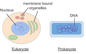
Compare prokaryotic and eukaryotic cells.
Prokaryotes have no membrane bound organelles, typically unicellular, simpler and smaller e.g. bacteria.
Eukaryotes have membrane bound organelles, more complex organisms, larger in size.
Both have DNA, prokaryotes can have ribosomes as they aren’t membrane bound.
What are the levels of organisation in an organism?
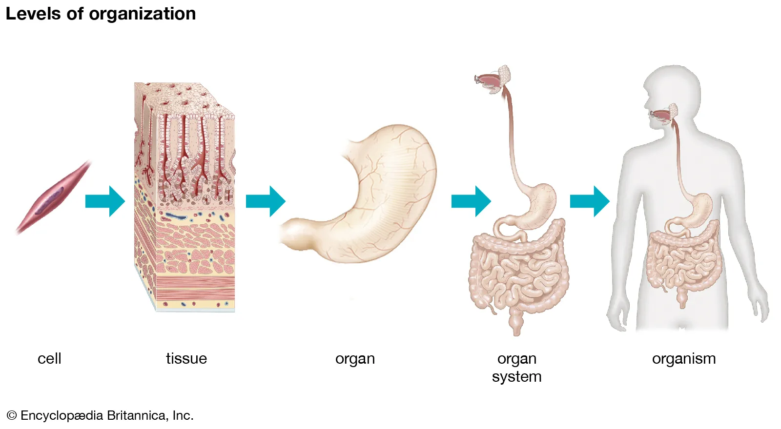
Cells → Tissues → Organs → Organ systems → Organism
What is the purpose of the plasma membrane?
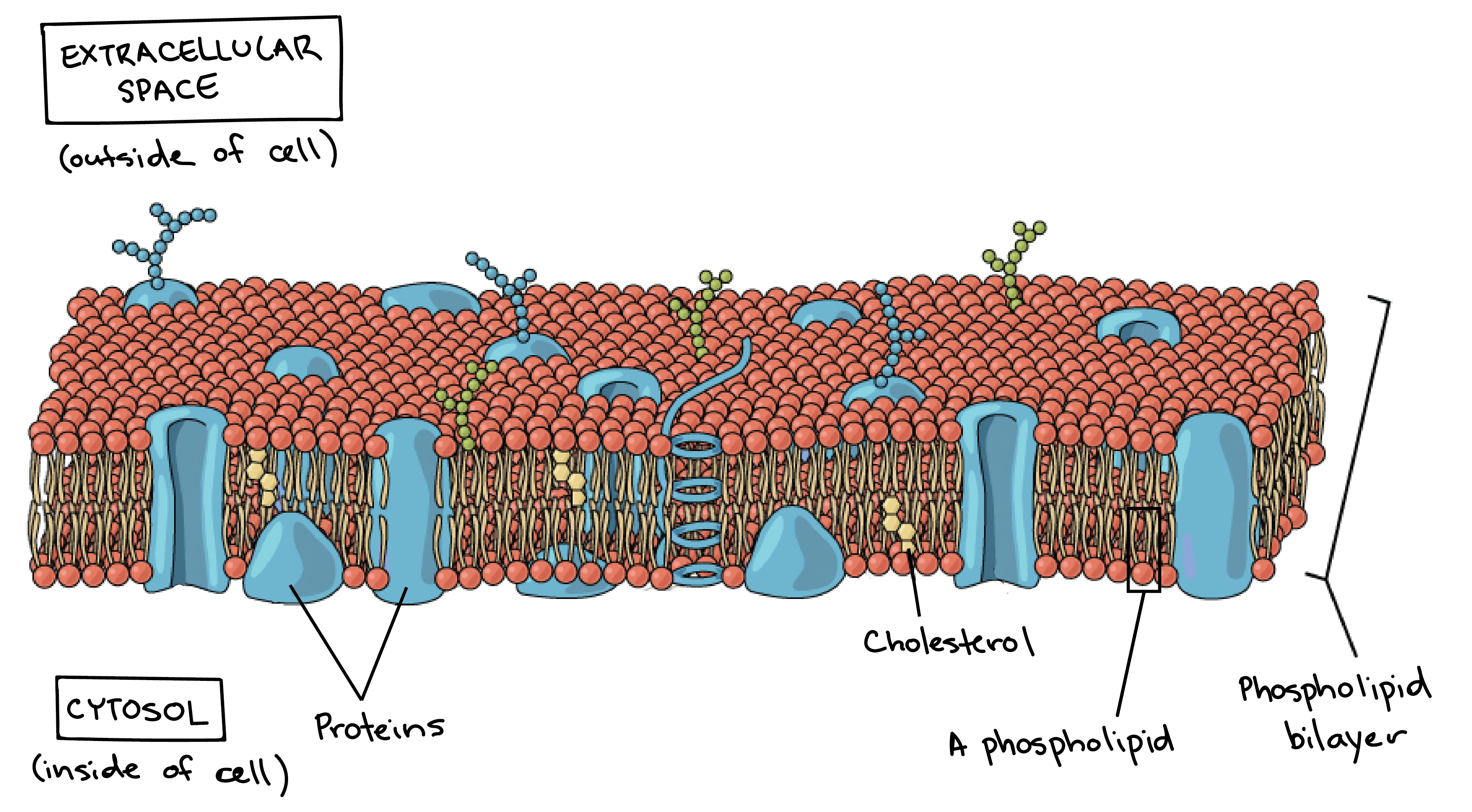
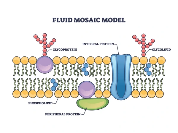
It is the boundary of the cell, and has 2 layers of fat and 1 layer of protein. It allows things in and out of the cell. Image of phospholipid bilayer.
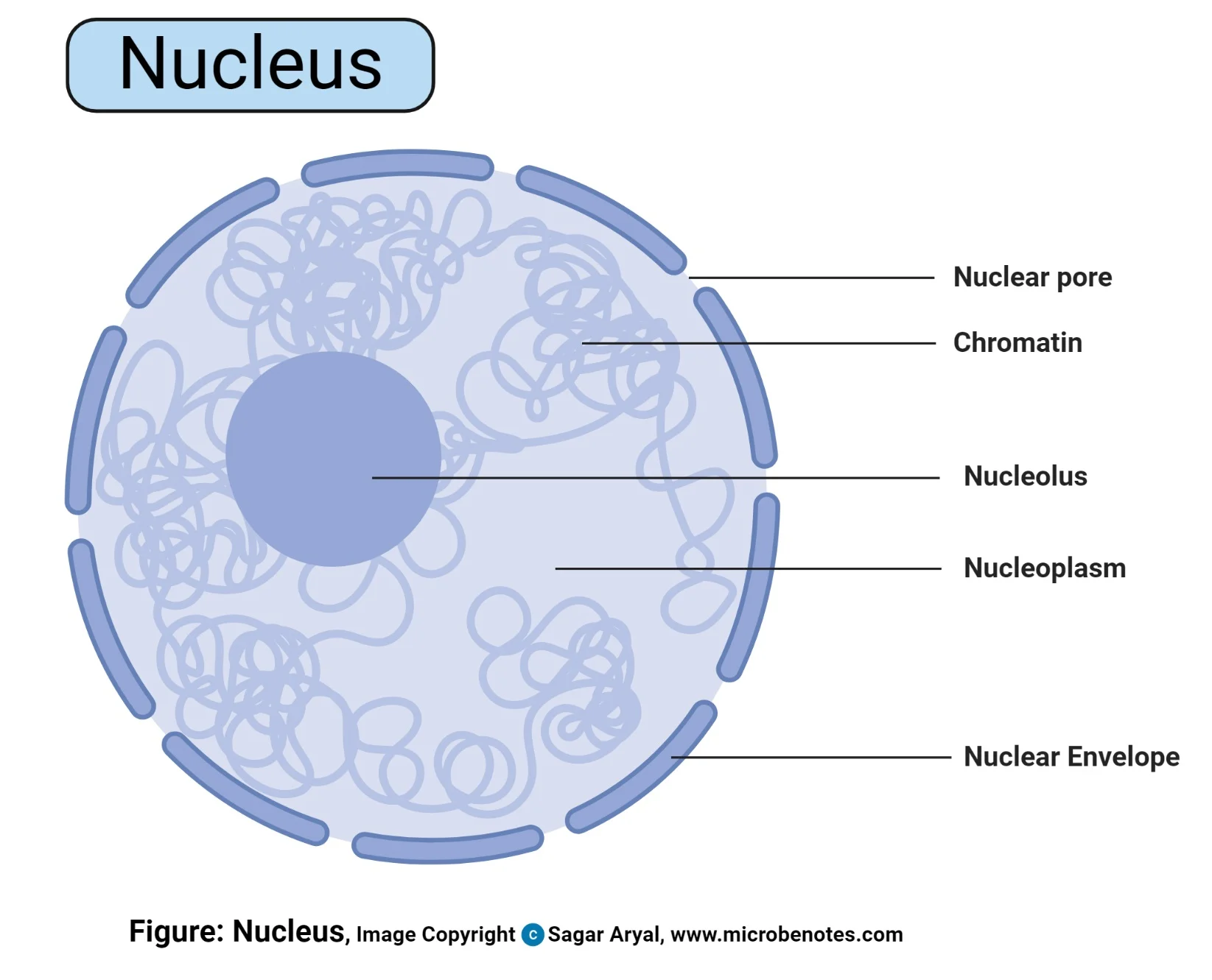
What is the nucleus?
A porous membrane containing DNA and chromatin, also containing the small round nucleolus. Image of nucleus only . Controls growth and reproduction.
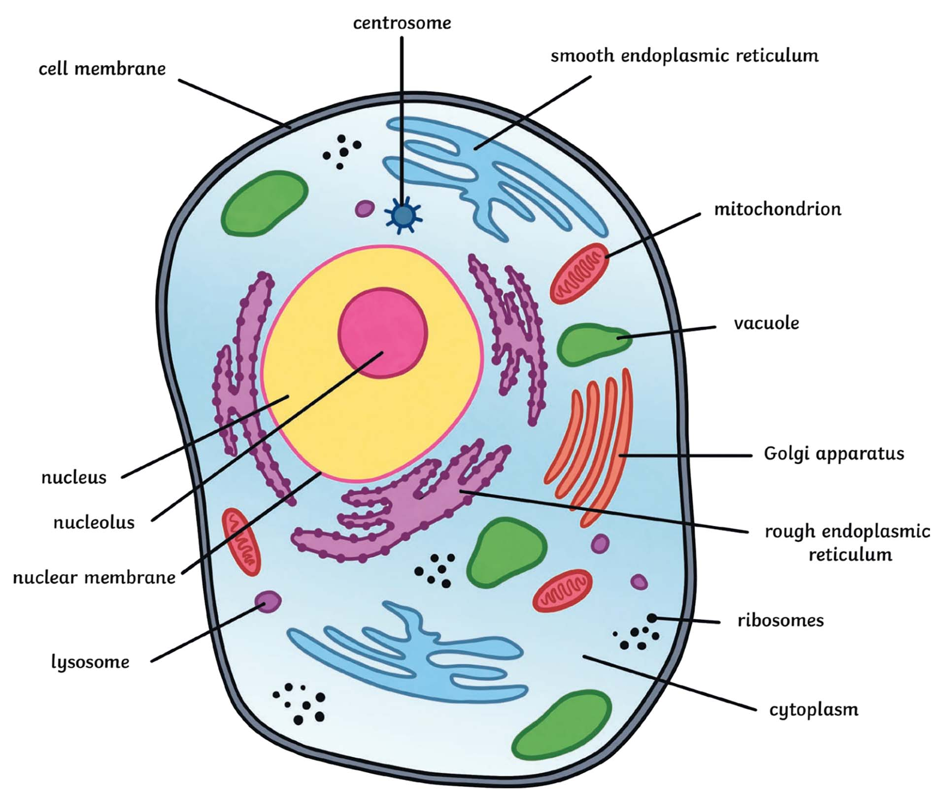
What are ribosomes?
They have no membrane, site of protein synthesis (create proteins), they are either free floating or attached to the Endoplasmic Reticulum, there are 2 subunits of ribosomes.
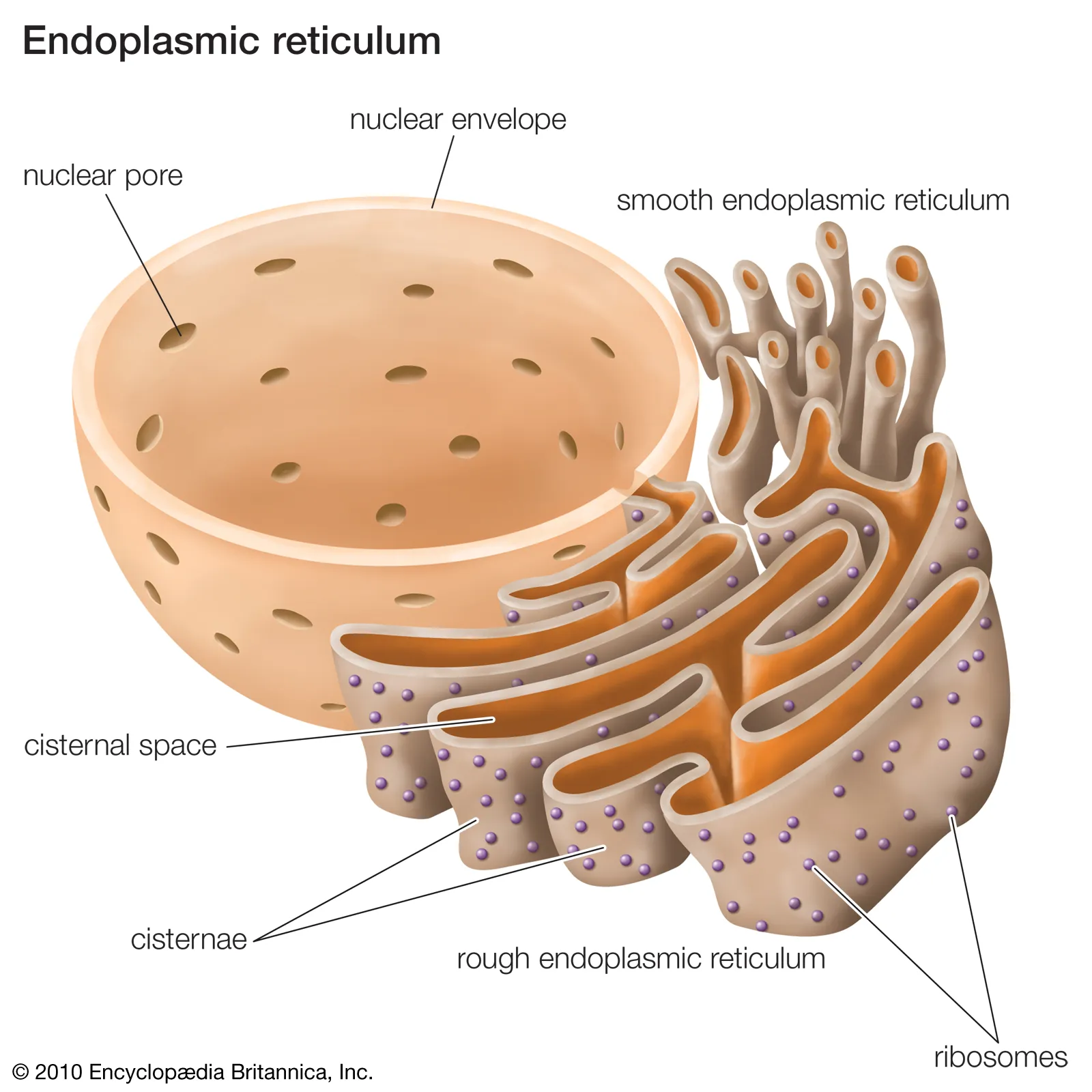
What is the SMOOTH endoplasmic reticulum?
The smooth ER is a single membrane, ribosome free and helps detox poisons, and creates and stores lipids & steroids. Image of nucleus and ER.
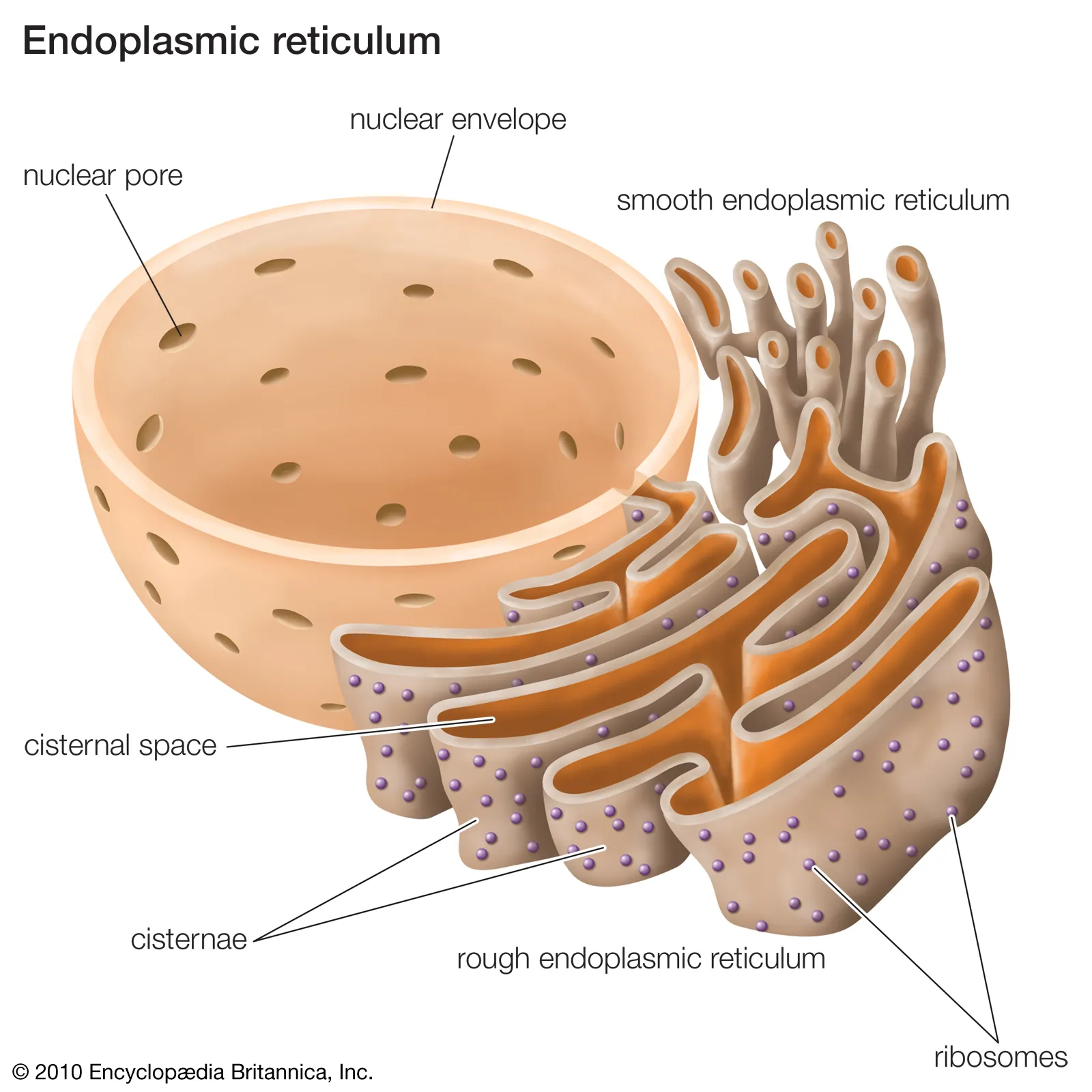
What is the ROUGH endoplasmic reticulum?
Contains ribosomes and releases newly made proteins from the cell. Gets it rough appearance from the ribosomes attached to its surface. Image of nucleus and ER.
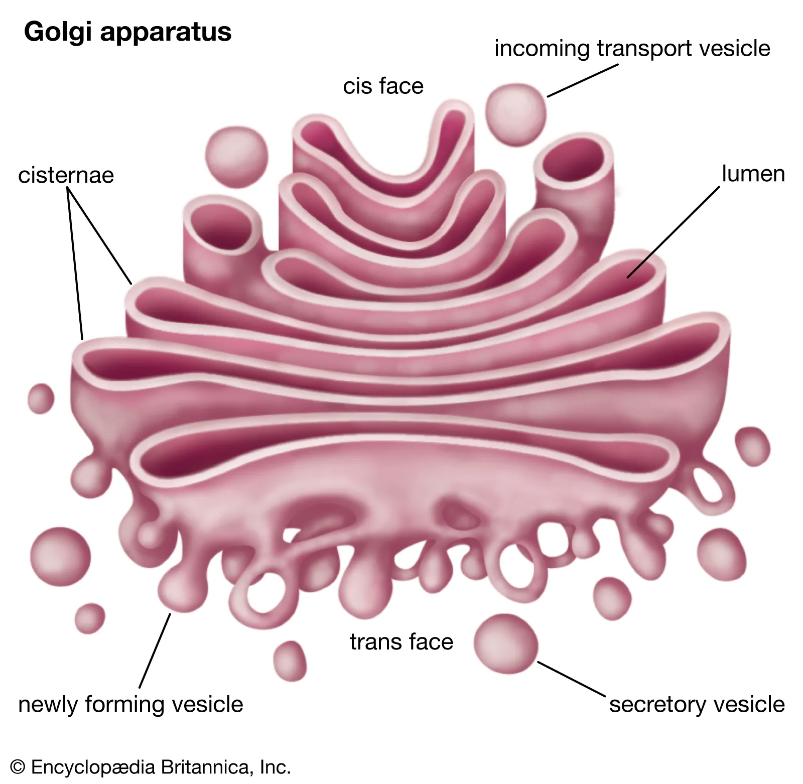
What is the golgi apparatus?
A stack of flattened sacs, which modifies and packages proteins and lipids from the ER, packaged into vesicles for transport to their final destinations. Makes molecules more complex.
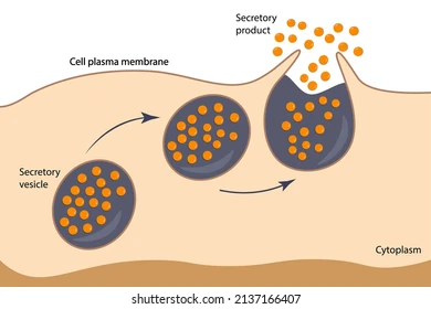
What is a secretory vesicle?
Membrane bound sacs that come from the golgi apparatus, and bind to the cell membrane exporting contents from the cell.
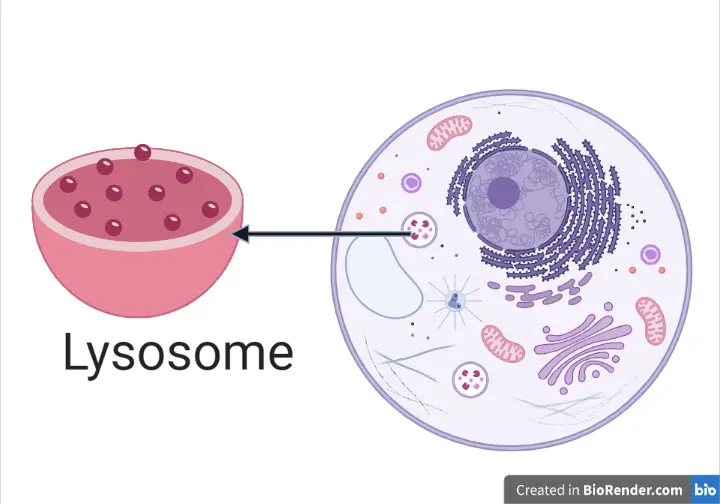
What are lysosomes?
They are organelles which recycle cellular debris, containing powerful enzymes to break down waste material. It has an acidic pH of 5, digesting particles in/outside of cells. Only animal cells. Image of full cell, pointing out lysosome.
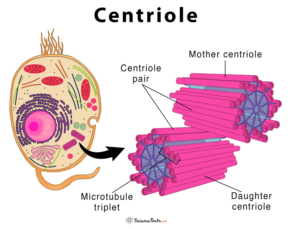
What are centrioles?
They are only found in animal cells, they are paired found together near the nucleus at right angles to eachother. Help build cilia and flagella (external attachments of cells e.g tails/hair), aids in cellular reproduction e.g mitosis. Image of full cell, pointing out centrioles.
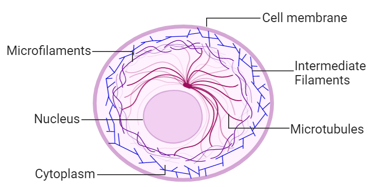
What is a cytoskeleton?
The framework of the cell, made up of microfilaments and microtubules. Provides structural support and helps movement of organelles. Image of full cell.
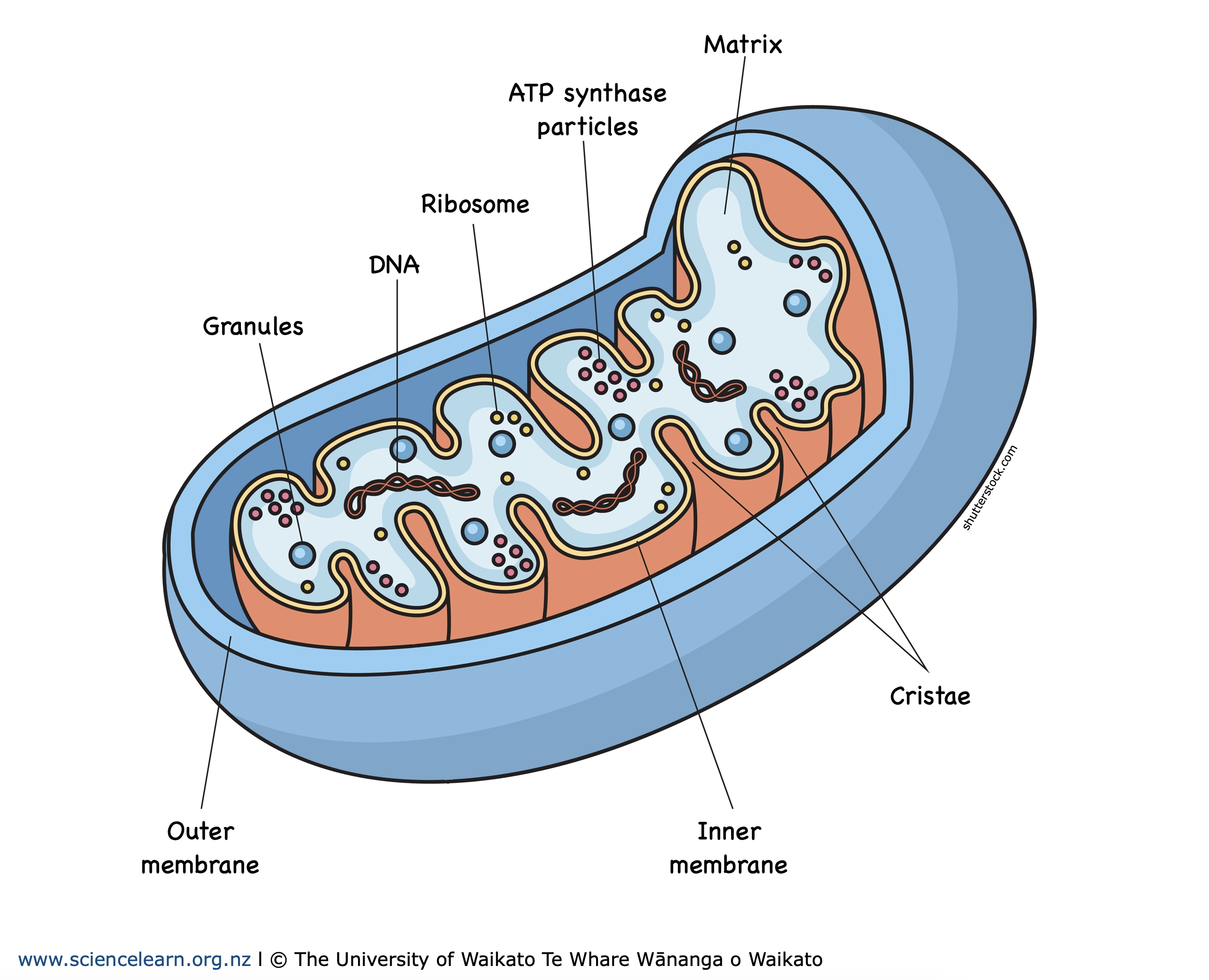
What is the mitochondria?
The powerhouse of the cell 😎🤙. It creates ATP (energy) via cellular respiration. It is double membraned and the size of a bacterium. It contains it’s own unique DNA, called mtDNA. Image of mitochondria.
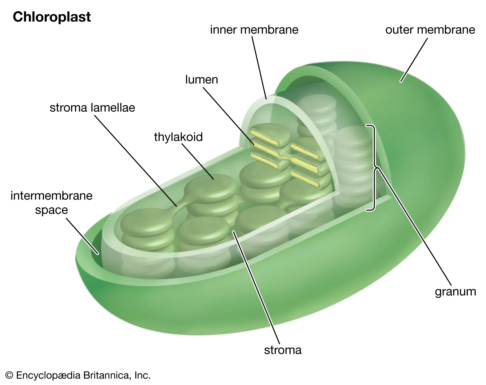
What are chloroplasts?
Double membraned organelles. Contains chlorophyll (green pigment in plants) and is the primary site of photosynthesis. Converts light energy into sugars. Image is of a chloroplast, not a full cell. Only in plant cells.
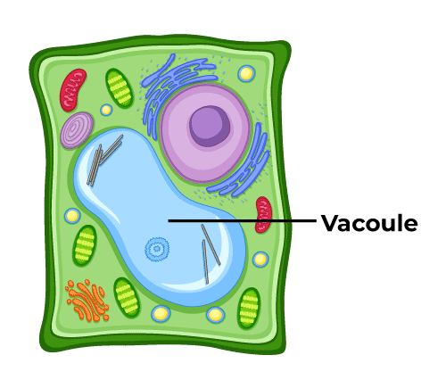
What are vacuoles?
Sacs that help store food and help cell retain water and maintain water balance. Found in animal and plant cells, larger in plant.
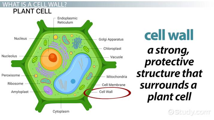
What is the cell wall?
Structural support surrounding plasma membrane. ONLY Found in plant, algae, fungi and bacteria. Provides strength and rigidity. In plants it’s made of cellulose, in fungi it’s made of chitin.
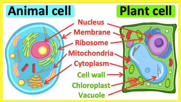
What organelles do plant cells have that animal cells don’t?
Chloroplasts, cell wall and large vacuoles.
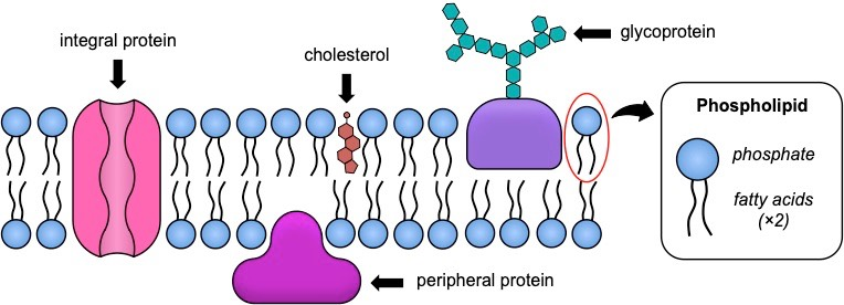
What is the phospholipid bilayer, the functions and the main components of it?
Plasma membrane, flexible and protective barrier.
Transport in and out of the cell (selectively permeable).
Allows for cell recognition, and provides anchoring sites for filaments of cytoskeleton.
It’s made of a hydrophilic phosphate head, two hydrophobic fatty acid tails, transport proteins that allow for larger molecules to pass through, and carbohydrates attached to the outer surface. Also fluid mosaic model (fluidity, looks like mosaic).
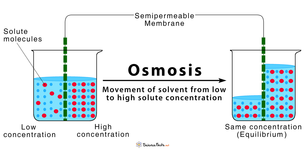
What is osmosis?
Specifically movement of water across semipermeable membrane, from a area of high water (solvent) concentration to an area of low water concentration to equalise concentration on both sides. Occurs without energy through natural concentration gradient.
What is an isotonic solution?
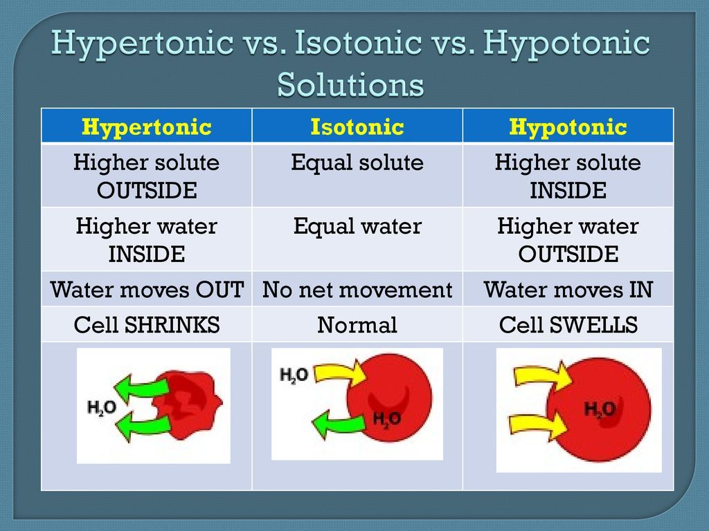
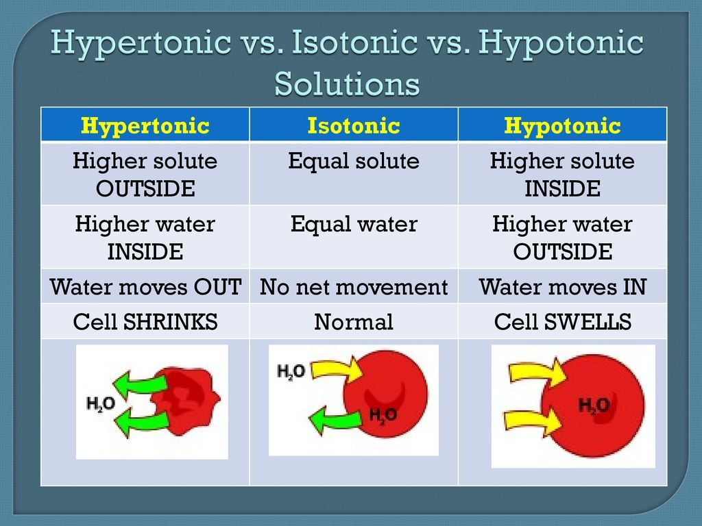
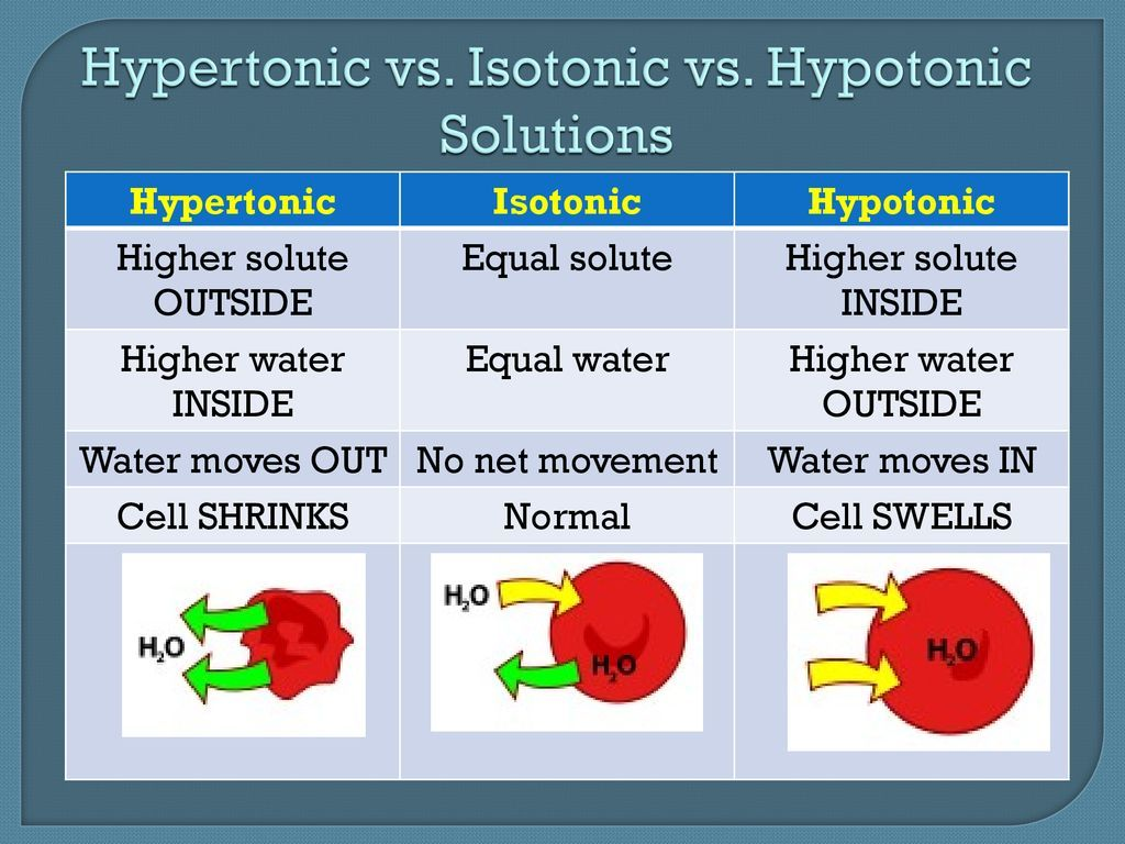
It is at an equilibrium, there is no movement of molecules across the membrane as the solute concentration is the same in the cell and outside of it thus no change in cell volume.
What is a hypotonic solution?
Movement into the cell, hypO, cell becomes swollen like an O. Water moves in to the cell, as theres less water inside, and it wants to move to a lower concentration via the natural concentration gradient.
What is a hypertonic solution?
It has movement out of the cell, the solvent/water moves out of the cell, as theres more water in the cell and it wants to move out to the lower concentrated environment, so the cell shrinks.
Compare passive and active transport.
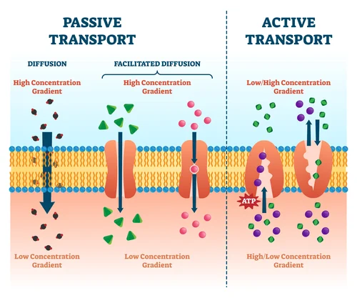
Passive transport
No energy (ATP) required
Particles move down the concentration gradient (high → low)
Includes diffusion, facilitated diffusion, osmosis
Active transport
Requires energy (ATP)
Particles move against the concentration gradient (low → high)
Uses carrier proteins or pumps
Compare simple and facilitated diffusion.
Simple
Passive
Moves down the concentration gradient
No ATP required
No transport proteins
Small, non-polar molecules (e.g. O₂, CO₂)
Facilitated
Passive
Moves down the concentration gradient
No ATP required
Requires transport proteins (channels or carriers)
Large, polar or charged molecules (e.g. glucose, ions)
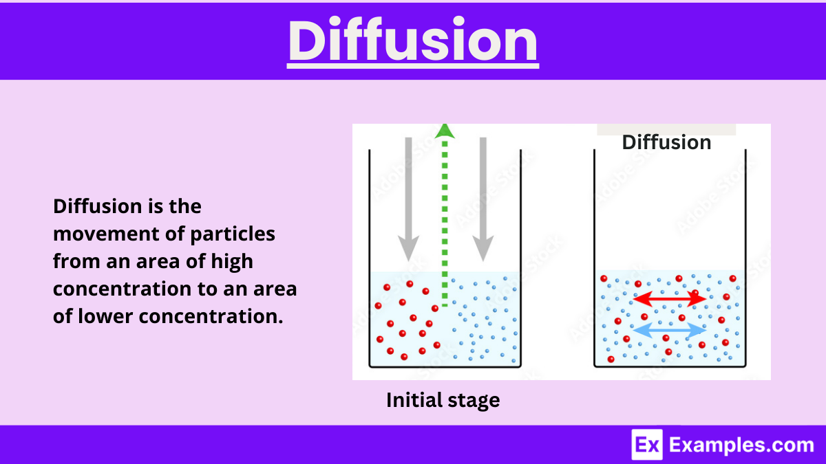
What is the role of cholesterol in the plasma membrane?
Regulates membrane fluidity and stability, preventing the membrane from becoming too rigid or too fluid. High temp = Less movement. Low temp = More movement.
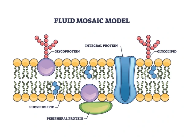
What is the role of glycoproteins in the plasma membrane?
(Proteins with carbohydrate chains) function in cell recognition, cell signalling and cell–cell communication. Similar to a lock and key mechanism.
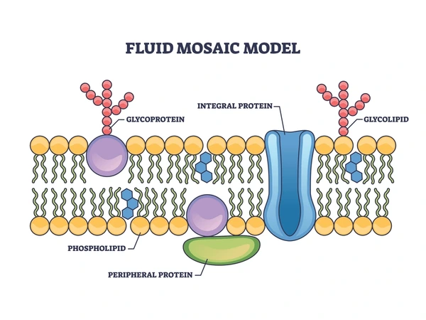
What is the role of proteins in the plasma membrane?
Embedded in the membrane, allow specific substances to cross the membrane. Assist in enzymatic activity, signal transduction, attaching to cytoskeleton and extracellular matrix.
Protein channels transport larger molecules.
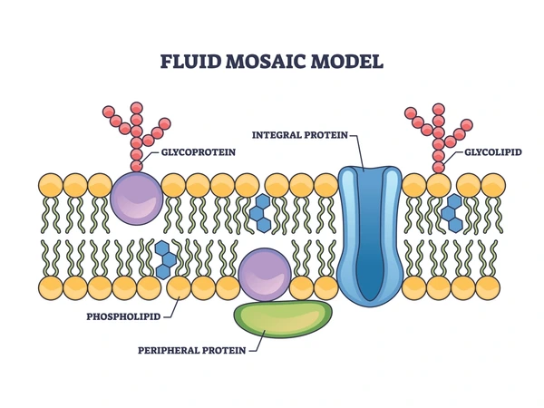
What is the role of phospholipids in the plasma membrane?
Form a bilayer with hydrophilic phosphate heads facing outwards and hydrophobic fatty acid tails facing inwards, creating a selectively permeable barrier. PHOSPHO-LIPID.
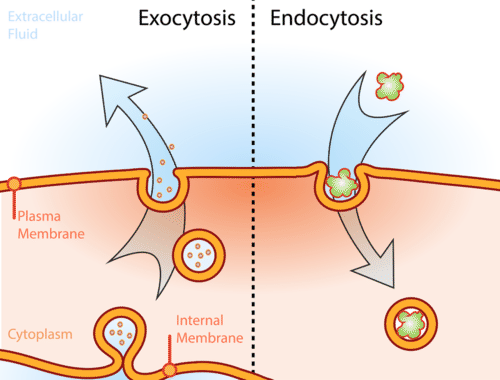
Compare endocytosis and exocytosis.
Exocytosis:
Substance exits, a vesicle containing the molecules fuses with the cell membrane and it’s expelled
Endocytosis:
Substance enters the cell, membrane engulfs it and transports it in.
Pinocytosis: Entry of extracellular fluids.
Phagocytosis: Entry of large particles e.g. cell debris/bacteria.
What is digestion?
The breakdown of food into smaller pieces/building blocks so it can be absorbed. Can be mechanical or chemical.
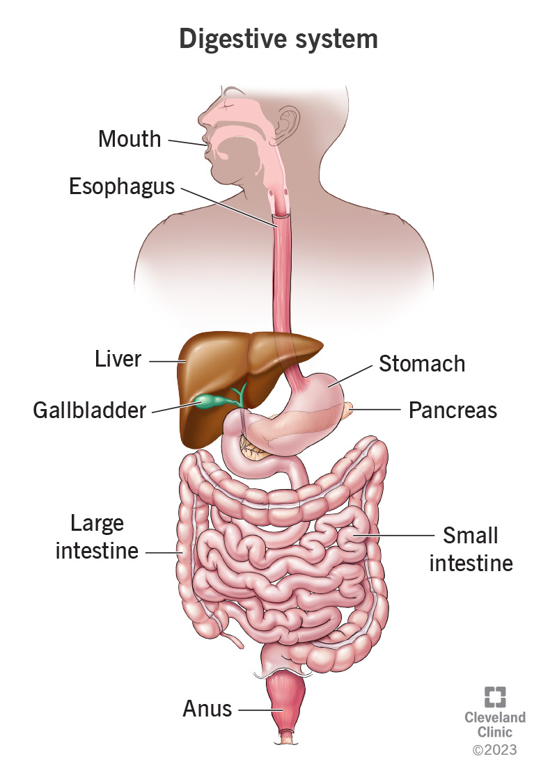
Compare mechanical and chemical digestion.
Mechanical digestion: Breaks food into smaller chunks. It occurs in chewing in the mouth, the stomach when it churns and the small intestine with the action of bile and peristalsis
Chemical digestion: Breaks food down into smaller organic compounds, into its building blocks via enzymes. They are then small enough to pass through the walls of the intestine to be used.

What is the function of amylase?
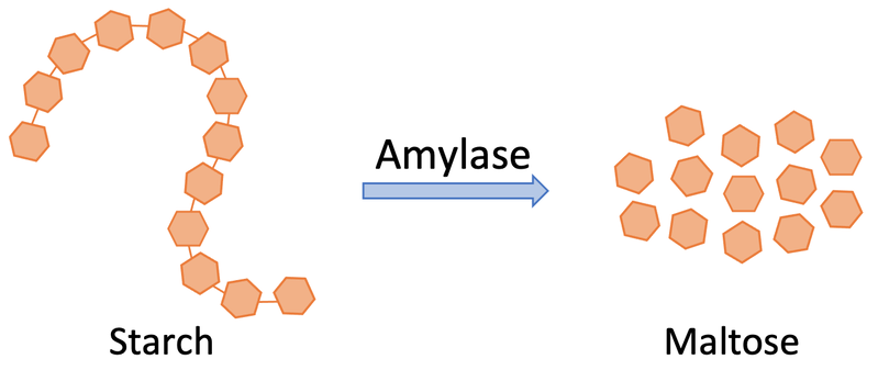
Enzyme which breaks down carbohydrates → Simple sugar e.g. starch → Glucose. Occurs in mouth and small intestine.
What is the function of protease?
Enzyme which breaks down protein → Amino acid. Occurs in stomach and small intestine.

What is the function of lipase?
Enzyme which breaks down lipids → fatty acids + glycerol.

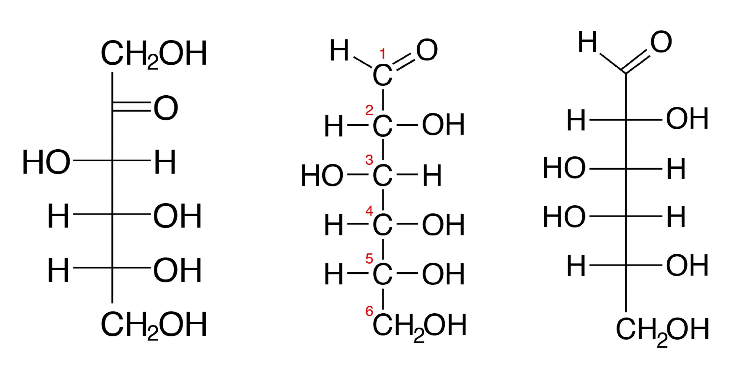
What is the structure of carbohydrates?
Made up of C, H and O. Made up of simple sugars and monosaccharides.
What is the structure of proteins?
Contains C, H, O and N. Made of amino acids. Forms structure like hair, muscle and cytoskeletons. Helps in enzymes, antibodies and clotting proteins.

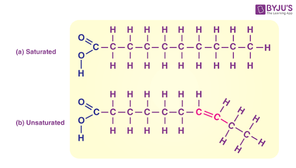
What is the structure of lipids?
Contains C, H and O. Made up of fatty acids and glycerol. Provides insulation and protection for organs.
What is the role of the mouth in the digestive system?
Teeth and tongue aid in mechanical digestion. Salivary glands produce enzymes (amylase), breaking down starch.

What is the role of the oesophagus in the digestive system
It connects the mouth to the stomach. Peristalsis, muscle contractions, force the food down.

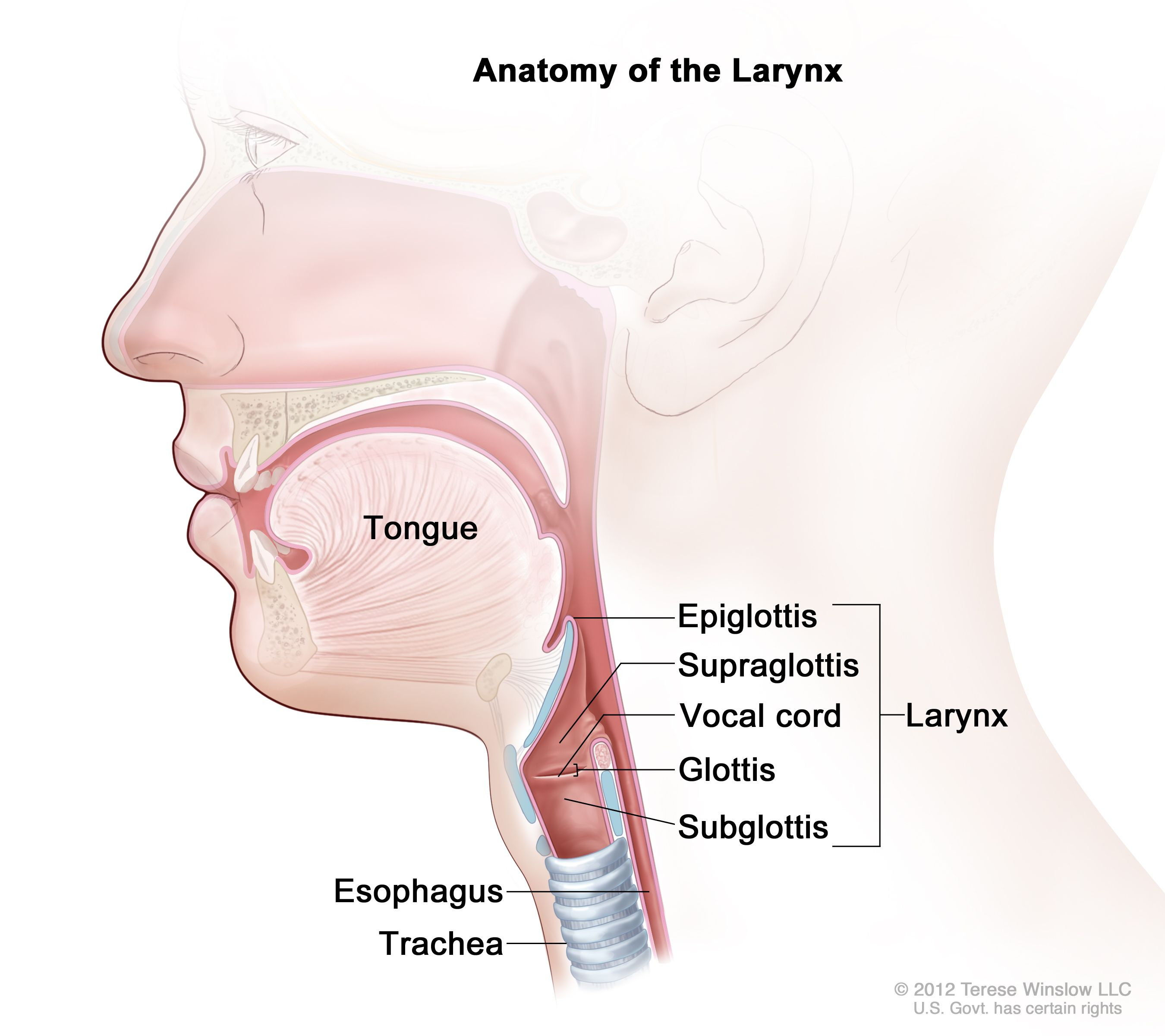
What is the role of the epiglottis?
It is a flap of skin that protects the trachea from food.
What is the role of the stomach in the digestive system and it’s main 2 sphincters? (Including gastric juice)
Acts as a storage sac for food, it stretches to hold alot of food. It churns food, protease digests protein in the stomach. Gastric juice contains protease and HCl, which works best in an acidic environment.
Cardiac sphincter at the top stops food from going up.
Pyloric sphincter opens only when liquids from stomach are ready to move to small intestine.

What is the function and structure of the small intestine: duodenum?
Food leaving the stomach enters the duodenum first, where the digestion of carbs, proteins and lipids are done.
It receives secretions from the liver, pancreas and the small intestine itself.
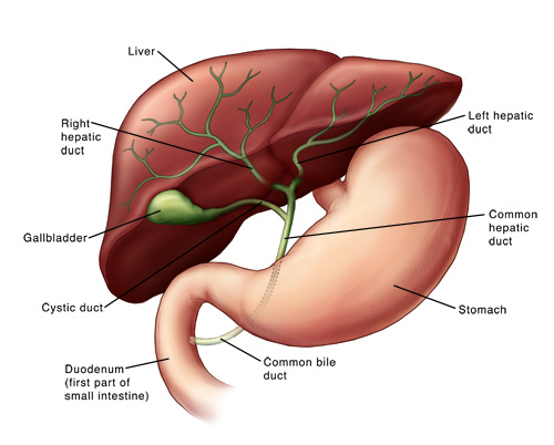
Gallbladder releases bile into small intestine.
Pancreatic juices and bile enter the small intestine through the common bile duct.

What is the function and structure of the small intestine: ileum?
The digested food in the small intestine is in liquid form and simpler molecules.
They can now enter cells, food moves via peristalsis along the ileum, the inner wall is lined with villi.
Digested food in the lumen of the intestine surrounds every villus. The molecules are so small they pass through the villus wall into each villus.
The villi provide immense surface area, and enzymes bound to the surface of epithelial cells break down peptides and carbohydrate molecules.

What is the function of the gallbladder in the DS?
Gallbladder stores bile. Connected to the liver. When you eat fat, the gallbladder squeezes bile into the small intestine.
What is bile?
Bile is a yellow-green substance made by the liver and travels to the small intestine through the bile duct. It’s job is to emulsify fats that have travelled through the stomach (break them down smaller). It is MECHANICAL, nothing has been alter chemically.

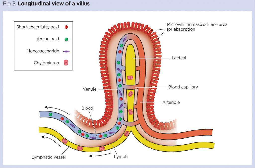
What is the structure of villi?
The villi provide immense surface area, and enzymes bound to the surface of epithelial cells break down peptides and carbohydrate molecules.
The dips of the intestinal glands (crypts of Lieberkuhn) secrete mucus and alkaline fluid.
Products broken down by enzymes in the surface of microvilli are absorbed into blood and lymph vessels.
Goblet cells produce mucus to protect epithelial cells from enzymatic digestion.
What is the role and structure of the large intestine?
It consists of the colon, rectum and an*s. Digested food is absorbed in the small intestine, most of the unabsorbed material is waste. Peristalsis moves the digestive waste into the LI, made up of materials we cant digest such as plant cellulose. Which is a tough material found in plants.
Bacteria that live in the large intestine also help breakdown undigested and unabsorbed materials. Important materials like vitamins dissolved in water must be returned back.
A solid waste is released, it can be stored in the rectum and released through defecation.

What is mitosis?
The process of one cell splitting into two identical daughter cells.
Interphase: Cell is just chilling.
Prophase: Preparing to divide, nucleus membrane starts to fade and chromosomes condense.
Metaphase: Spindle fibres appear and line them up in the middle.
Anaphase: Spindle fibres pull them away towards each side of the cell.
Telophase: Two new nuclei membranes form, cell starts pinching.
Cytokinesis: Cytoplasm splits, two new cells form.

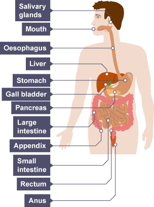
What is the process of digestion?
Mouth → Oesophagus → Stomach → Small intestine → Large intestine → Rectum → Left!
Mechanical and chemical digestion in mouth → Mechanical in oesophagus → Mechanical and chemical in the stomach → Mechanical and chemical in the small intestine and large → Only mechanical in rectum.
Accessory organs aid in chemical digestion by releasing substances.
Differentiate between unipotent, multipotent, pluripotent and totipotent stem cells.
Stem cells are cells that can change into other cell types and self-renew.
Totipotent: Can totally turn into anything
Pluripotent embyronic: Can turn into anything but placental
Multipotent: Only a few cell types, can change into closely related cell types, adult stem cells are multipotent.
Unipotent: Only one cell, but self-renewable.

How are specialised cells structurally suited to their function in body systems?
Size and shape → affects surface area to volume ratio (SA:V), influencing exchange efficiency.
Organelle composition → number and type of organelles reflect the cell’s role (e.g. more mitochondria for energy-demanding cells).

Why is surface area to volume ratio important in specialised cells?
SA: Supply. V: Needs.
A high SA:V ratio increases the rate of diffusion and exchange across membranes. Cells involved in absorption or gas exchange are often small or folded to maximise surface area. If the SA and V are too close to each other, there may not be enough supply to support the cell.

Why is cell size limited by surface area-to-volume ratio and diffusion rate?
As a cell increases in size, its volume increases faster than its surface area, reducing its surface area-to-volume (SA:V) ratio.
A lower SA:V ratio limits the rate at which substances can diffuse into and out of the cell.
If diffusion cannot supply nutrients and remove wastes fast enough, the cell cannot function efficiently, limiting its maximum size.

Describe the function of an enzyme, and compare the induced-fit and lock-and-key models of enzyme function.
To accelerate ROR, substrate joins the enzyme at the active site, which forms enzyme substrate complex.
In the induced fit model: The substrate doesn’t fit, so the enzyme changes slightly to accomodate which lines up/stresses the bonds in the substrate. It is general and slower than L&K.
In the lock and key model: The shape of the substrate is exact to the active site. It fits like a lock and key, the enzyme substrate complex is formed and products are released. It is specific and faster.

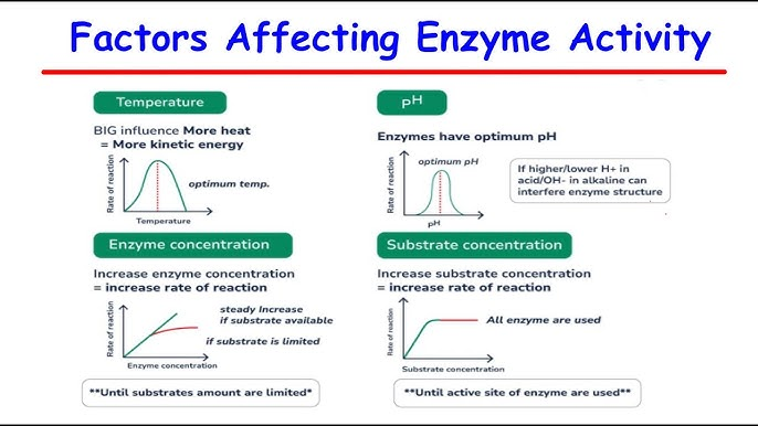
What are 5 factors affecting enzymatic activity?
Temperature: Weakens hydrogen bonds, distorting the 3D structure and denaturing enzymes.
pH: Enzymes have an optimal pH condition, specific to the type of enzyme.
Cofactors: Some need cofactors to be active, which are non-proteins. Permanently attaches cofactors are prosthetic groups, temporarily attached are coenzymes.
Enzyme concentration: As enzyme concentration goes up, ROR goes up linear. More enzymes can react with substrate.
Substrate concentration: As it goes up, ROR goes up, assuming a fixed amount of enzymes it will reach a plateau.
How do the 2 inhibitors stop enzyme activity?
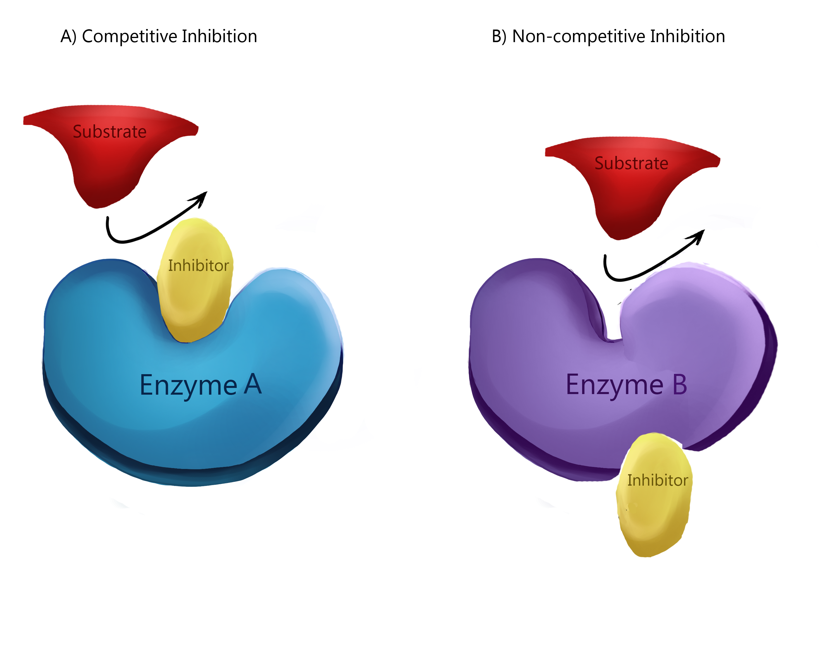
Competitive inhibitors impede the active site by having the same active site config, so it competes for access to the active site.
Non-competitive inhibitors are molecules that attach to a different site of the enzyme, and the molecular forces cause the active site to change.
What is excretion, and the organs of excretion?
The term used for the removal of metabolic waste.
Skin: Small amounts of urea, water, mineral salts.
Lungs: CO2 and water.
Large intestine, rectum and an*s: Organs of egestion, remove undigested substance.
Kidneys: Nitrogenous wastes (mainly urea), mineral salts, excess vitamins and water.

What is metabolism?
The sum total of all complex chemical reactions going on in the body.
What is the role of the liver in the excretory system?
Excess amino acids from proteins are broken down in the liver. The NH2 is initially converted to ammonia which is toxic. The ammonia → To urea. In birds and reptiles is uric acid.

What is the function of the kidneys in the excretory system?
Filter blood to remove metabolic wastes, particularly urea (a by-product of protein metabolism).
Regulate water balance (osmoregulation) by adjusting how much water is reabsorbed back into the bloodstream.
Control ion concentrations (e.g. sodium, potassium, chloride) to maintain electrolyte balance.
Help regulate pH of the blood by controlling hydrogen ions and bicarbonate levels.
Produce urine, which excretes excess water, salts and nitrogenous wastes.
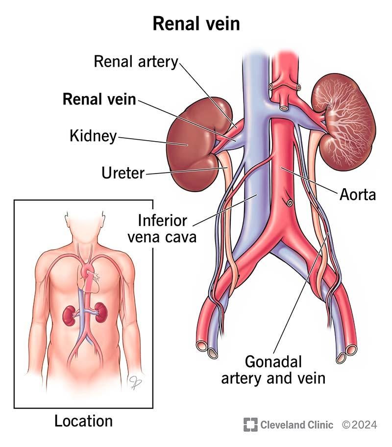
What is the order of blood flow from the heart to the kidneys.
Blood travels to the kidneys from the heart via the aorta, which branch into the left and right renal arteries. The blood is filtered in kidneys and leaves via renal veins. Blood enters the inferior vena cava and goes back to heart. Heart → Aorta → Renal arteries → Kidneys → Renal veins → Inferior vena cava → Heart.
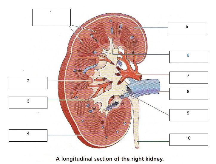
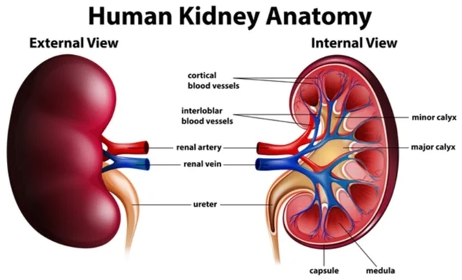
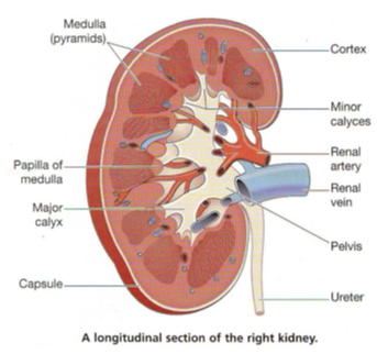
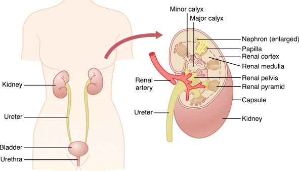
Label the internal structures of the kidney.
What is the simple pathway of waste within the excretory system.
Nephron → Collecting tubule → Pelvis → Ureter → Bladder.

Label the structure of the nephron.
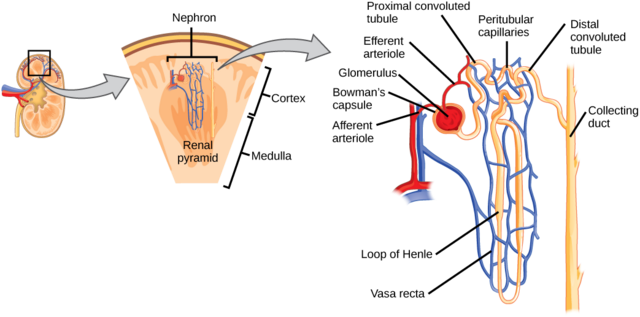
What are the three stages to removing wastes from the blood and explain them.
Filtration: Urea is filtered out of the blood. Can also filter glucose, salts etc. Oxygenated blood goes to the glomerelus → Urea goes to Bowman’s capsule. It is like a C sucking it all out from the glomerulus.
Reabsorption: After fluid leaves Bowman’s capsule it passes through tubule, what’s needed is reabsorbed into the blood capillaries. Some what like absorbed like water, sodium, potassium, glucose, hormones etc. What’s leftover → Pelvis → Ureter → Bladder. OSMOREGULATION OCCURS HERE, BECAUSE THE WATER REAPSORBTION WILL CHANGE.
Secretion: The pH of urine is managed by release of ions, waste is removed. Tubules push down urine to ureters, into the bladder.

What is the circulatory system, and it’s organs and function?
Cardiovascular and Lymphatic system = Circulatory System.
Circulates blood and lymph through the body.
Heart, blood vessels, blood, lymph, and the lymphatic vessels and glands.
It carries oxygen, nutrients, and hormones to cells, and removes waste products, like carbon dioxide.
Arteries carry blood away from the heart
Veins carry blood back to the heart.

Describe the CVS and lymphatic system.
CVS: made up of heart, lungs and blood vessels. Lungs supply blood with oxygen and remove CO2.
Lymphatic system: Defence against pathogens, aid transport in liqui dna matter. Only in one direction, returns to blood through walls of small blood vessels.
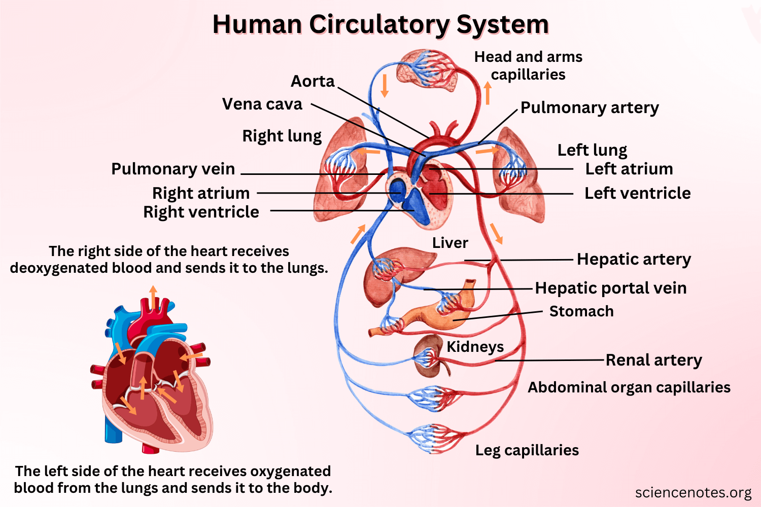
What are the 3 types of circulation?
Pulmonary: Carries oxygen-depleted blood away from the heart, to the lungs and back to the heart.
Systemic: Carries oxygenated blood away from heart to other parts of the body.
Coronary: Provides heart with oxygenated blood so it can function.

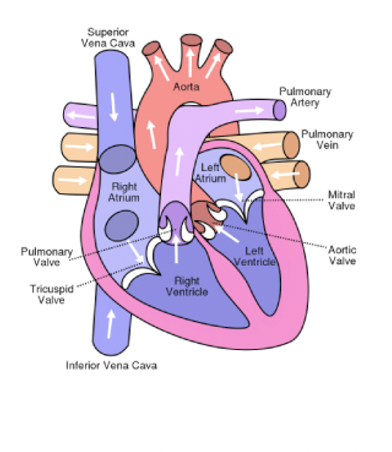
Describe the heart, what is the order of blood flow in the heart?
Movement of blood through heart is controlled by contraction of heart muscle, valves open and close at right time. 4 Chambers, two top ATRIUM and two bottom VENTRICLES.
Right side receives de-oxygenated blood from major veins of the body, and pumps blood into the lungs.
Left side receives oxygenated blood from lungs, and pumps it into the body through the aorta.
Right side → Lungs → Left side → Body
Body → Vena cava (superior & inferior) → Right atrium → Tricuspid valve → Right ventricle → Pulmonary valve → Pulmonary artery → Lungs → Pulmonary veins → Left atrium → Mitral (biscupid) valve → Left ventricle → Aortic valve → Aorta → Body.
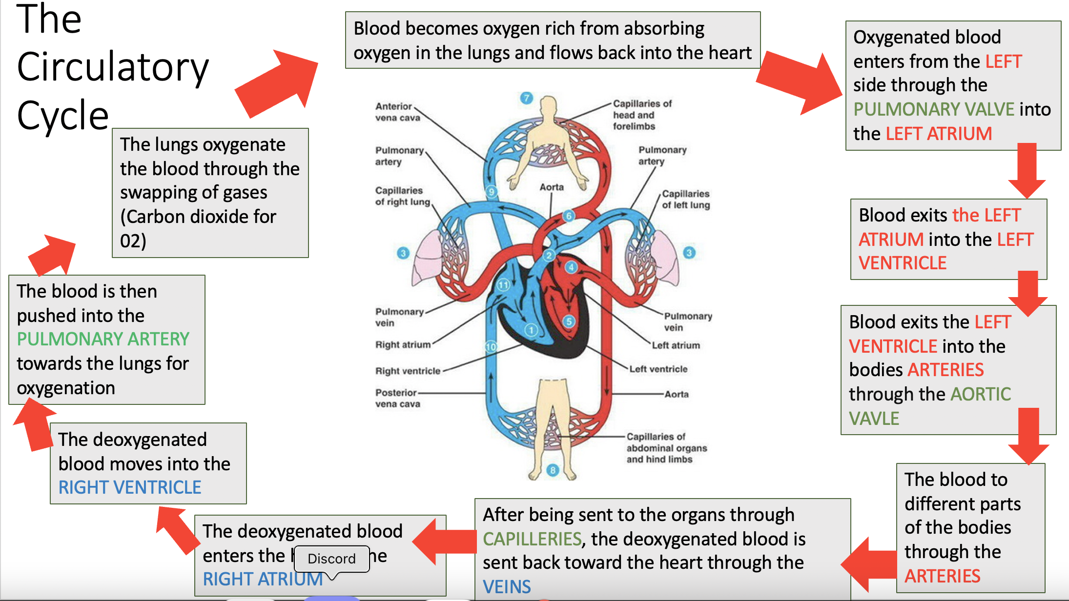

Label the parts of this heart.
What are the major functions of blood? (Name at least 3)
Transport of gases, transport of nutrients, waste removal including gas, regulation of body temperature, transport of hormones, prevents infection, prevents blood loss.
What are the cellular components of blood, and describe them.
Red blood cells: Anucleated, bi-concave disc shapes, lifespan 90-120 days, made in bone marrow. Contains protein called haemoglobin, forms oxyhemoglobin with oxygen. Specialised to carry O2, some carry CO2.
White blood cells: Contains nucleus, lifespans few days-several years. Made in bone marrow. Many different types, not as many wbc as red blood cells. Produce antibodies to protect body from infection. Attack virus and cancerous cells. Some engulf other cells called phagocytes.
Platelets: Formed from fragments of bone marrow cells. Pieces of cells, not whole cells. Do not have a nucleus. Help blood clot formation and tissue repair. Life span is few days if not used.

What are non-cellular components of blood?
Plasma: Includes wastes and water, proteins and nutrients.
Over 90% water, yucky yellow colour. Suspended in water are waste such as CO2 and urea, nutrients, specialised proteins and hormones.
Compare arteries, veins and capillaries.
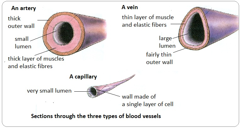
Arteries: Carry blood away from heart. Have thick muscular elastic walls. DO NOT HAVE VALVES!!! Pulmonary artery carries blood to lungs. Aorta carries blood to rest of body.
Veins: Carry blood to the heart. Thinner walls than arteries. Diameter of lumen is larger to get as much blood back to the heart as possibile. Muscle contraction assists, veins have valves to prevent back-flow of blood.
Capillaries: Connected to smallest arteries, only one cell thick, no muscle layer. O2 and nutrients can easily pass out of capillaries to surrounding cells. CO2 and other wastes can pass from cell into capillary. So tiny only a red blood cell can fit. Where diffusion happens.