Cardiovascular System - Physiology
 Call Kai
 Learn
Learn Practice Test
 Spaced Repetition
 Match
Match Flashcards
 Knowt Play
1/42

There's no tags or description
Looks like no tags are added yet.
Name | Mastery | Learn | Test | Matching | Spaced | Call with Kai | Chat |
|---|
No analytics yet
Send a link to your students to track their progress
43 Terms
Electrical Pathway:
Sinoatrial (SA) node: pacemaker near the right atrium; initiates each normal heartbeat (“sinus rhythm”)
atrial myocardium: depolarisation ripples through atrial muscle tissue via gap junctions
atrioventricular (AV) node: the only electrical connection between atria and ventricles
AV bundle (Bundle of His): receives impulse from AV node
Right and Left bundle branches: direct signal down toward the heart’s apex
Purkinje fibres: fast conduction network spreading up through ventricular walls for simultaneous ventricular contraction
Functional Significance:
AV node delay: conducts very slowly to create a brief delay, allowing the atria to finish contracting and filling the ventricles before ventricular systole begins
gap junctions: electrically couple cardiac cells so a wave of depolarisation spreads rapidly through the entire tissue from a small group of pacemaker cells
what is the step-by-step pathway of electrical conduction through the heart, and why are the and AV nodes functionally significant
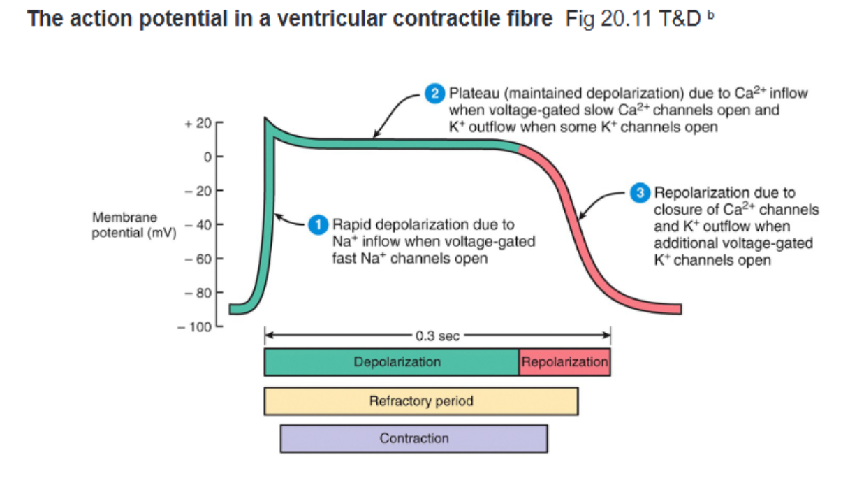
Rapid Depolarisation (Upstroke):
mechanism: voltage-gated fast Na+ channels open upon reaching threshold → fast Na+ influx (same channel type as in nerve APs)
Plateau:
mechanism: depolarisation opens voltage-gated Ca2+ channels → large Ca2+ influx (triggers calcium release from sarcoplasmic reticulum), inward Ca2+ influx is balanced by outward K+ efflux, keeping membrane potential flat
contraction: Ca2+ influx directly drives myofibril contraction (explaining why contraction lags slightly behind electrical activity)
protection: long plateau creates an extended refractory period, preventing tetany and re-entry to ensure one-way excitation
Repolarisation:
mechanism: delayed rectifier voltage-gated K+ channels open while Ca2+ channels close → rapid K+ efflux repolarises the cell
Timing and Coordination: Total AP duration is ~0.3 seconds at rest, variations in plateau duration across cells ensure the entire ventricle repolarises and relaxes in a coordinated fashion to allow refilling
what are the three main phases of a contractile myocyte action potential, their ionic mechanisms, and their functional significance

unstable resting potential: lacks a true resting phase; the membrane potential spontaneously and gradually depolarises (“pacemaker potential”) between beats, drifting up toward threshold
action potential trigger: once threshold is reached, fast Na+ channels open → fires a full action potential to trigger a heartbeat
intrinsic rate: isolated SA nodes fire spontaneously at ~90-100 bpm (resting HR is lower due to parasympathetic tone)
physical size: the SA node is small, measuring roughly 1-2 cm long by 2-3 mm wide
what are the defining characteristics of a pacemaker cell action potential in the SA node, including its resting potential, intrinsic rate, and dimensions
Feature | Somatic | Autonomic |
|---|---|---|
Sensory input | Somatic & special senses | Interoceptors (internal sensing) |
Control | Voluntary – cerebral cortex | Involuntary – limbic system, brainstem, hypothalamus, spinal cord |
Effectors | Skeletal muscle | Smooth muscle, cardiac muscle, glands |
Neurons | One-neuron pathway | Two-neuron pathway (pre- and post-ganglionic) |
Neurotransmitter | Acetylcholine (ACh) | Pre-ganglionic (both branches): ACh |
how do the somatic and autonomic nervous systems differ across sensory input, control, effectors, neuronal pathways, and neurotransmitters
Parasympathetic Branch:
origin: brainstem/medulla
structure: long pre-ganglionic fibre traveling via Cranial Nerve X (Vagus nerve) to a ganglion right next to/on the target organ; short post-ganglionic fibre
rest and digest
Sympathetic Branch:
origin: medulla (spinal origin region)
structure: short pre-ganglionic fibre synapsing in sympathetic chain ganglia near the spinal column; long post-ganglionic fibre travelling to target organ
fight or flight/alarm
Dual Innervation: most visceral organs receive input from both sympathetic and parasympathetic branches to balance function involuntarily (“self-governing”)
what are the anatomical differences between the parasympathetic and sympathetic pathways and what is dual innervation
dual innervation: both ANS branches directly innervate the SA node
resting tone: parasympathetic (vagal) input dominates at rest, acting as a constant “brake” to hold HR at ~50-70 bpm (well below the SA node’s intrinsic ~90-100 bpm rate)
moderate HR increase: accomplished by withdrawing parasympathetic tone (HR rises up toward ~90-100 bpm)
high HR increase: accomplished by activating sympathetic drive on top of parasympathetic withdrawal (HR an reach up to 200 bpm during intense exercise/stress)
how does the autonomic nervous system regulated resting heart rate, and how do parasympathetic vs. sympathetic adjustments alter HR
receptors: both sympathetic and parasympathetic receptors are present on the same SA node pacemaker cells
sympathetic mechanism: noradrenaline (NE) binds receptors → speeds up spontaneous pacemaker depolarisation → threshold reached sooner → faster HR
parasympathetic mechanism: acetylcholine (ACh) binds receptors → slows down spontaneous pacemaker depolarisation → threshold reached later → slower HR
what neurotransmitters and cellular mechanisms do the sympathetic and parasympathetic systems use to alter heart rate at SA node pacemaker cells
Sympathetic System:
Neurotransmitter: Noradrenaline (NE)
Effect on HR: Increases (↑)
Effect on Inotropy: Increases (↑ positive inotropy / force of contraction)
Parasympathetic System:
Neurotransmitter: Acetylcholine (ACh)
Effect on HR: Decreases (↓)
Effect on Inotropy: Decreases (↓ negative inotropy / force of contraction)
compare the sympathetic and parasympathetic systems regarding their neurotransmitters and overall effects on heart rate and contractility (inotropy)

P Wave:
Represents: Atrial depolarisation.
Cellular Event: Atrial contractile cells depolarise.
P–R (P–Q) Segment (Flat/Isoelectric):
Represents: Conduction delay through the AV node.
Cellular Event: AV node conducts slowly, allowing atrial contraction to finish and ventricles to fill before ventricular depolarisation starts.
QRS Complex:
Represents: Onset & rapid depolarisation of the ventricles.
Cellular Event: Wave travels down the septum to the apex, then back up via the Purkinje network ("down-then-up" spread causes large positive/negative deflections). Fast and large to generate coordinated force for systemic ejection.
S–T Segment (Flat/Isoelectric):
Represents: Ventricular cells sitting in the plateau phase.
Cellular Event: Membrane is depolarised, but inward (Ca2+) and outward (K+) currents are balanced (net current = 0). Ca2+ influx actively drives ventricular contraction.
T Wave:
Represents: Ventricular repolarisation.
Cellular Event: K+ efflux repolarises ventricular myocytes.
what do the P-wave, P-R segment, QRS complex, S-T segment, and T wave represent on an ECG, and what underlying cellular events drive each
Definition: The volume of blood pumped by the heart per minute.
Equation: Cardiac Output (CO)=Heart Rate (HR)×Stroke Volume (SV)
what is cardiac output and its equations
SV = End Diastolic Volume (EDV) - End Systolic Volume (ESV)
End-Diastolic Volume (EDV): 120–140 mL (blood volume at end of filling)
End-Systolic Volume (ESV): 50–70 mL (blood volume remaining after contraction)
Stroke Volume (SV): 50–100 mL/beat (volume ejected per beat)
what is the formula for stroke volume (SV)
Preload: The stretching force on heart muscle prior to contraction (filling/EDV)
Contractility (Inotropy): The intrinsic force of contraction at a given preload and afterload
Afterload: The pressure the heart must overcome to eject blood during contraction
what are the three primary physiological determinants of stroke volume
Definition: The force that stretches cardiac muscle prior to contraction (reflects ventricular fullness right before contraction).
Core Relationship: ↑ Diastolic filling→↑ EDV→↑ SV
Clinical Proxy: Ventricular volume is difficult to measure directly due to irregular cardiac geometry. Because pressure and volume track together, LV end-diastolic pressure (LVEDP) is measured as a direct practical proxy for preload.
what is preload and why is end-diastolic pressure used as its clinical proxy instead of volume
Venous Return & Volume: Blood loss/dehydration drops preload; increased venous return increases it.
Filling Time: High heart rates shorten diastolic filling time →↓ preload.
Respiratory Pump: Deep inspiration draws blood toward the heart →↑ preload.
Compliance: Stiff/scarred ventricles (e.g., post-MI) resist filling →↓ preload.
Venous Tone & Posture: Venoconstriction shifts blood back to the heart (↑ preload); sudden standing causes pooling in lower limbs (↓ preload).
Atrial Fibrillation: Loss of coordinated atrial kick eliminates final ventricular filling →↓ preload.
what key factors increase or decrease preload

Law Statement: Left ventricular stroke volume increases as end-diastolic volume increases due to myocyte stretch causing a more forceful systolic contraction.
Key Phrase: "More in = more out" (length-dependent activation).
Mechanisms:
Stretch increases filament sensitivity to Ca2+ (pulls actin-myosin closer).
Stretch opens stretch-gated Ca2+ channels, increasing Ca2+ influx for stronger contraction.
Graphical Effect: ↑ Venous return moves operating point up the SV vs. LVEDP curve (↑ SV).
what is the Frank-Starling Law of the Heart, its key phrase, and its proposed mechanisms
Definition: The intrinsic performance/force of contraction of the heart at a given preload and afterload.
Preload vs. Contractility: Preload relies on muscle stretch (filling volume), whereas contractility is independent of stretch and reflects the heart's baseline contraction force.
Cellular Drivers: Modulated by factors that alter ion gradients—especially intra-myocyte Ca2+:
Sympathetic Stimulation: Increases contractility (positive inotropy).
Increased [Ca2+]: Boosts contractile force.
Inotropic Drugs & Ion Imbalances: Can increase or decrease contractility.
Clinical Significance: Key therapeutic target for managing conditions like heart failure.
what is cardiac contractility (inotropy), how does it differ from preload, and what cellular factors drive changes in it
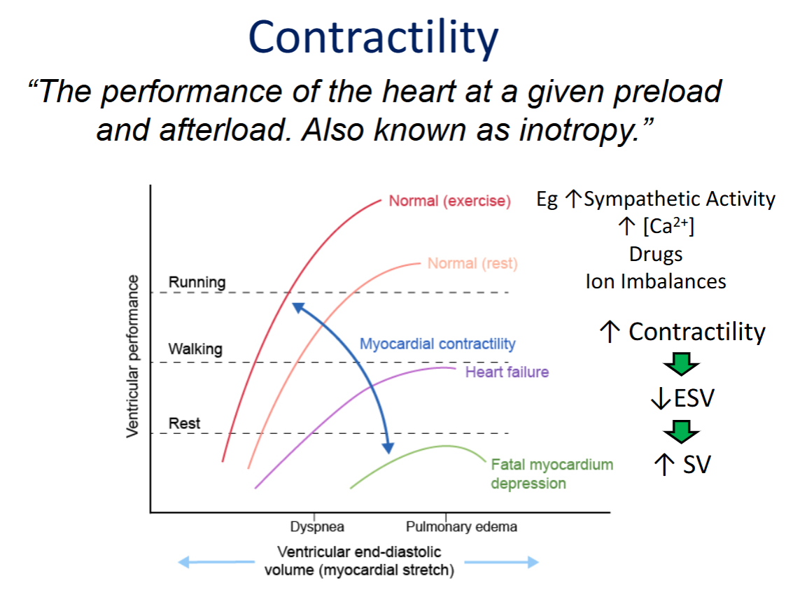
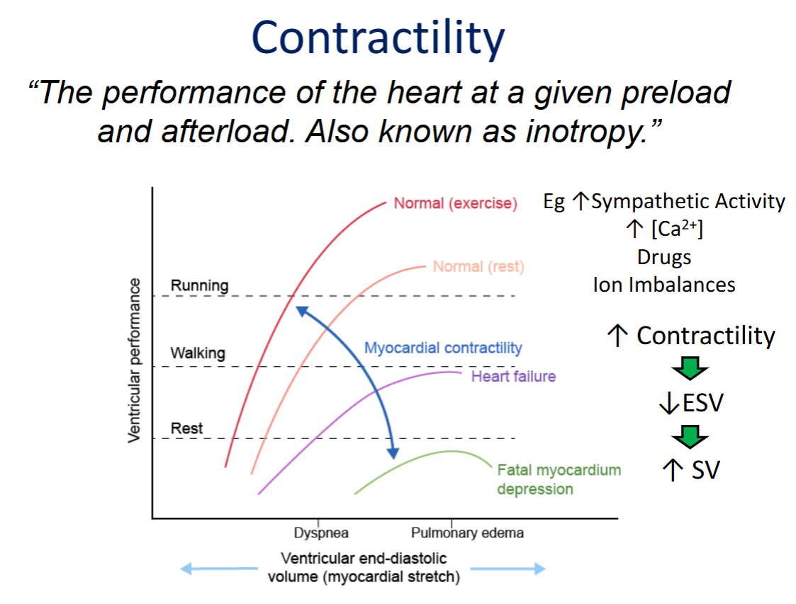
Graphical Representation: Changes in contractility shift the entire Frank-Starling curve up or down (creating a family of curves).
Increased Contractility (e.g., Exercise):
Shifts curve UP → same preload yields a more powerful contraction.
Results in ↓ ESV and ↑ SV.
Decreased Contractility (e.g., Heart Failure / Myocardial Depression):
Shifts curve DOWN → same preload yields a weaker contraction.
Results in ↑ ESV and ↓ SV.
how are changes in contractility represented on the Frank-Starling curve, and how do they affect ESV and SV
Definition: The amount of pressure the heart must generate/exert to eject blood during ventricular contraction.
Isovolumetric Contraction Phase: Ventricular pressure rises, but blood cannot leave because aortic pressure is higher.
Ejection Trigger: Once ventricular pressure exceeds aortic pressure, the aortic valve opens and ejection begins.
what is afterload, and what must occur during isovolumetric contraction for ejection to begin
Causes of ↑ Afterload: Hypertension, valve pathology, aortic plaques/atherosclerosis, and aortic stenosis (narrowing).
Effects of ↑ Afterload:
Heart works harder to eject blood → ↑ ESV and ↓ SV.
Shifts SV vs. LVEDP curve down and to the right.
Effects of ↓ Afterload:
Blood ejects more easily → ↓ ESV and ↑ SV.
Shifts SV vs. LVEDP curve up and to the left.
what causes increased afterload, and how do shifts in afterload affect ESV, SV, and the Frank-Starling curve
Primary Drivers:
Preload: Primarily determines End-Diastolic Volume (EDV) by controlling ventricular filling.
Afterload & Contractility: Primarily determine End-Systolic Volume (ESV) by controlling how much blood remains after contraction.
Integrated Dynamic Control: In everyday physiological responses (e.g., exercise), HR, preload, contractility, and afterload all adjust simultaneously, granting a powerful range for modulating total cardiac output.
how do preload, afterload, contractility, and heart rate work together to determine cardiac output
Formula: EF (%)=SV/EDV
Normal Resting Value: ∼55–70% (meaning ∼30–40% of blood remains in the ventricle after each contraction, which is entirely normal).
Effect of Exercise/Training: Increases overall beat size (larger EDV and SV) but does NOT change EF—EF is a fixed structural/functional feature rather than a fitness metric.
Pathology: Can drop drastically in severe disease states (e.g., dropping to ∼14% in end-stage heart failure prior to transplantation).
what is ejection fraction (EF), its equation, its normal resting range, and how does it respond to exercise vs. severe disease
Definition: The force exerted by circulating blood against the walls of the arteries as the heart pumps.
Equation: BP=CO×TPR
Total Peripheral Resistance (TPR): The net amount of constriction or "squeeze" throughout the systemic blood vessels.
Vasodilation: Decreases (↓) TPR.
Vasoconstriction: Increases (↑) TPR.
what is the definition of blood pressure (BP), what is its underlying equation, and what does total peripheral resistance (TPR) represent
Normal Value: Approximately 120/80 mmHg.
Measurement Location: Measured in a large elastic artery close to the heart (e.g., the brachial artery).
what is normal systemic blood pressure, and where in the circulation is this specific value measured
Highest Pressure: Near the heart in the large elastic arteries.
Progressive Fall: Pressure drops continuously along the pathway:
Large Arteries → Arterioles → Capillaries → Venules → Veins → Vena Cava.
Significant Drop Site: A major pressure drop occurs across the capillaries, which is critical for regulating capillary exchange and preventing tissue damage.
how does blood pressure change as blood travels from the heart through the entire
Locations & Purpose:
Aortic Arch: Measures pressure directly at the source (as blood leaves the heart).
Carotid Sinus: Measures pressure going to the brain (protects against cerebral ischemia).
Detection Mechanism: Arterial wall stretch opens ion channels to generate an electrical signal proportional to blood pressure.
Tonic Activity: Baroreceptors have a steady baseline firing rate at rest and modulate firing rate up or down (acting like a dial rather than an on/off alarm).
where are arterial baroreceptors located, why are they placed there, and how do they detect blood pressure changes
When BP Rises (↑ BP):
Firing: ↑ Baroreceptor firing to the medulla.
Reflex: ↑ Parasympathetic (Vagus/CN X), ↓ Sympathetic.
Effect: ↓ HR, ↓ Contractility, and Vasodilation (↓ TPR).
Net Result: ↓ CO and ↓ TPR→ BP drops back to normal.
When BP Falls (↓ BP):
Firing: ↓ Baroreceptor firing to the medulla.
Reflex: ↓ Parasympathetic, ↑ Sympathetic.
Effect: ↑ HR, ↑ Contractility, and Vasoconstriction (↑ TPR).
Net Result: ↑ CO and ↑ TPR→ BP rises back to normal.
how does the arterial baroreflex arc respond to an increase vs a decrease in blood pressure to restore homeostasis
Definition: The baseline level of constriction maintained by blood vessels at rest.
Neurotransmitter & Receptors: Sympathetic nerves continuously release noradrenaline (NE) onto α-receptors on vascular smooth muscle.
Control Mechanism:
↑ NE release / ↑ Signal rate: Vessel constricts.
↓ NE release / ↓ Signal rate: Vessel dilates.
Analogy: Operates like a volume knob set in the middle at rest, allowing for easy adjustment up (constriction) or down (dilation).
What is vascular tone, which neurotransmitter and receptors regulate it, and how does sympathetic signal rate control vessel diameter
transcytosis:
mechanism: endocytic vesicles carry large, lipid-insoluble molecules (e.g., insulin) across capillary endothelial cells via facilitated, tightly regulated transport; used by a very small number of molecules
diffusion:
mechanism: passive solute movement down concentration gradients; continuous process, but relatively slow on its own
bulk flow/filtration:
mechanism: passive, rapid movement of fluid carryigng dissolved substances; driven by the balance of hydrostatic and osomotic pressure gradients across the capillary wall
what are the three mechanisms of capillary exchange, and how each function to move substances across the capillary wall
Blood Hydrostatic Pressure (BHP): ∼35 mmHg (arterial end) →∼16 mmHg (venous end) | Favours Filtration (pushes fluid out).
Interstitial Fluid Osmotic Pressure (IFOP): ∼1 mmHg | Favours Filtration (pulls fluid out).
Blood Colloid Osmotic Pressure (BCOP): ∼26 mmHg (constant) | Favours Reabsorption (pulls fluid in).
Interstitial Fluid Hydrostatic Pressure (IFHP): ∼0 mmHg | Favours Reabsorption (pushes fluid in).
what are the four Starling forces that determine capillary bulk flow, their abbreviations, typical values, and whether they favour filtration or reabsorption
Why BCOP Stays Constant: Plasma proteins (mainly albumin) are too large to cross the capillary wall under normal conditions, remaining in the vessel to exert a steady, constant inward "pull."
Why BHP Falls: It follows the physical pressure gradient generated by the heart, running "downhill" from the arterial end (∼35 mmHg) to the venous end (∼16 mmHg)
Why does Blood Colloid Osmotic Pressure (BCOP) remain constant along the capillary while Blood Hydrostatic Pressure (BHP) falls
Formula: NFP=(BHP+IFOP)−(BCOP+IFHP)
Arterial End:
Calculation: (35+1)−(26+0)=+10 mmHg
Net Effect: Filtration (fluid moves out of capillary to deliver O2 and nutrients).
Venous End:
Calculation: (16+1)−(26+0)=−9 mmHg
Net Effect: Reabsorption (fluid moves back into capillary to pick up CO2 and waste).
Key Transition: BHP drops from 35→16 mmHg along the capillary while BCOP stays constant at 26 mmHg, shifting the net force from pushing fluid out to pulling fluid back in.
What is the formula for Net Filtration Pressure (NFP), and how do the pressures, values, and net effects differ at the arterial vs. venous ends of a capillary
Daily Volume Balance:
Filtered Out: ∼20 L/day
Reabsorbed: ∼17 L/day (∼85% reabsorption efficiency).
Why Values Don't Match: Arterial filtration force (+10 mmHg) slightly outweighs venous reabsorption force (−9 mmHg).
Lymphatic Role: The remaining ∼3 L/day of leftover interstitial fluid is passively drained by the lymphatic system and returned to the circulation using hydrostatic/osmotic gradients.
What is the daily fluid filtration vs. reabsorption balance across capillaries, and what role does the lymphatic system play
Anatomical Layout: Lymph vessels sit directly alongside capillary beds in a parallel arrangement (arteriole on one side, venule on the other, lymph vessel in the middle).
Porosity: Lymph vessels are more porous than standard blood capillaries.
Fenestrations: Their large openings (fenestrations) allow large macromolecules (e.g., plasma proteins) to enter, not just fluid
How are lymphatic vessels arranged anatomically relative to capillary beds, and how does their porosity compare to blood capillaries
Fluid Clearance: Clears the leftover ~3 L/day of interstitial fluid that is not reabsorbed at the venous end of capillaries.
Waste Clearance: Drains waste macromolecules that are too large to re-enter blood capillaries.
Circulatory Return: Returns this collected fluid (lymph) back into the venous circulation to maintain blood volume
What is the main physiological function of the lymphatic system regarding interstitial fluid balance
The Exception: The brain lacks conventional lymphatic drainage despite being the most metabolically active organ.
Alternative System: It relies on a specialized clearance pathway known as the glymphatic system to clear interstitial fluid and metabolic waste.
What major organ lacks conventional lymphatic vessels, and how does it manage fluid/waste clearance instead
Hypertension (↑ Arterial BHP): Shifts the balance toward filtration by pushing more fluid out on the arterial side, making full reabsorption harder.
Kidney Disease (↓ BCOP): Protein loss in urine (proteinuria) lowers plasma protein concentration, reducing the inward osmotic "pull."
Heart Failure (↑ Venous BHP & ↑ IFOP): Fluid retention and venous stretching increase venous pressure and capillary permeability, strongly favouring fluid buildup in tissues.
Long-Haul Travel (Local Pressure): Venous compression from sitting limits venous return, causing local blood pooling and increased venous pressure in the lower legs.
How do hypertension, kidney disease, heart failure, and long-haul travel each alter capillary pressure gradients to produce oedema
Problem with "In Series": Each organ would deplete a portion of the ~35 mmHg pressure gradient (e.g., dropping to 16 mmHg), leaving insufficient pressure for downstream organs to undergo capillary exchange.
Solution ("In Parallel"): All organs receive the same ~35 mmHg arterial pressure gradient independently, ensuring equal and adequate driving pressure for exchange regardless of changes in other organs.
Circuit Analogy: Functions like a parallel electrical circuit where pressure drop equals voltage drop, blood flow equals current, and vascular resistance equals electrical resistance (F=ΔP/R).
Why is the systemic circulation arranged "in parallel" rather than "in series"
Relationship: Velocity is inversely proportional to cross-sectional area (Velocity ∝ 1/Cross-Sectional Area).
Aorta: Small area (∼3–5 cm²) → fast blood velocity.
Capillaries: Massive total area (∼5000 cm², ∼1000× greater than aorta) → blood slows dramatically (∼0.1 cm/sec).
Functional Importance: Slow velocity gives necessary time for diffusion, filtration, and reabsorption to occur across the capillary wall.
This is why capillary beds are so anatomically dense — corrosion casts (e.g. of the brain) show vessels packed so tightly there's barely room for anything else. Result: every cell in the body is within ~2 cells of a capillary.
How does the total cross-sectional area of capillaries compare to the aorta, and why is blood flow velocity in capillaries so slow
Strategy 1: Increase Cardiac Output (↑ CO)
Mechanism: Increases HR and/or SV.
Drawback: Energetically expensive (forces the heart to work harder overall).
Strategy 2: Redirect Existing Cardiac Output (Preferred)
Mechanism: Localized vasoconstriction and vasodilation.
Advantage: Energetically "cheaper" and more efficient; used continuously in everyday life without requiring total flow changes.
What are the two main strategies the body uses to increase blood flow to a specific organ, and which strategy is preferred
Redirection Example: If total CO is fixed at 4 L/min across 4 organs (1 L/min each), constricting the GI supply vessel to 0.25 L/min during exercise frees up 0.75 L/min to redistribute to active organs (1.25 L/min each). Total CO remains 4 L/min.
Local vs. Systemic Control: Local redirection constricts one vascular bed while dilating another.
Effect on BP/TPR: Constriction in one region offset by dilation elsewhere does not necessarily increase TPR or systemic BP—it depends entirely on whether the net global resistance changes.
How does local blood flow redirection work (with an example), and how does it differ from systemic BP/TPR changes
Pressure Gradient (ΔP): Flow∝ΔP (roughly linear relationship).
Vessel Radius (r): Flow∝r4 (flow is proportional to the fourth power of the radius).
Key Takeaway: A small change in vessel radius causes a massive change in blood flow.
What are the two key proportional relationships described by Poiseuille's Law regarding blood flow, pressure gradient, and vessel radius
Two Modifiable Variables:
Pressure Gradient (ΔP): Altered via cardiac output or overall vascular squeeze (only has a linear/proportional effect).
Vessel Diameter (r): Altered via arteriole constriction or dilation.
Why Radius is Preferred: Because Flow∝r4, small adjustments to arteriole diameter provide exceptionally powerful and precise local control over blood flow without needing to alter systemic pressure gradients
What two variables can the body modify to control blood flow to an organ, and why does it predominantly rely on adjusting vessel diameter
Definition & Location: Small bands of smooth muscle located at the entrance to individual capillaries branching off a metarteriole.
Function when Relaxed (Normal): Allows blood to flow freely through the entire capillary bed.
Function when Contracted: Constricts to reduce or redirect blood flow away from that specific capillary bed (e.g., diverting flow away from the gut to active muscles during exercise)
What are precapillary sphincters, where are they located, and how do they function when relaxed versus contracted