Circulatory System Diagram | Quizlet
1/99
There's no tags or description
Looks like no tags are added yet.
Name | Mastery | Learn | Test | Matching | Spaced | Call with Kai | Chat |
|---|
No analytics yet
Send a link to your students to track their progress
100 Terms
What system is responsible for circulating and transporting nutrients, O2, CO2, hormones, and red blood cells throughout the body?
circulatory system
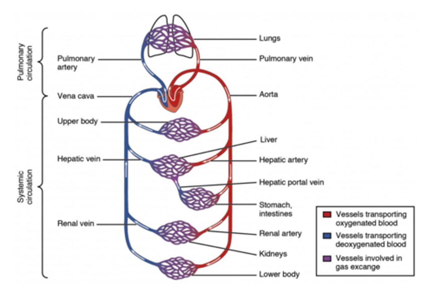
What is the path of blood flow from the heart to tissues?
aorta ->
arteries ->
arterioles ->
capillaries

What is the path of blood flow from the tissues to the heart?
capillaries ->
venules ->
veins ->
superior/inferior vena cava

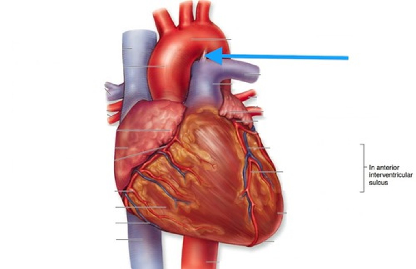
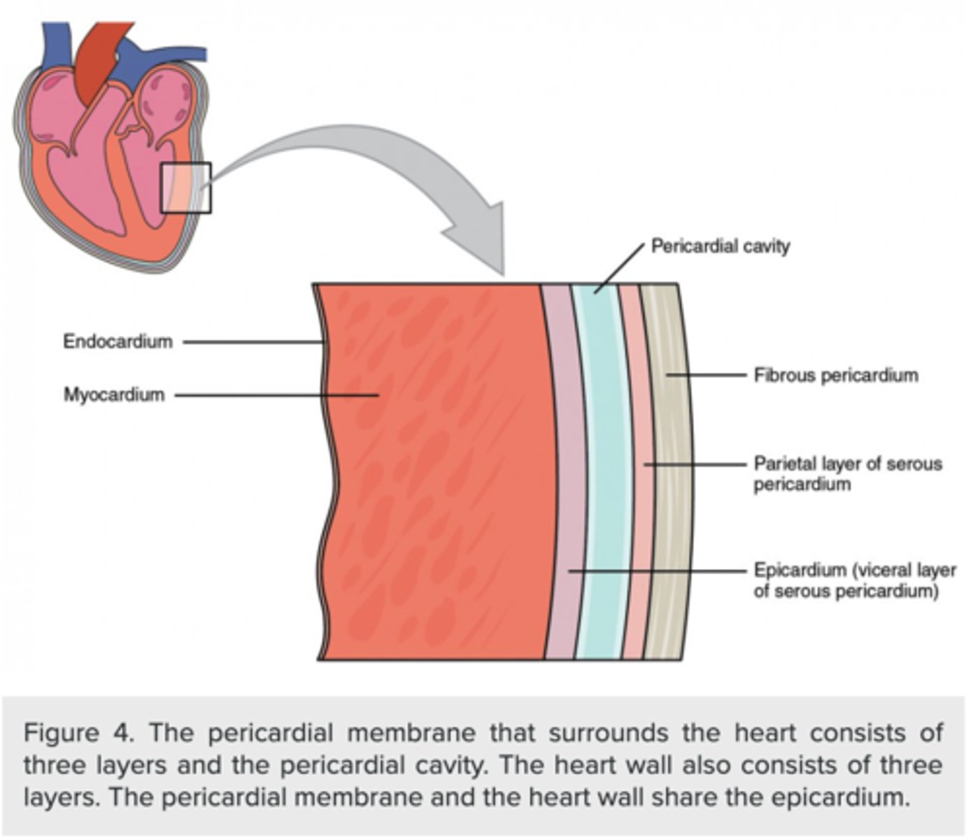
what is the fluid-filled sac that surrounds the heart in order to protect and lubricate it for proper function?
pericardium
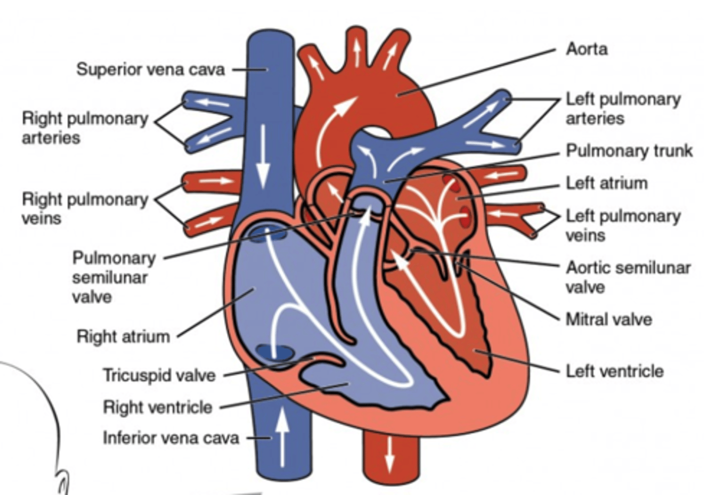
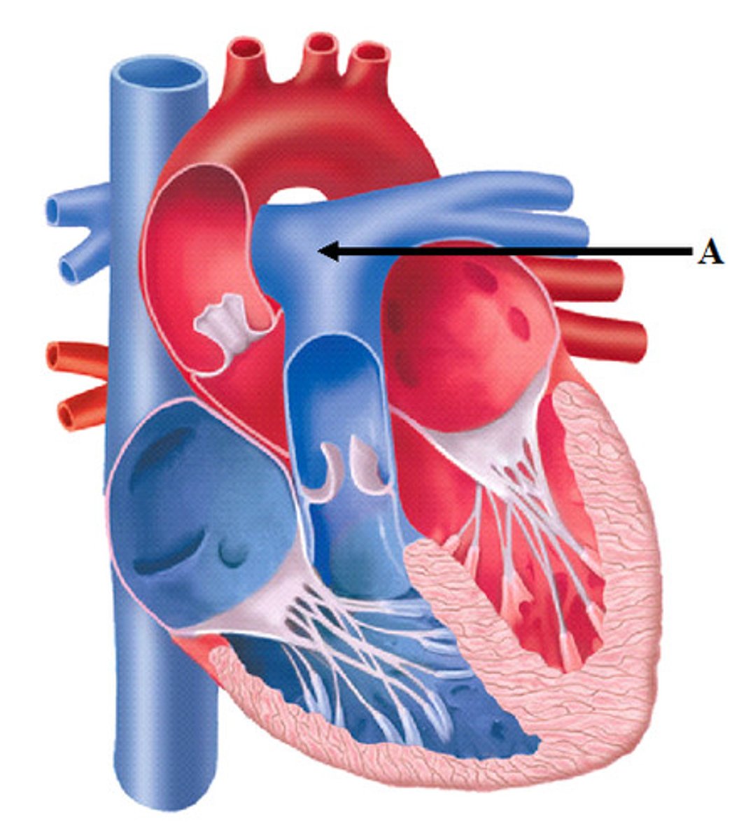
What is the heart chamber where deoxygenated blood enters via the superior and inferior vena cava?
right atrium

What is the heart chamber where blood enters from the right atrium and exits through the pulmonary artery?
right ventricle

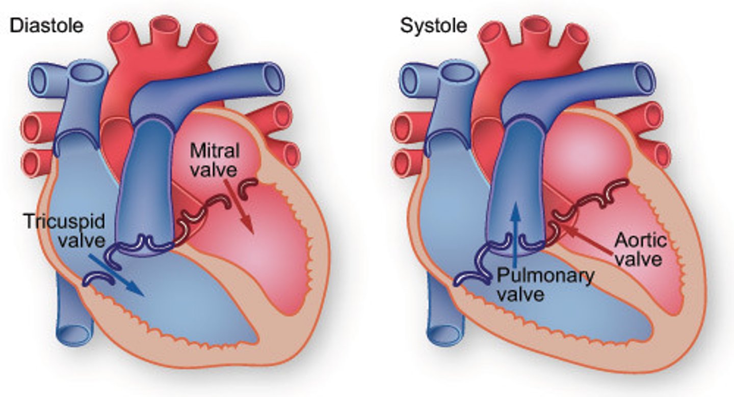
From what valve does blood blood enter the right ventricle?
right AV valve (atrioventricular valve)
(AKA: tricuspid)

Through what valve does blood exit the right ventricle?
pulmonary semilunar valve

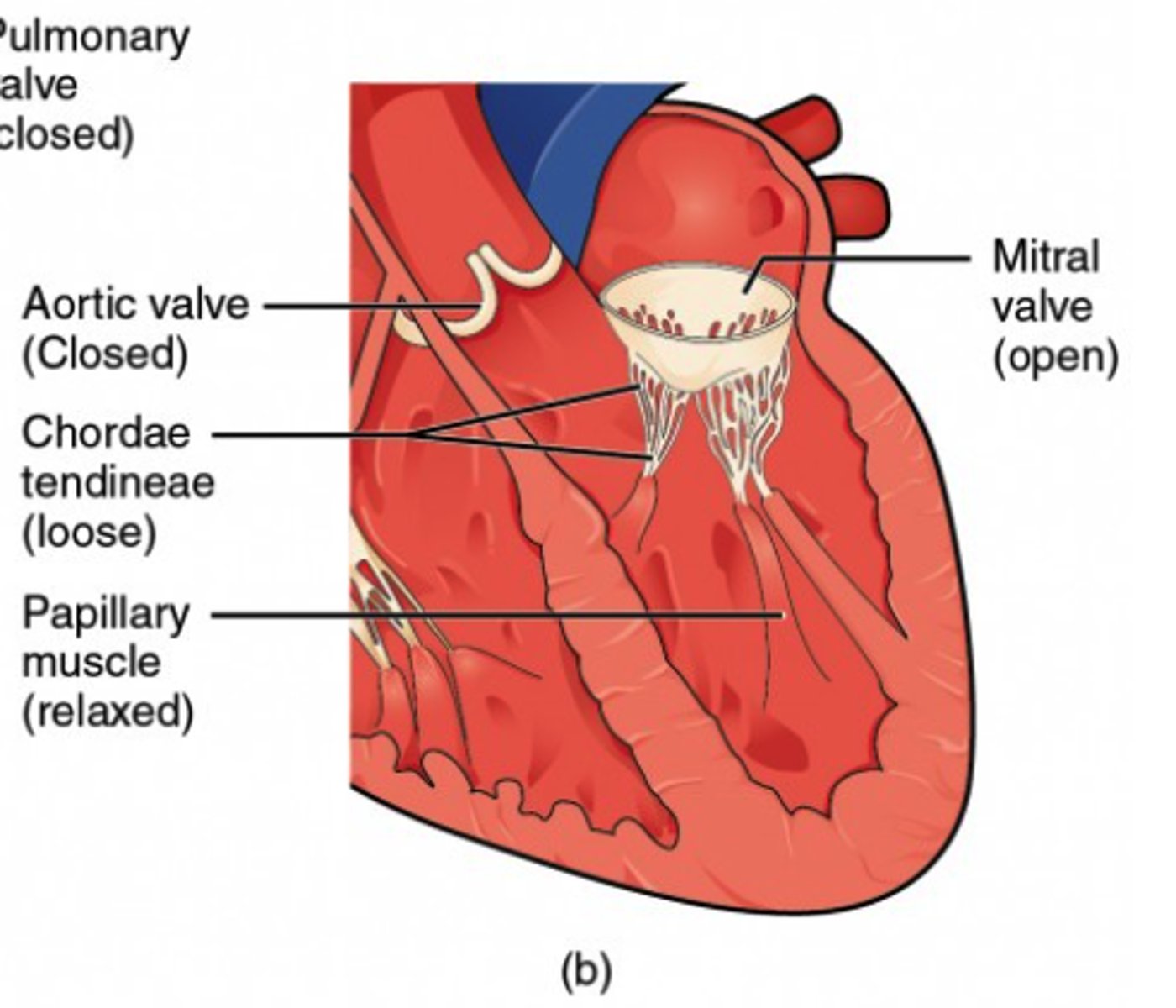
When does the right AV valve close?
when the right ventricle contracts
(Note: prevents back flow into right atria - it produces the "lub" sound)
When does the pulmonary semilunar valve close?
when the right ventricle relaxes
(Note: prevents back flow into right ventricle - it produces the "dub" sound)
What describes the blood pathway from the right side of the heart to the lungs, ending at the left side of the heart?
pulmonary circuit
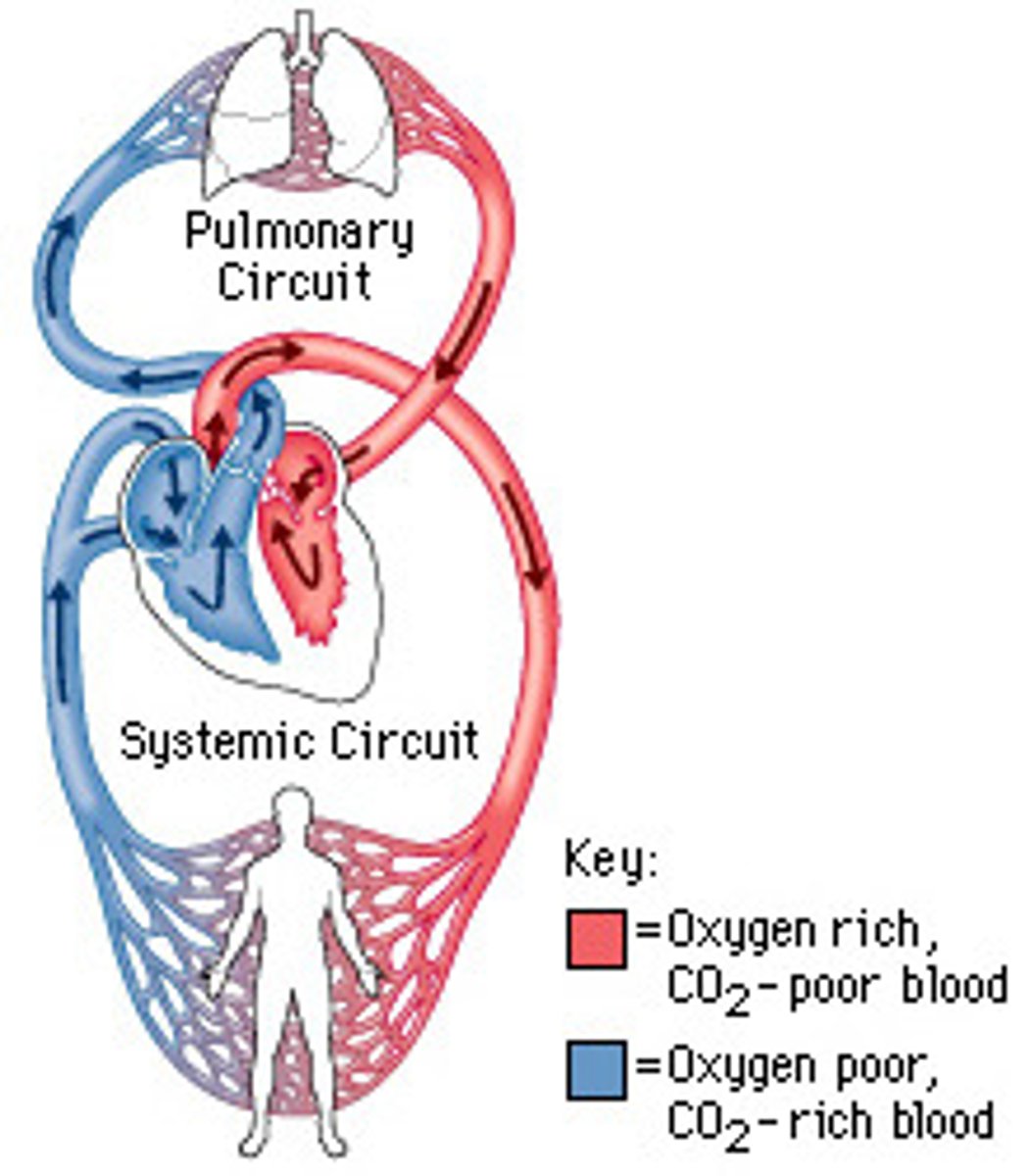
What is the path of blood flow in the pulmonary circuit?
right ventricle -> pulmonary arteries -> arterioles -> capillaries of the lungs -> venules -> veins -> pulmonary veins -> left atrium
What describes the blood pathway from the left side of the heart to the right side of the heart?
systemic circuit

What is the heart chamber where blood enters from the pulmonary veins?
left atrium

What is the heart chamber where blood enters from the left atrium and exits through the aorta?
left ventricle

What valve does the blood enter the left ventricle?
left AV valve
(AKA: mitral/bicuspid valve -
prevents back-flow
into the left atrium )

What valve does the blood exit the left ventricle?
aortic semilunar valve
(Note: prevents back-flow
into left ventricle)

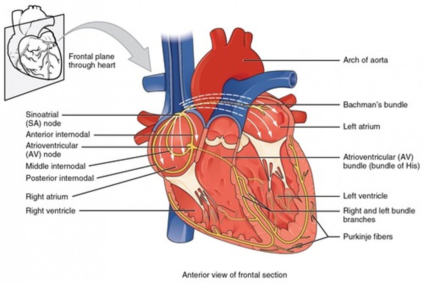
Where is the SA node located?
upper wall of the right atrium
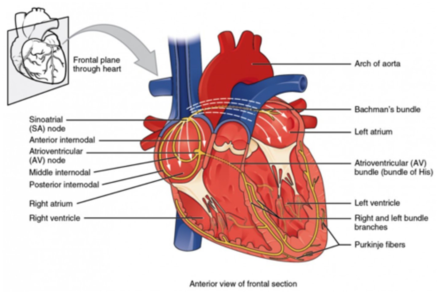
Of what units is the SA node composed?
specialized cardiac muscle cells
What does the SA node initiate first when it starts a heart contraction?
initiate the contraction of both atria

What is the SA (sinoatrial node) node also known as?
pacemaker of the heart
What is the path of the SA node's impulse?
SA node (atrial contraction) ->
AV node ->
Bundle of His ->
purkinje fibers (ventricular contraction)
Why is the impulse briefly delayed at the AV node?
to allow the atria to completely empty, allowing ventricles to fill with blood.
How does the impulse travel through cardiac muscles?
gap junctions
Is the pace of the SA node faster or slower compared to the normal heartbeat?
faster
What slows down the SA node pace to stimulate the heart at a comfortable pace?
vagus nerve (parasympathetic nervous system)
(Note: the point of vagus nerve innervation is to allow the heart rate to be both increased and decreased- creates a sort of midpoint)
Where is the AV node located?
lower wall of the right
atrium/interatrial septa

When the ventricles contract, what structures is blood forced through?
pulmonary arteries and aorta
What structures are attached to cardiac valves and force them closed during systole?
papillary muscles and
chordae tendinae
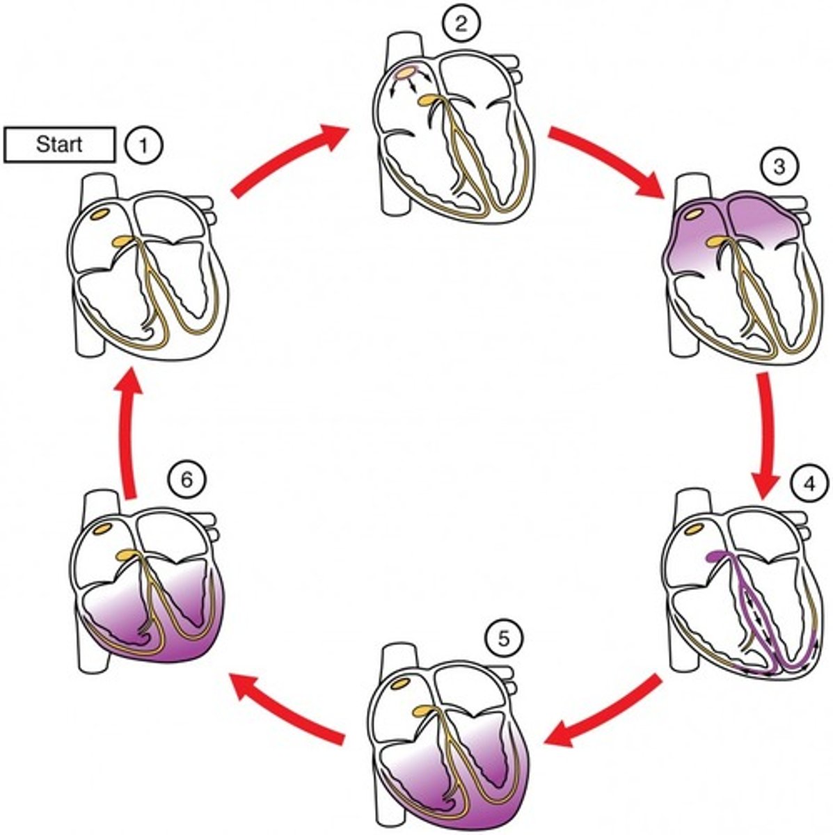
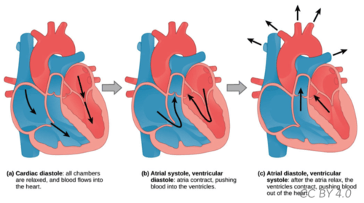
What is diastole?
filling of the heart
What is systole?
contraction of the heart

Which ventricle is thicker?
left
(Note: it is thicker b/c
it pumps blood to extremities)
What are the two semilunar valves?
aortic and
pulmonary
(Note: prevent back
flow into ventricles)
What are the two atrioventricular valves?
- tricuspid/right AV valve
- bicuspid/left AV valve

What force causes blood to move through the arteries?
hydrostatic pressure
Blood pressure drops as it reaches the _______, and reaches zero in the ________.
capillaries; venules
What are the three processes that push blood through the veins?
1. pumping of heart/movements of skeletal muscle
2. expansion of atria during each beat
3. falling pressure in chest due to breathing
(Note: veins have thinner walls and less smooth muscle compared to arteries, so they need these factors to move blood through)
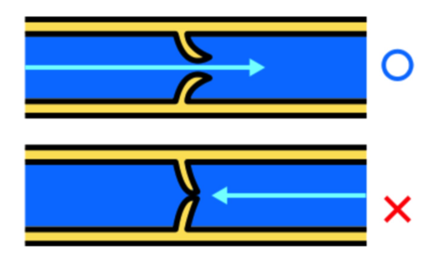
What structures prevent back flow in veins?
valves
(Note: because the
pressure is so low,
valves in the central
veins are necessary.
Otherwise, blood would
collect in the extremities)
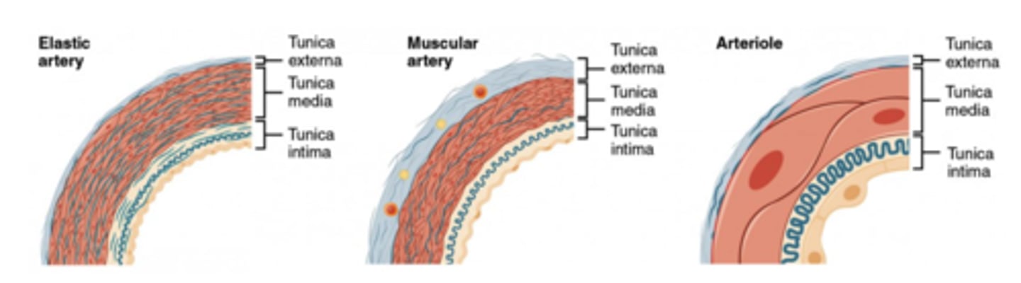
What are thick-walled, muscular, elastic vessels that pump oxygenated blood away from the heart?
arteries
What branch of the nervous system typically innervates arteries?
sympathetic nervous system
What is the one exception to the rule that arteries alway pump oxygenated blood away from the heart?
pulmonary arteries
transport deoxygenated
blood away from heart to lungs
Do large arteries have more or less smooth muscle (per volume) than medium-sized arteries?
less
Are larger arteries more or less affected by the sympathetic nervous system than smaller arteries?
less
(Note: allows for the constriction of medium sized arteries to reroute blood)
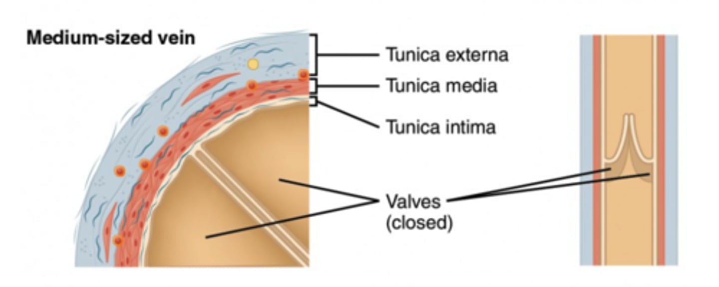
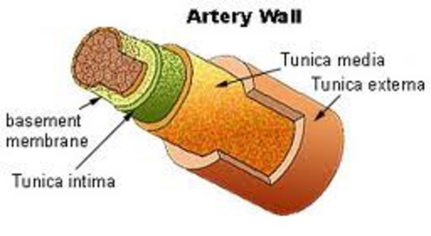
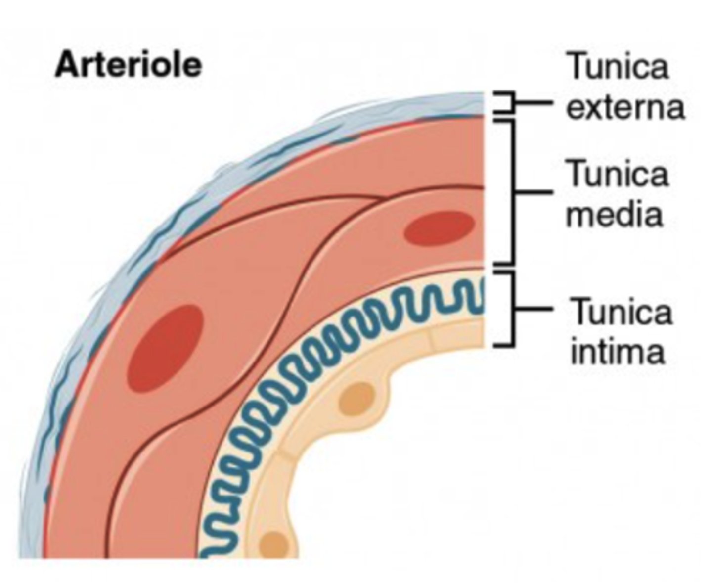
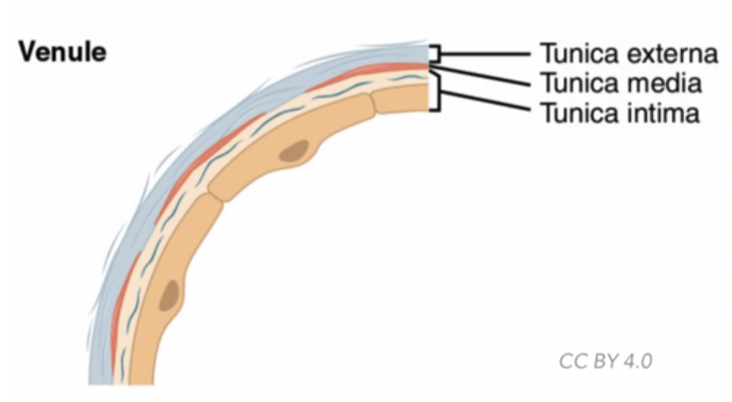
What are the three layers (tunics) of arteries?
1. endothelial lining (inner)
2. smooth muscle and elastic tissue (middle)
3. Connective tissues (outer)
(Note: the technical names are tunica
intima, tunica media, and tunica extrema,
respectively)
What are very small vessels wrapped in smooth muscle, and constrict or dilate to regulate blood pressure or reroute blood?
arterioles

What vessels are a major determinant of blood pressure as they have the greatest resistance to blood flow
arterioles
What vessels have the smallest diameter and have a single layer of endothelial cells?
Capillaries
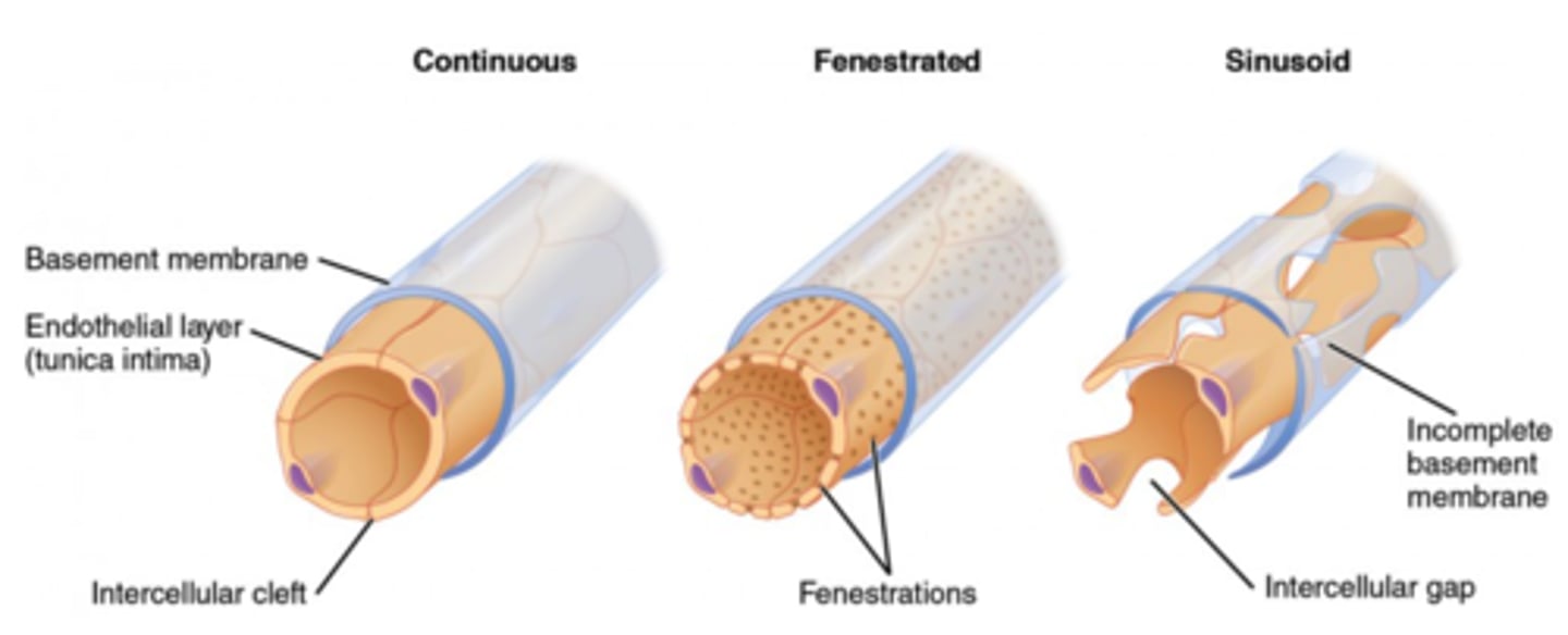
Why are capillaries single layered?
allows gas, nutrients, enzymes,
hormones, and waste to diffuse

What are the four methods for materials to cross the capillary wall?
1. endo/exocytosis (protein)
2. Diffusion through capillary cell membrane (O2/CO2)
3. movement through fenestrations
4. movement through space between cells (ions)
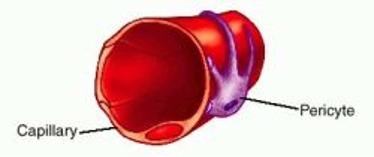
What are the contractile cells you will sometimes see around the the capillaries and venules throughout the body?
pericytes
With what substance are the capillaries technically exchanging nutrients and waste?
interstitial fluid
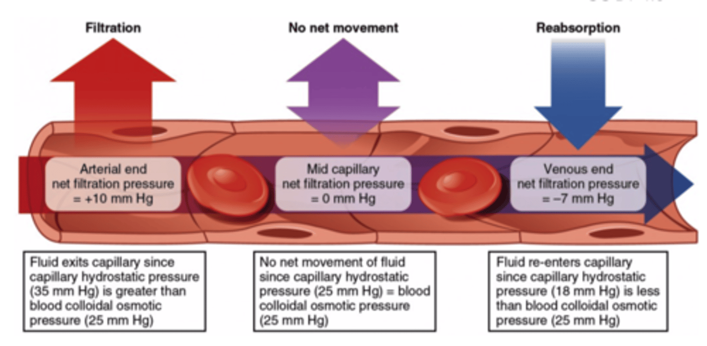
What are the two factors to consider to know where what kind of exchange occurs in capillaries?
1. The blood hydrostatic pressure (pressure of blood pushing out) - out
2. The blood colloid osmotic pressure (proteins in blood pull water into capillary) - in
(Note: tissues also has these pressures, but they are negligible)
What kind of exchange dominates on the arterial end of a capillary?
filtriation
(Note: Blood hydrostatic
pressure overcomes Blood
colloid osmotic pressure)

What kind of exchange dominates at the venous end of a capillary?
reabsorption
(Note: Blood colloidal
osmotic pressure
overcomes the other forces)
What are small blood vessels that lead back to veins and are very thin and porous?
venules
what are larger vessels that carry deoxygenated blood back to the heart?
veins
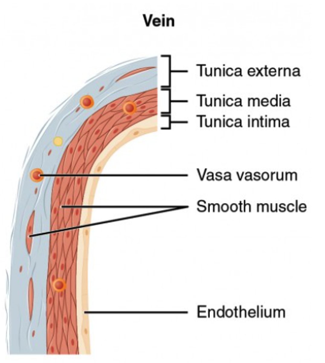
Why do some larger veins have valves?
prevent back-flow specifically
from fighting the force of gravity
(Note: one-way valve)
How do veins compare to arteries in cross sectional area?
veins are much larger (about 4x)
(Note: capillaries contain total cross sectional area greater than arteries and veins combined)
What is blood velocity inversely proportional to?
total cross sectional area
(Note: smaller blood vessels = slower blood velocity)
Why does blood pressure drop from the aorta to the capillaries?
increased resistance and decreased vessel diameter
Given the formula for blood pressure:
Blood pressure = cardiac output*total peripheral resistance, why does pressure decrease going from the aorta to the capillaries?
the blood pressure before the narrower vessel is higher, BUT, the pressure after the constriction is lowered.
(Note: by the time we hit the venules/veins, the original source of blood pressure/flow is essentially gone.
What vessels have the greatest resistance to flow?
arterioles
(Note: high ability to constrict)

At any given time, where is most blood located?
veins/venules/venus sinuses
What is the system that is an open secondary circulatory system that transports excess interstitial fluids?
lymphatic system
How does lymph move through the lymphatic system?
contraction of adjacent muscles, and the smooth muscle in some of the walls of the larger lymph vessels.
How are proteins and large particles that cannot be taken up by capillaries removed?
lymphatic system
(allows the lymphatic system to monitor blood for infections)
Where does the lymphatic system transport absorbed fat from the small intestines?
to the blood
What are the two major lymphatic ducts in the shoulder region that prevent back flow?
1. thoracic duct
2. right lymphatic duct
(Note: empty into the left and right subclavian vein, respectively. Joins blood as plasma)
What cells exist in the lymph nodes that filter the lymph and serve as immune response centers?
leukocytes
(Note: swollen glands during a sickness are lymph nodes filled with white blood cells)
What are the primary central lymphoid organs?
1. thymus
2. bone marrow
(Note: replenish specific immune cells)
What immune cells does the thymus replenish?
T-Cells
(Note: think T = Thymus)
What immune cells does the bone marrow replenish?
B-cells
(Note: think B = Bone marrow)
What are the primary peripheral lymphoid tissues?
1. tonsils
2. adenoids
3. lymph nodes
4. appendix
5. peyer's patches (in small intestine)
6. spleen
(Mnemonic: TALAPS)
What do peripheral lymphoid tissues do with immune system cells?
they house immune system cells, but they cannot replenish them
The thymus technically doesn't make new T-cells, but T-cells _______ there so it houses fresh ones.
mature
What volume of blood does the human body carry?
4-6 liters
How many liters of blood does the heart pump per day?
~7000 L
What is the ratio of liquid (plasma) components of blood and cellular components of blood?
55% liquid (plasma) and 45% cellular
What is an aqueous mixture of nutrients, salts, gases, wastes, hormones, and blood proteins?
plasma
What substance is the same as plasma minus any clotting factor components?
blood serum
What are red blood cells (RBCs) also called?
erythrocytes
What do RBCs transport?
oxygen
(Note: via hemoglobin)
What reaction do RBCs catalyze?
conversion of CO2 to H20 and H2CO3
(Note: via carbonic anhydrase)
How do RBCs maximize hemoglobin content?
they lack nucleus and organelles
RBCs do not undergo ______.
mitosis
What substance enables RBCs to resist strong shearing forces?
spectrin
What is the process of blood clotting?
1. Formation of platelet plug
2. release thromboplastin
3. conversion of prothrombin to thrombin
4. conversion of fibrinogen to fibrin
5. clot formation
How is the platelet plug formed?
platelets contact exposed collagen of injury and induce other platelets to form a plug
What is thromboplastin called?
clotting factor
What structures release thromboplastin?
both platelets and damaged tissue
What is the function of thromboplastin?
converts inactive plasma protein prothrombin to thrombin (active form)
What is the function of thrombin?
thrombin converts fibrinogen to fibrin
How is the blood clot formed?
fibrin threads coat damaged area and trap blood cells
What is a blood clot that forms in a vessel abnormally?
thrombus
(Note: can cause heart attack - if in the heart - or stroke - if in nervous tissue in brain)
In fetal circulation blood from the placenta flows to the fetus through what vessel?
the umbilical vein
In fetal circulation, what are the two paths the blood can go after the umbilical vein.
1. ductus venosus
2. liver/portal vein
(Note: 1/2 of blood goes to each route
In fetal circulation, what is the path of blood if the blood goes to the ductus venosus?
ductus venosus -> inferior vena cava -> right atrium -> right ventricle -> ductus arteriosis -> aorta
In fetal circulation, what is the path of blood if it goes to the liver/portal vein
liver/portal vein -> right atrium -> foramen ovale -> left atrium -> left ventricle -> aorta
What is the function of the ductus venosus?
allows fetal blood flow
to bypass the liver
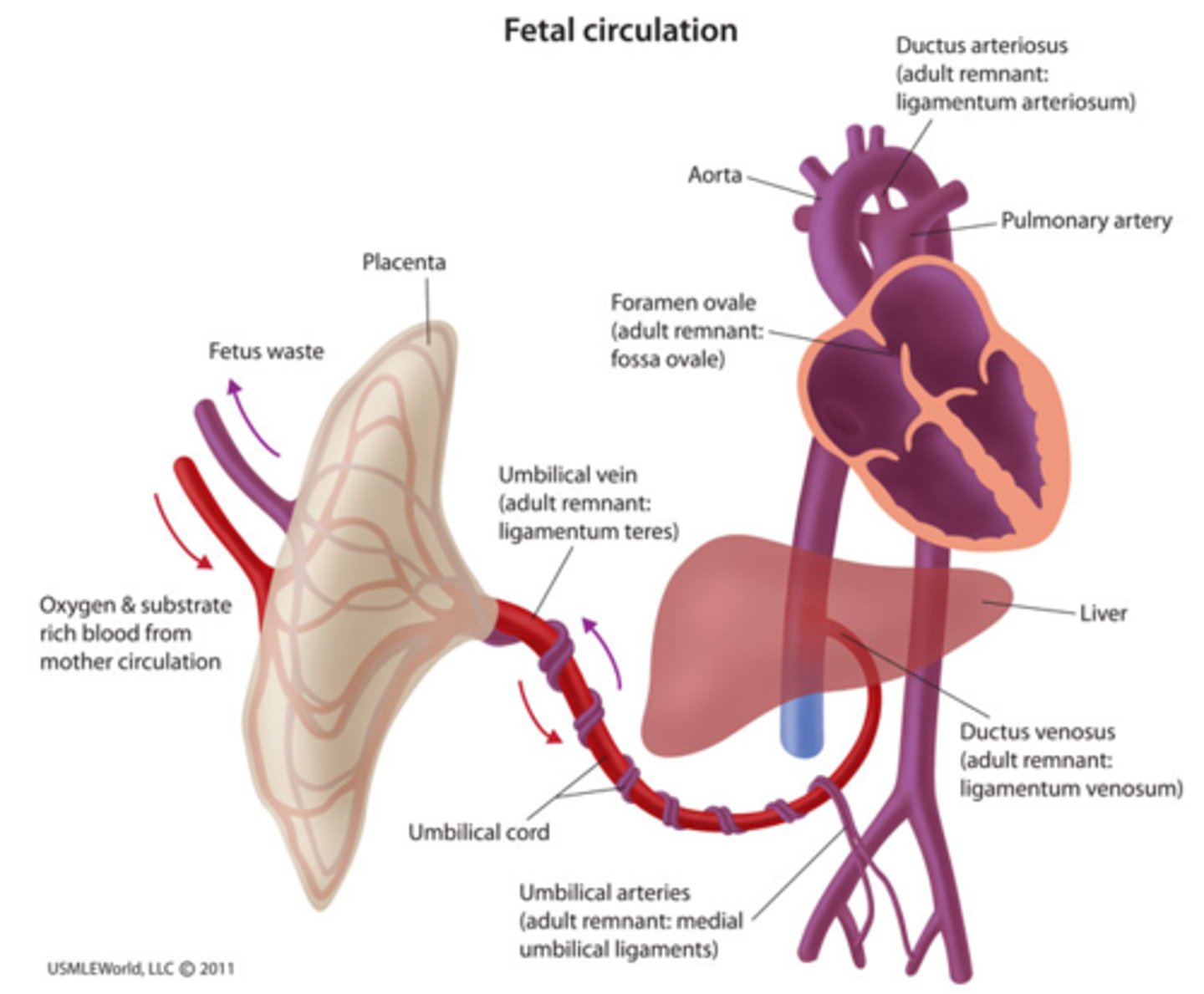
What is the function of ductus arteriosus?
it conducts some blood
from the pulmonary artery
to the aorta (bypassing the
lungs/fetal pulmonary circulation)