Biochem Lecture 2: Protein function
 Call Kai
 Learn
Learn Practice Test
 Spaced Repetition
 Match
Match Flashcards
 Knowt Play
1/34

There's no tags or description
Looks like no tags are added yet.
Name | Mastery | Learn | Test | Matching | Spaced | Call with Kai | Chat |
|---|
No analytics yet
Send a link to your students to track their progress
35 Terms
Ligand
a molecule bound reversibly by a protein
Binding site
the site of the protein that binds to the ligand, it is complementary in size, shape, charge, and hydrophobic/hydrophilic character
Conformational change
shift in 3D shape of a protein when a ligand binds, makes the site more complementary to the ligand, permitting tighter binding
Induced fit
the structural adaptation that occurs between the protein and ligand
Function of Hemoglobin
transports oxygen from lungs to other parts of the body
structure of Hemoglobin
No amino acids can bind to O2 (oxygen is poorly soluble in water) so Hb needs a Heme group. Contains four polypeptide chains (2a, 2b), each subunit has a heme group which can bind to oxygen
Wyman Linkage relationship
if the ligand specifically binds to one of the two states, then equilibrium between those two states will be shifted in favor of the state with bound ligand with the the increase of ligand concentration
T and R states
T-state→ oxygen is absent, low affinity for oxygen. O2 binding triggers a conformational change from T to R. R state→ the pocket narrows between the beta subunits, high affinity for oxygen
Allosteric protein
one in which binding of a ligand to one site affects the binding properties of another site in the same protein
Hill Plots
Hill coefficient nH, nH>1 indicates positive cooperatively
How does Carbon monoxide poisoning occur?
CO has higher affinity for Hb than O2, and Hb will get trapped in the high-affinity state and cannot release the bound O2 into tissues
Hb transport
transports 40% of the total H+ and 15-20% of the CO2 formed in the tissues to the lungs and kidneys. The binding of H and CO2 to Hb is inversely related to the binding of oxygen
Bohr effect
the effect of pH and Co2 concentration on the binding and release of O2 by Hb. (at a low pH (high H+) and high CO2 concentration of peripheral tissues, the affinity of Hb for oxygen decreases as H+ and CO2 are bound, and O2 is released to the tissues. In the capillaries, as CO2 is excreted and the blood pH consequently rises, the affinity of Hb for oxygen increases and the protein bind more O2 for transport to the peripheral tissues)
Antibodies/ immunoglobins Ig
heart of the immune response, the soluble proteins which bind to bacteria, viruses, or large foreign molecules and target them for destruction. Make up 20% of blood protein, humans are capable of producing 100million antibodies with distinct binding specifications
Antigen
any molecule or pathogen capable of eliciting an immune response
Five classes of antibodies
IgA, IgG, IgD, IgE, IgM. IgA found in saliva, tears, and mother’s milk), IgG is the major class of antibody
mAbs
monoclonal antibodies, newly approved innovator drugs. Generally very safe drugs because of their target selectivity, avoids unnecessary exposure to and consequently activity in nontarget organs
Murine Ab
-momab, full mouse antibody
Chimeric Ab
-ximab, some mouse some human. The human Fc region renders a longer in vivo half-life than the parent murine mAb
Humanized Ab
-zumab, mostly human with slivers of mouse. Human mAbs except the CDRs in the variable domain
Human Ab
-mumab, full human

IgG overall structure
contains four polypeptide chains: two large (heavy chains) and two light chains, linked by noncovalent disulfide bonds (Cys residues). The heavy chains interact at one end, and then branch to interact separately with the light chains, forming a Y shaped molecule. Each branch has a single antigen-binding site.
IgG domains
heavy and light chains are made of identifiable domains: constant, sequence and structure constant between IgG’s. Variable domains= with varying amino acid sequences
IgG structure
all beta sheet protein, carbohydrates do not affect the IgG structure and function but stabilize the IgG structure
Antigenic determinant or epitope
each antigen-binding binds only a particular molecular structure within the antigen. Upon binding a conformation change occurs
Phagocytosis
the cellular process by which a cell engulfs and digests large solid particles, such as bacteria, dead cell debris, or foreign matter
When IgG binds to an antigen
activates macrophages to engulf and destroy the invader, receptors on the macrophage surface recognize and bind the Fc region of the IgG
Epitope
an individual molecular structure within an antigen to which an individual antibody binds
Muscle contraction
Interaction of two motor proteins: actin and myosin. Proteins are arranged in filaments that undergo transient interactions and slide past each other to bring about contraction, chemical energy is derived from ATP hydrolysis. Actin and myosin make up 80% of muscle mass
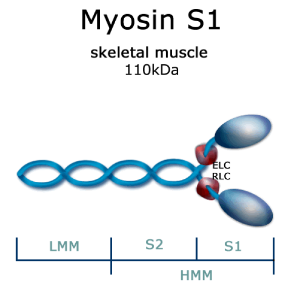
Myosin structure
6 subunits; tow heavy chains, four light chains. Heavy chains are arranged at their C terminus as extended alpha helices, wrapped around each other in fibrous, left-handed coil. It’s N terminus, each chain has a large globular domain containing an ATP hydrolysis site. The myosin head (globular domain) is the motor domain that makes muscle contraction possible

Thick filaments
Muscle cells where myosin aggregates to form structures. Several hundred myosin molecules are arranged with their fibrous tails associated to form a long bipolar structure
Globular and fibrous actin
forms F-actin, every G-acin monomer binds ATP and hydrolysis it to ADP, which helps in the assembly of the filaments. F-actin together with other proteins form thin filaments
actin interacting with myosin
each actin monomer in the thin filament can bind tightly and specifically to one myosin head group. 1) ATP binds to myosin head, causing dissociation from actin 2) as tightly bound ATP is hydrolyzed, a conformational change occurs, ADP and Pi remain associated with the myosin head 3) Myosin head attaches to actin filament, causing release of Pi, 4) Pi release triggers a power stroke, a conformational change in the myosin head that moves actin and myosin filaments relative to one another. ADP is released in the process.
A and I bands, Z disks
A and I: arise from the thick and thin filaments
Z disk: made up of other proteins, is the structure serving as an anchor to which the thin filaments are attached.
Muscle contraction occurs by the slides of the thick and thin filaments past each other so that the Z disks in neighboring I bands draw closer together
Motor proteins convert chemical energy into
kinetic energy