Human Anatomy Exam 5
 Call Kai
 Learn
Learn Practice Test
 Spaced Repetition
 Match
Match Flashcards
 Knowt Play
1/175

There's no tags or description
Looks like no tags are added yet.
Name | Mastery | Learn | Test | Matching | Spaced | Call with Kai | Chat |
|---|
No analytics yet
Send a link to your students to track their progress
176 Terms
pubic crest
ridge just anteromedial to the pubic tubercle that meets the pubic symphysis - attachment point for transverse abdominis m. and rectus abdominis m.
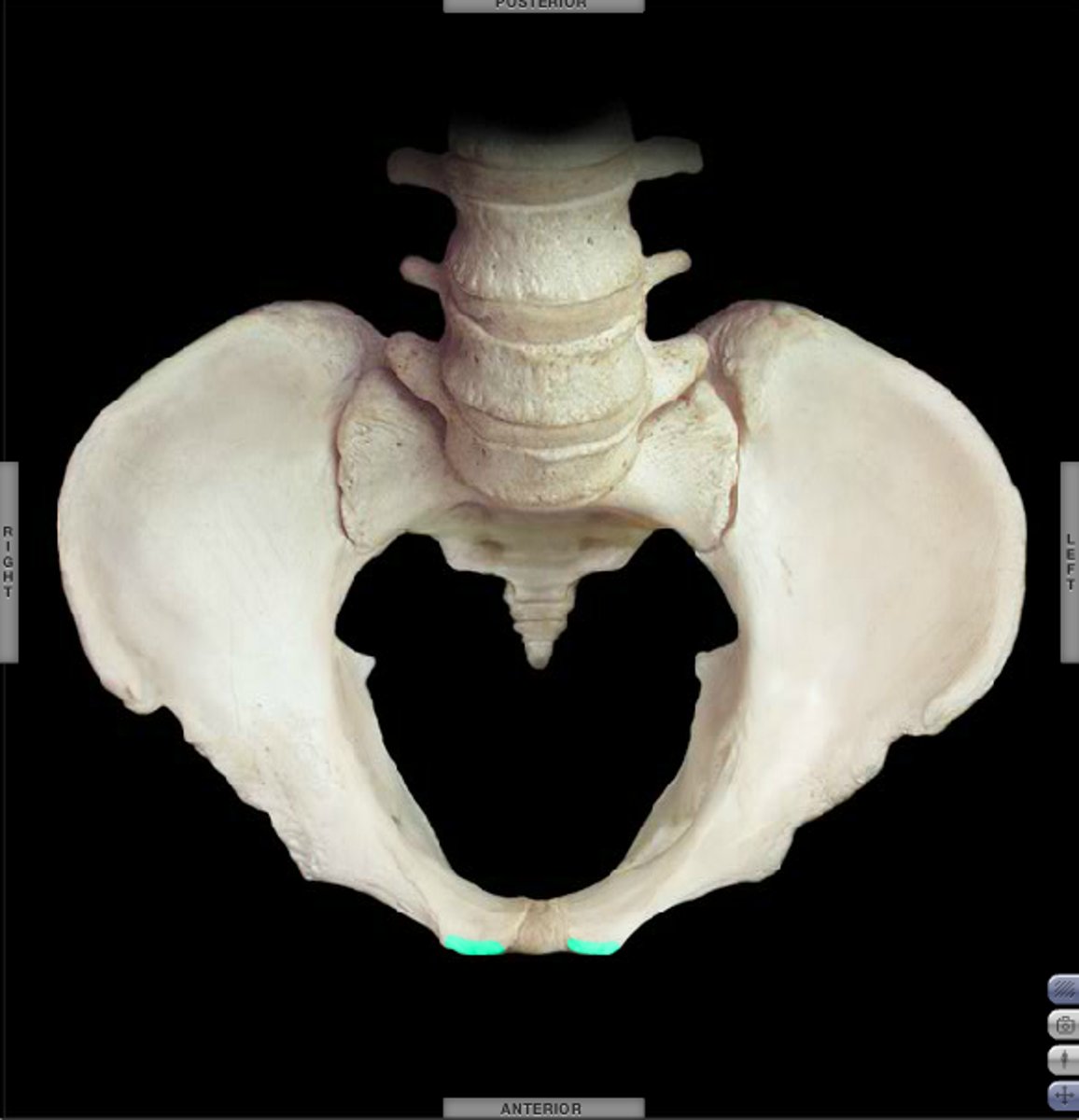
pubic tubercle
anterior prominence superior to the pubic symphysis, lateral end of pubic crest - attachment point for inguinal l. (from ASIS) and external abdominal oblique m.
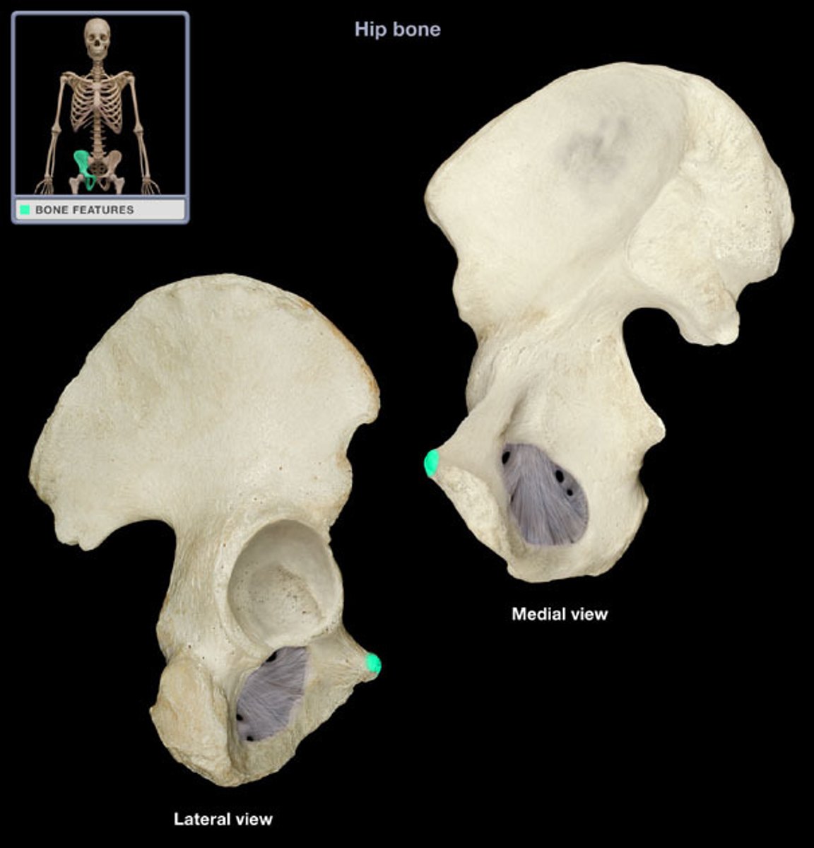
pectineal line of pubis (pecten pubis)
ridge on superior ramus of pubis - attachment point for psoas minor and conjoint tendon of transverse abdominis m. and internal abdominal oblique m.
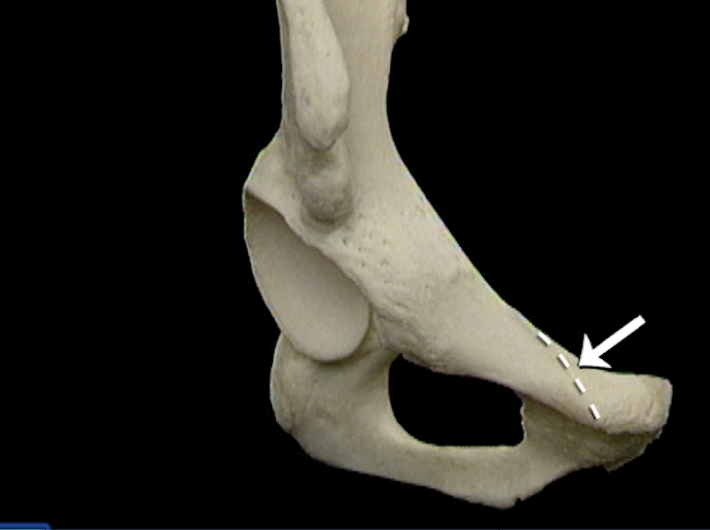
iliopubic eminence
junction between of the ilium and the pubis - point of convergence for iliopsoas on their way to the lesser trochanter of the femur
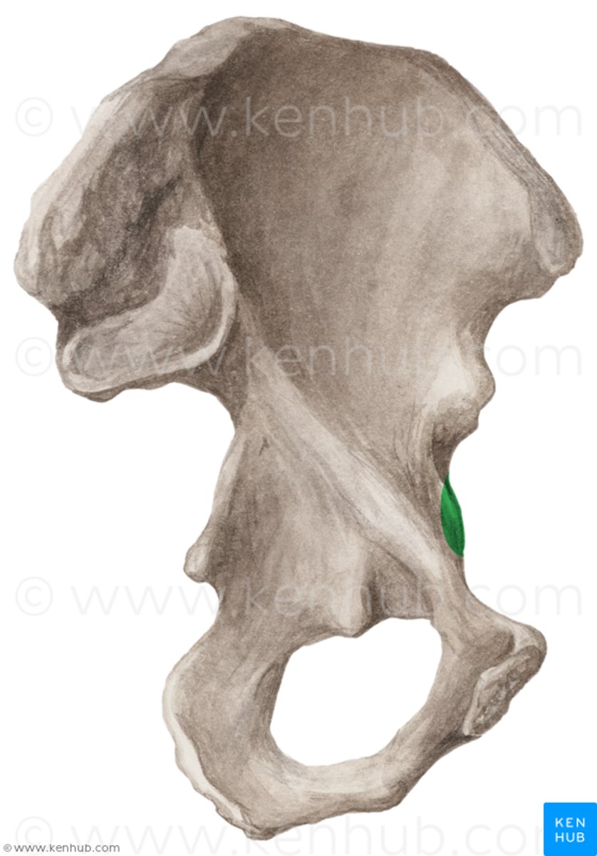
abdominal planes that create the REGIONS of the abdomen
midclavicular - vertical down middle of clavicles to iliopubc eminences
subcostal - horizontal under the level of the ribs (level of L2 vertebra)
transtubercular - horizontal through tubercle of iliac crests (level of L5 vertebra)

abdominal planes that create the QUADRANTS of the abdomen
transumbilical - horizontal through belly button (between level of L3 and L4 vertebrae)
median plane - vertical down midline
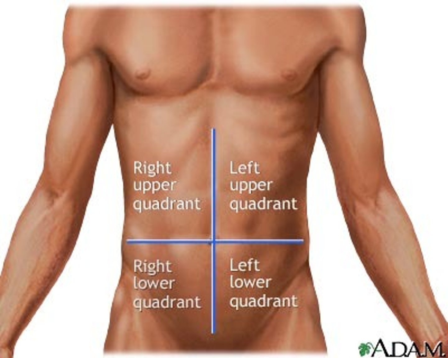
9 abdominal regions
left hypochondriac, epigastric, right hypochondriac, left lateral (lumbar), umbilical, right lateral lumbar), left inguinal, pubic (hypogastric), right inguinal
contents of right upper quadrant (RUQ) (10)
main function: biliary system
- liver (right lobe)
- gallbladder
- stomach (pylorus)
- duodenum (parts 1-3)
- pancreas (head)
- right suprarenal gland
- right kidney
- right colic (hepatic) flexure
- ascending colon (superior part)
- transverse colon (right half)
contents of left upper quadrant (LUQ) (10)
- Liver (left lobe)
- spleen
- stomach
- jejunum and proximal ileum
- pancreas (body and tail)
- left kidney
- left suprarenal gland
- left celiac (splenic) flexure
- transverse colon (left half)
- descending colon (superior part)
contents of right lower quadrant (RLQ) (10)
connects large/small intestines
- cecum
- appendix
- most of ileum
- ascending colon (inferior part)
- right ovary
- right uterine tube
- right ureter (abdominal part)
- right spermatic cord (abdominal part)
- uterus (if enlarged)
- urinary bladder (if very full)
contents of left lower quadrant (LLQ) (8)
connects alimentary canal to excretory system
- sigmoid colon
- descending colon (inferior part)
- left ovary
- left uterine tube
- left ureter (abdominal part)
- left spermatic cord (abdominal part)
- uterus (if enlarged)
- urinary bladder (if very full)
lateral cutaneous n. of T7-T9
cutaneous n. to anterior and lateral aspects of abdominal wall
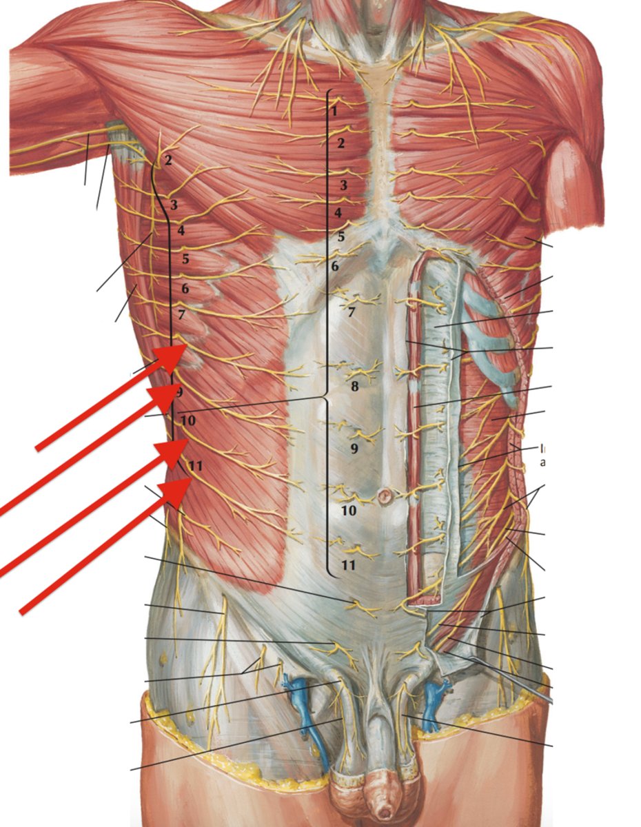
thoracoabdominal n.
nerve root levels: T7-T11
made up of intercostal n. 7-11
function: muscular to anterior abdominal wall
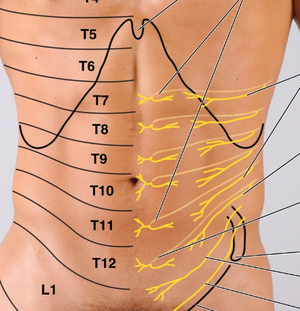
subcostal n.
nerve root level: T12
from the lumbar plexus --> through lateral arch
function: muscular to external oblique m. and portions of other abdominal mm. (along with thoracoabdominal n.); cutaneous to pubic region
(#1 in image)

iliohypogastric n.
nerve root level: L1
path: lumbar plexus --> thru medial arch --> into external oblique m. and transverse abdominis m. --> cutaneous to lateral gluteal and suprapubic regions
(#2 in image)

ilioinguinal n.
nerve root level: L1
path: lumbar plexus --> thru medial arch --> pierce internal oblique m. --> through inguinal canal --> cutaneous to skin at root of penis and upper scrotum (male) and skin to mons pubis and labia majora (female)
(#3 in image)

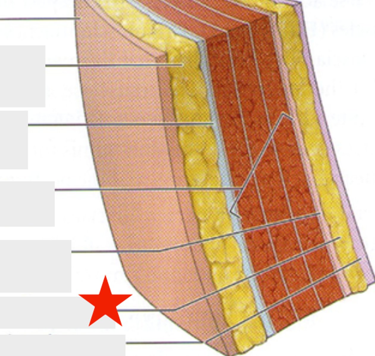
superficial fascia of the anterior abdomen
two layers of subcutaneous connective tissue
Camper's fascia: fatty layer - matrix that holds fatty deposit in place
Scarpa's fascia: deeper layer - membranous, creates firmness
investing fascia of the anterior abdomen
superficial/intermediate/deep
superficial - anterior to external oblique m.
intermediate - anterior to internal oblique m.
deep - anterior to transverse abdominis m.
transversalis fascia
final layer of investing fascia of the anterior abdomen and outside layer of the abdominal cavity
deep to transverse abdominis m.
Extraperitoneal fat
deepest fat layer of the anterior abdomen (deep to transversalis fascia) - can increase in size and contribute to visceral fat collection
Why compression of abdominal viscera important
there is not as much support/muscular contraction on the abdomen as there is on the back
compression --> increases intra-abdominal (intrathecal) pressure --> increased stability
special structures of the rectus abdominis
linea alba - midline structure connecting right and left rectus abdominis m.
linea semilunaris - curved edges around the muscle
transverse tendinous separations (6-pack) for contraction without widespread bundling of muscle
pyramidalis m. - specialized portion of muscle to taut the linea alba

rectus sheath
investing layer of fascia that surrounds the rectus abdominis m. - allows it to interconnect and separate its function, defines superior and inferior action of abs

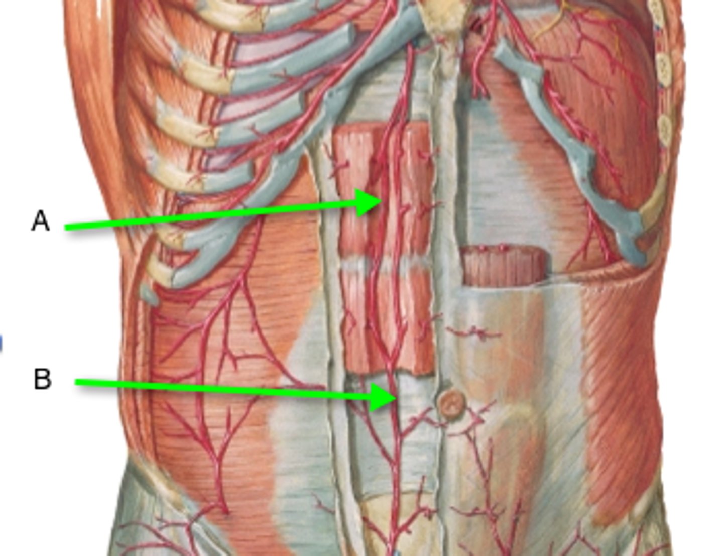
epigastric arteries
superior - branch off internal thoracic a. --> travels b/w rectus sheath and rectus abdominis m.
inferior - branch off external iliac a. --> travels b/w transversalis fascia and rectus abdominis m. on posterior side
both feed the rectus abdominis m.
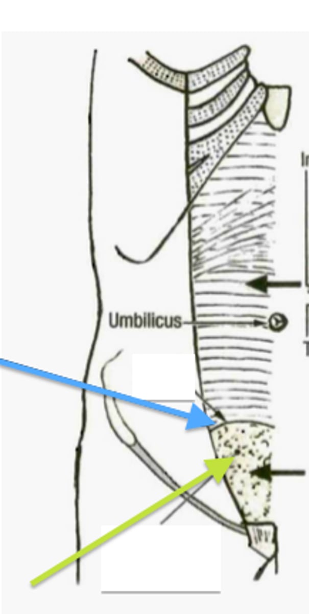
arcuate line
structure of the rectus sheath below the umbilicus - represents movement of the rectus sheath to invest certain muscles above it and certain muscles below it
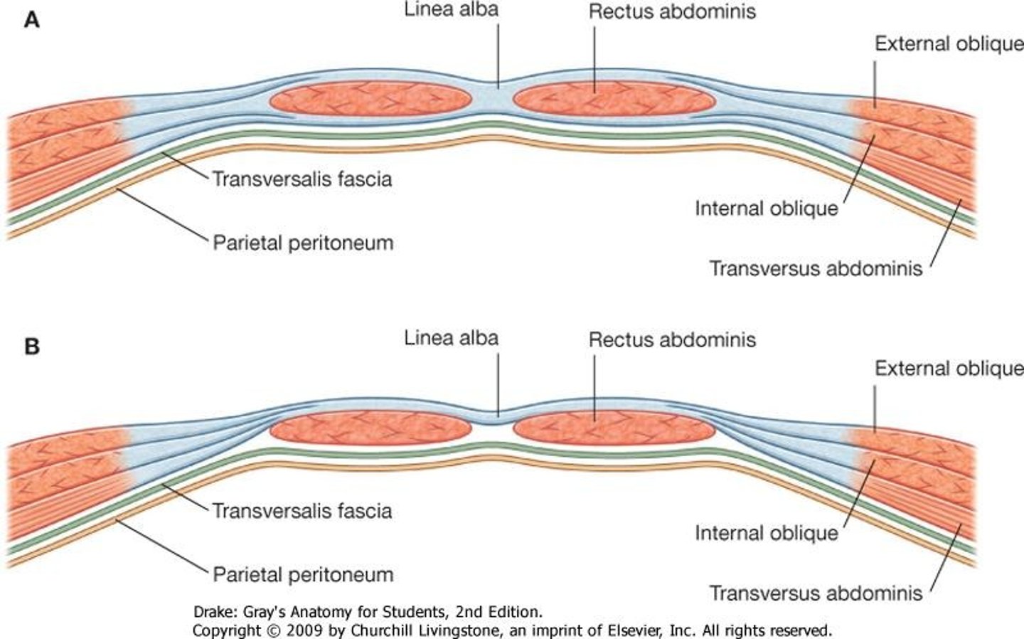
structure/function of the abdominals ABOVE the arcuate line
anterior to rectus abdominis m. - external oblique, internal oblique
posterior to rectus abdominis m - internal oblique, transverse abdominis, transversalis fascia
abdominal muscles have independent function, more rotations occurs here
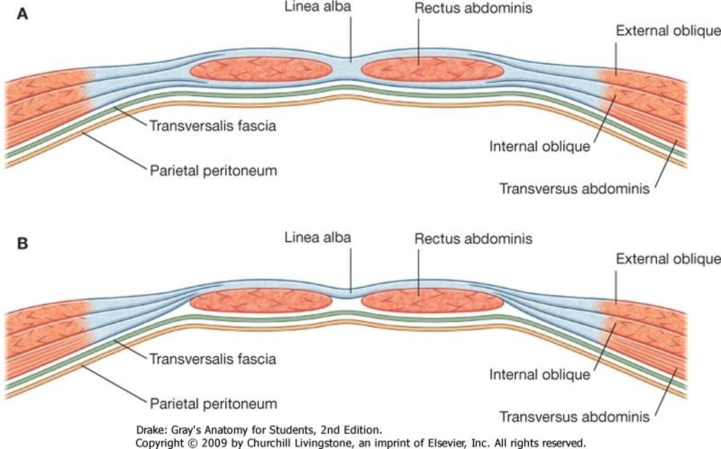
structure/function of the abdominals BELOW the arcuate line
anterior to rectus abdominis - external oblique, internal oblique, transverse abdominis
posterior to rectus abdominis - transversalis fascia
abdominal muscles work together, more compression/flexion occurs here
specialized actions of the abdominal muscles
expiration - collectively compress abdomen --> force diaphragm upwards
rotation - internal oblique (ipsilateral) + external oblique (contralateral)
boundaries of inguinal canal
roof - internal oblique m., transverse abdominis m.
floor - inguinal l., lacunar l. (both created by external abdominal oblique fascia)
posterior wall - transversalis fascia
anterior wall - aponeurosis of internal and external obliques
medial wall - falx inguinalis ("conjoint tendon" - blending of internal oblique and transverse abdominis fascia)
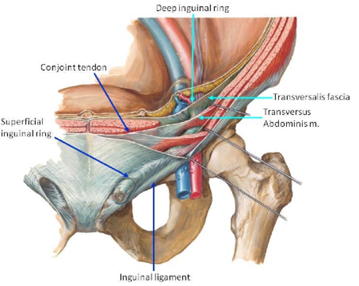
contents of the inguinal canal
ilioinguinal n., spermatic cord (male), round l. of uterus (female)
spermatic cord
collection of male reproductive structures: cremaster m., pampiniform complex of veins, vas deferens, testicular a., genital branch of genitofemoral n.)
travel from the internal pelvic cavity --> through inguinal canal --> scrotum
function: reproduction
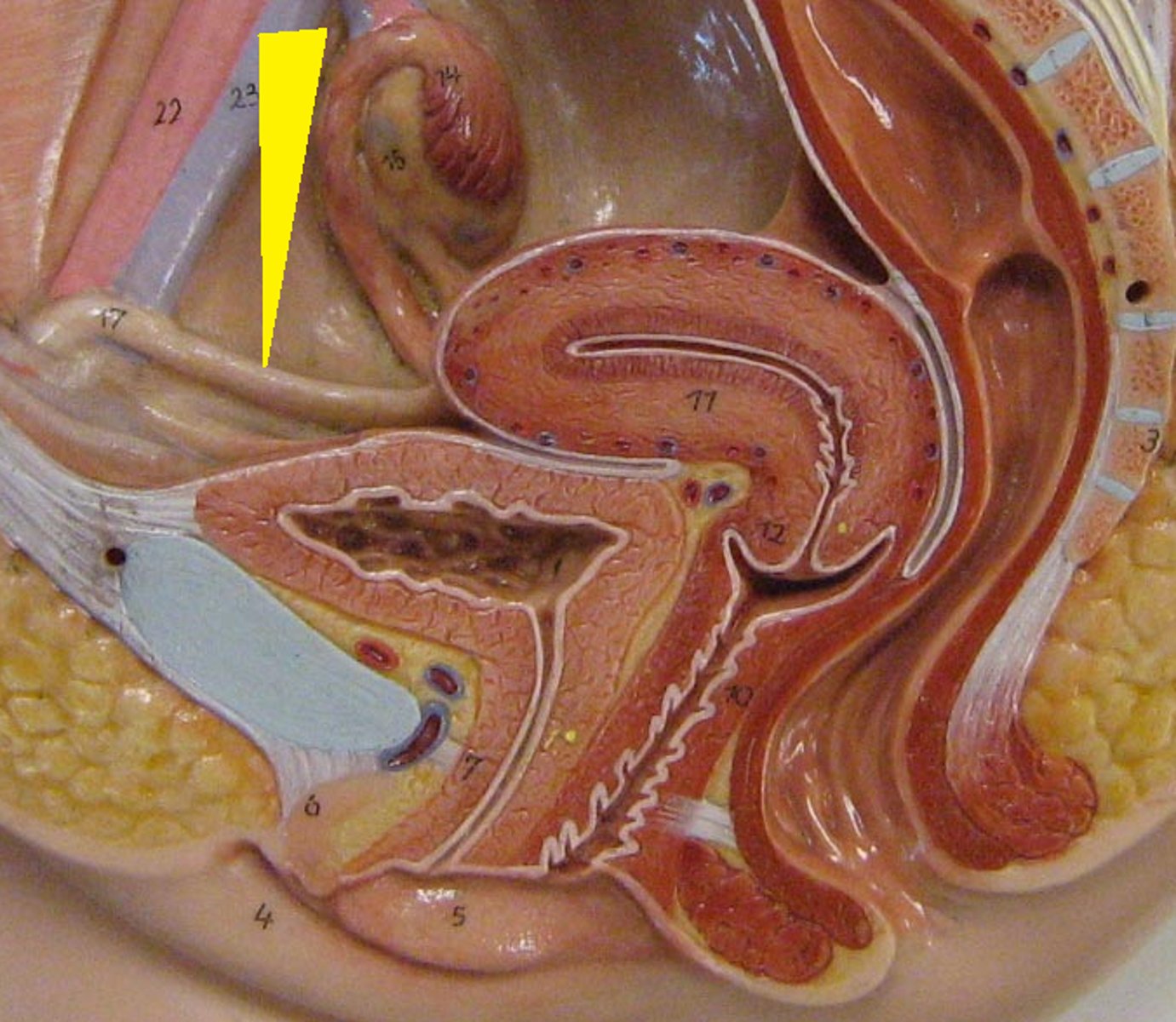
round l. of the uterus
l. in females that attaches to lateral walls of the uterus --> turns around and attaches to lateral walls of pelvis --> through superficial ring of inguinal canal --> attaches to pubic bone
function: anchors uterus
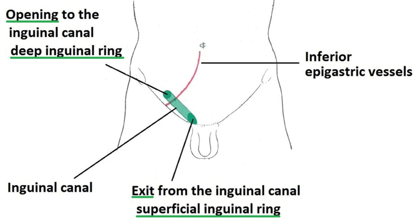
openings in the inguinal canal
deep inguinal ring - opening in transversalis fascia, entrance to inguinal canal (spermatic cord enters)
superficial inguinal ring - where spermatic cord exits to go to the scrotum/ round l. of uterus passes through
specialized functions of external oblique fascia (5)
- anterior layer of rectus sheath (above AND below arcuate line)
- inguinal l. (double fold)
- lacunar l.
- pectineal l. (supports lacunar l.)
- external portions of spermatic fascia
specialized functions of internal oblique fascia (4)
- anterior layer of rectus sheath (above AND below arcuate line)
- posterior layer of rectus sheath (above arcuate line ONLY)
- falx inguinalis (w/ transverse abdominis fascia)
- cremaster m. and fascia
specialized functions of transverse abdominis fascia (3)
- anterior layer of rectus sheath (below arcuate line ONLY)
- posterior layer of rectus sheath (above arcuate line ONLY)
- falx inguinalis (w/ internal oblique fascia)
specialized functions of transversalis fascia (2)
- deep inguinal ring
- internal spermatic fascia
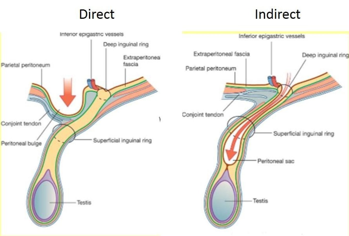
indirect inguinal hernia
small intestine herniates into the inguinal canal through the deep inguinal ring --> exits through the superficial inguinal ring --> descends into the scrotum
occurs LATERAL to inferior epigastric v.,a.
congenital, more common, occurs passively over time
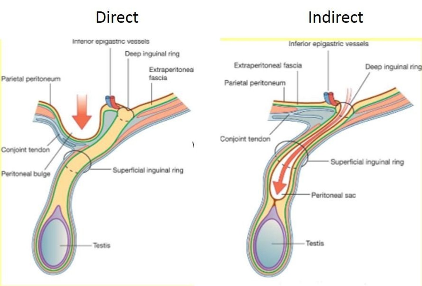
direct inguinal hernia
small intestine herniates into the inguinal canal by piercing through muscles and fascia --> exits the superficial inguinal ring
occurs MEDIAL to inferior epigastric v.,a.
acquired through strain
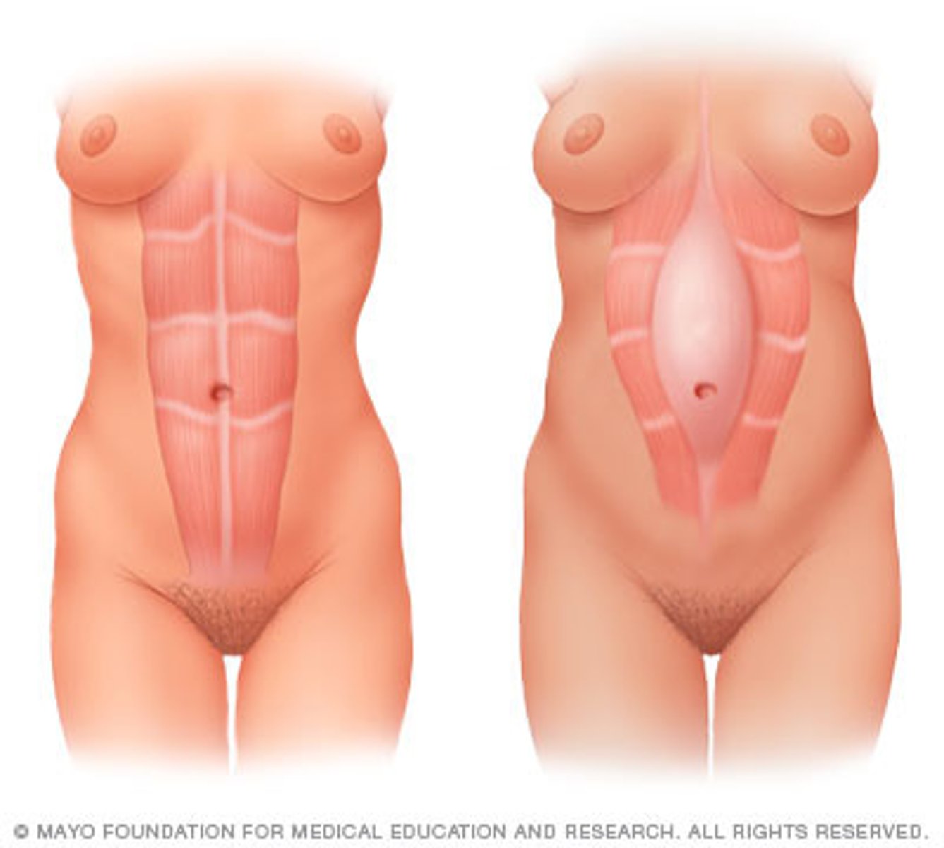
abdominal ptosis/diastasis recti
a lot of visceral fat (or in pregnancy) --> separation of rectus abdominal m. --> loss of compression/stabilization from musculature
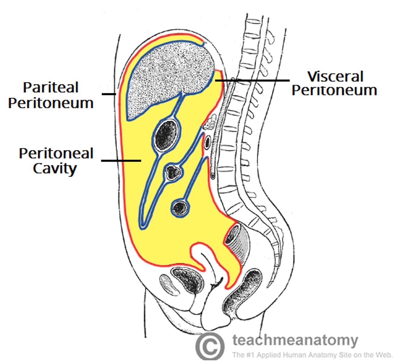
peritoneum
Double-layered membrane surrounding the abdominal organs
parietal - lining of abdominal wall
visceral - covering the organs
cavity - potential space between layers (closed in males, communication w/ external structures occurs in females)
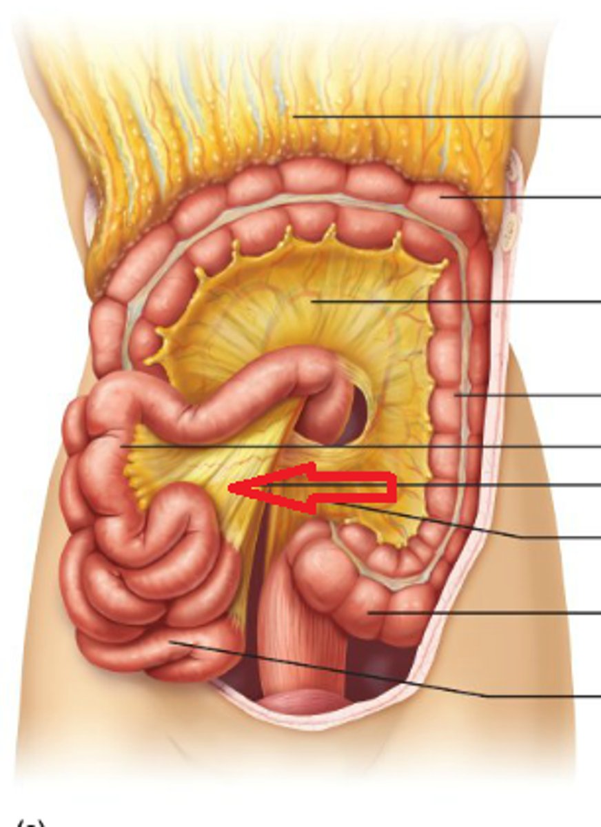
mesentery
double layer of visceral peritoneum found mainly on intestines - "shrink wrap" that holds structures together, carries VANs, may be mobile w/ the organ
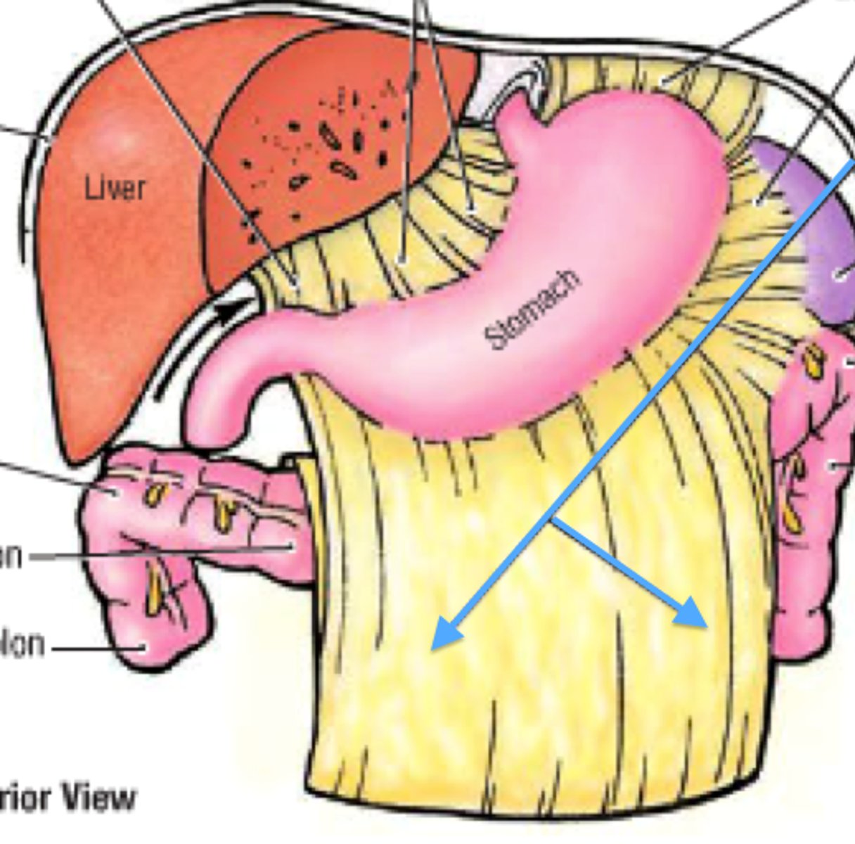
greater omentum
double layer of peritoneum that connects the greater curvature of the stomach to the musculature of the abdominal wall (specialized transverse mesocolon attaches large intestine to posterior abdominal wall)
lesser omentum
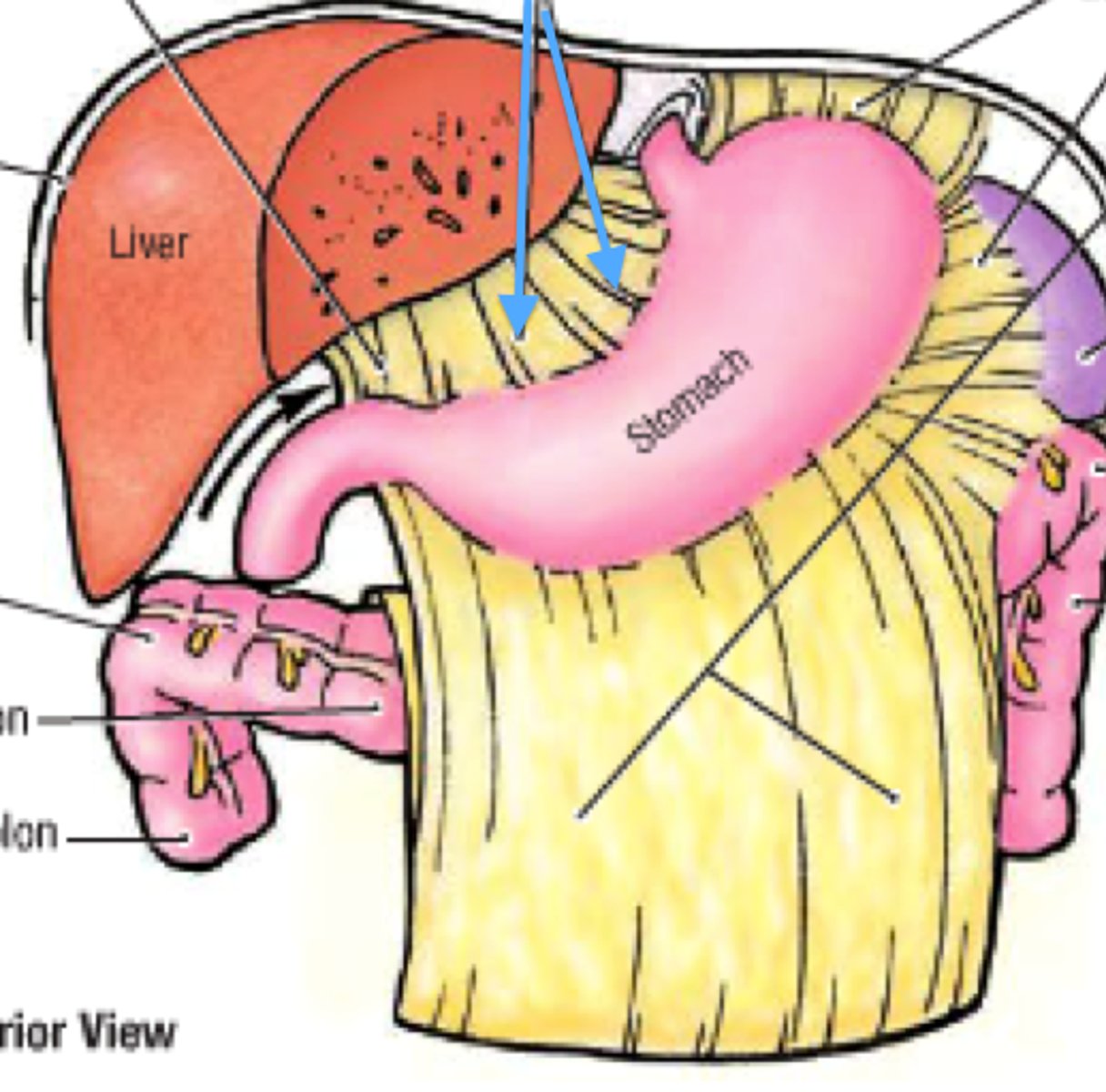
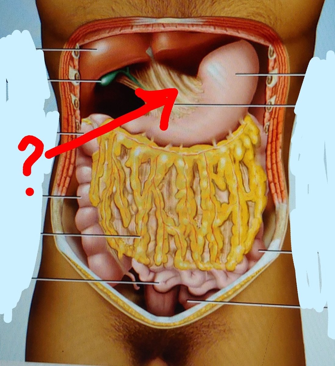
double layer of peritoneum that connects the lesser curvature of the stomach to the liver, envelopes the porta hepatis
contains 2 ligaments: hepatogastric l. (liver--> lesser curvature of the stomach) and hepatoduodenal l. (liver--> superior duodenum)
peritoneal ligaments
double layers of the peritoneum that connect organs with other structures
peritoneal recess
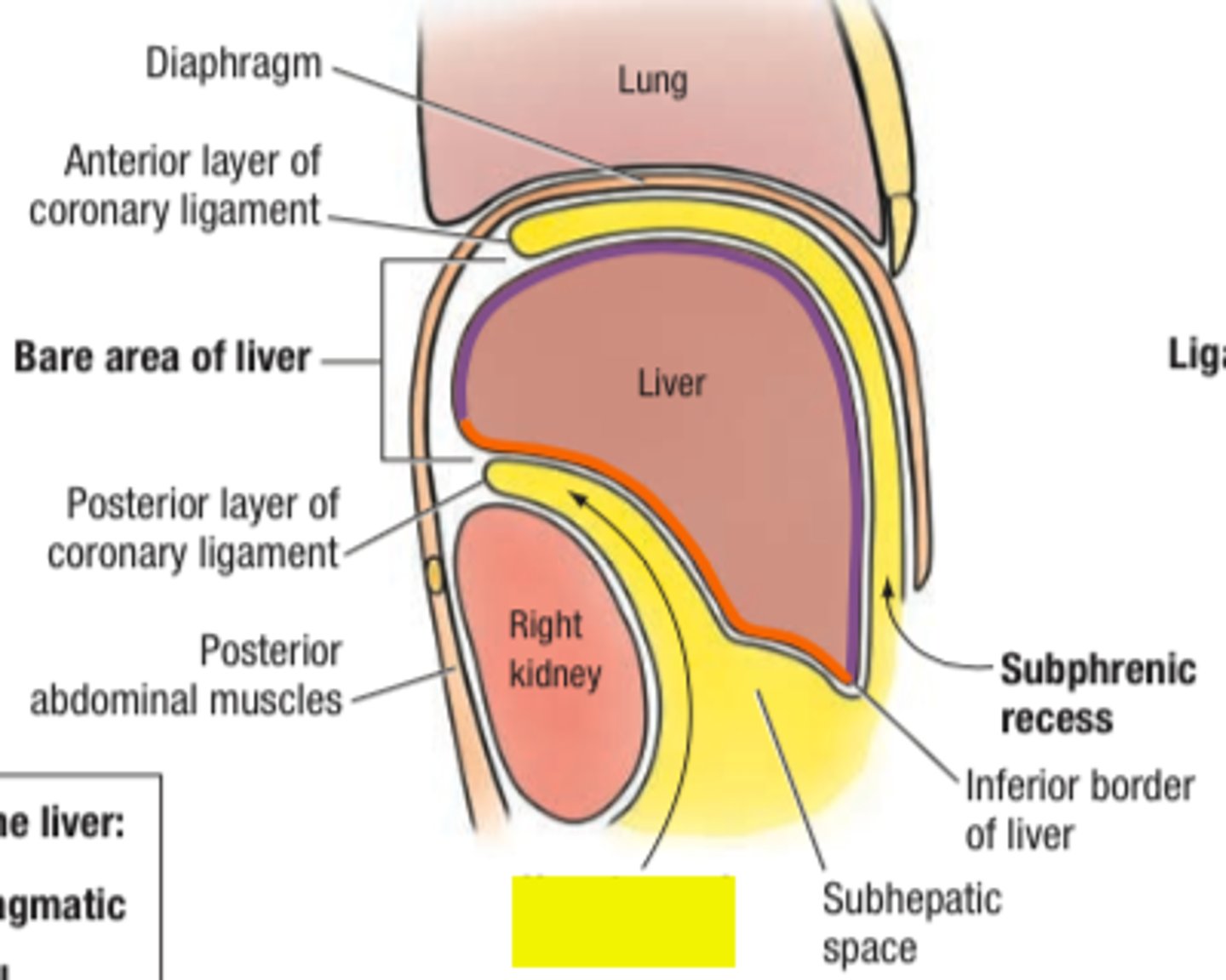
pouch in the peritoneum created by folds - several around the stomach to allow for expansion (ex. gastrophrenic recess)
peritoneal fold
a reflection of peritoneum that is raised from the body wall by an VAN (ex. lesser omentum)
celiac trunk
very superior branch off the abdominal aorta, supplies the foregut
branches: R/L gastric a., R/L gastroepiploic (gastro-omental) a., duodenal a., hepatic a., splenic a.
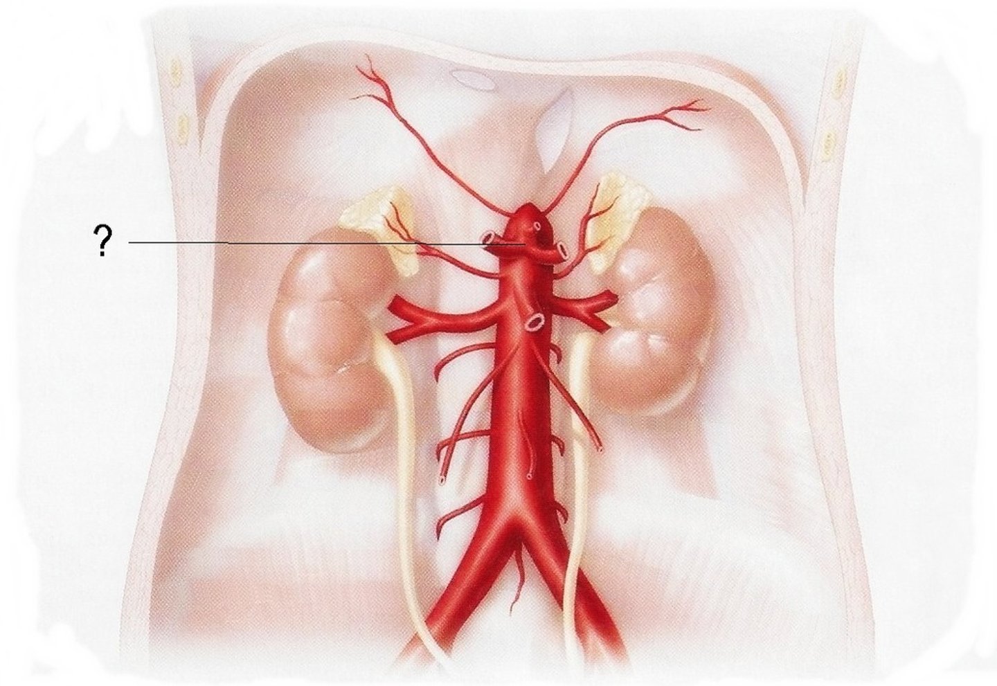
superior mesenteric a.
branch off abdominal aorta, blood supply to midgut
branches: colic branches to jejunum and ileum, 1 branch to duodenum (distal to bile duct)
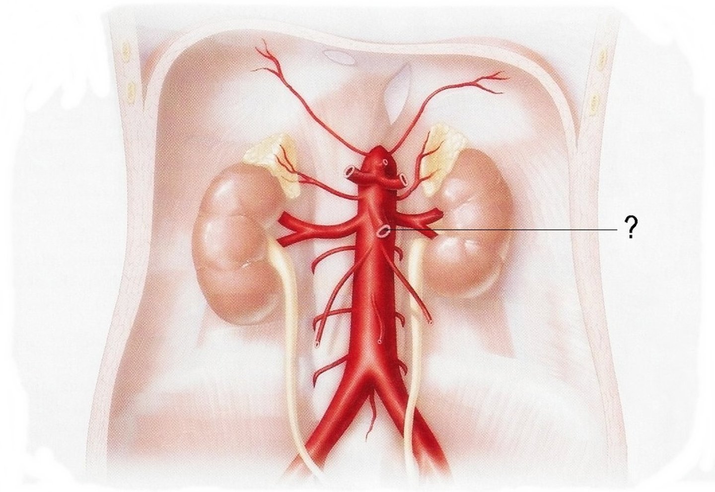
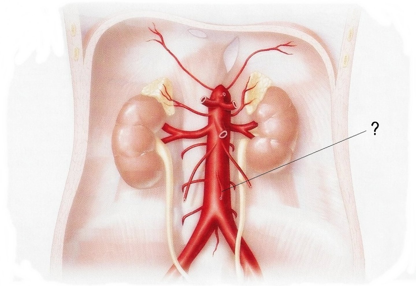
inferior mesenteric a.
branch off abdominal aorta, feeds hindgut
branches: colic and sigmoidal branches to the large intestine, rectal branches to the rectal canal
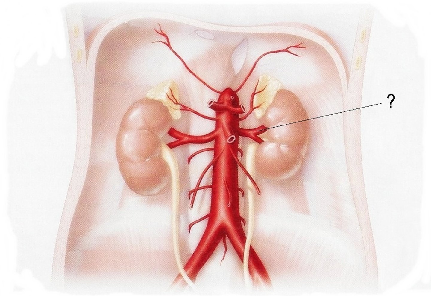
renal a.
branch off abdominal aorta --> blood supply to the kidneys
portal v.
nutrients/blood from the intestines/ other digestive structures (SMV, IMV, celiac v., and their tributaries contribute) travel through this structure to the liver for initial processing --> blood then picked up by inferior vena cava from the liver to go back to central circulation
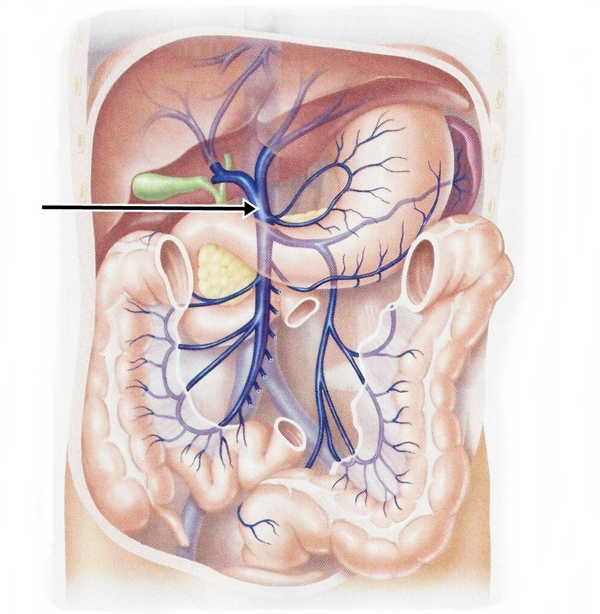
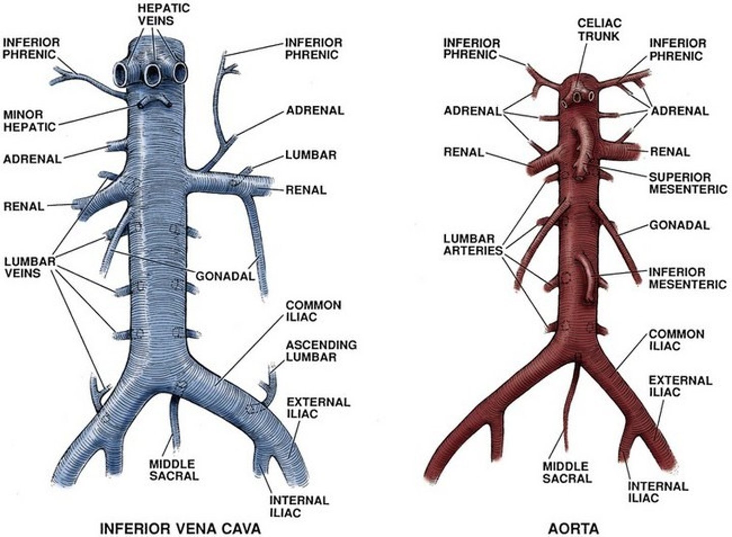
paired vessels of abdomen
all vessels are paired EXCEPT if they are digestive in nature - portal v. takes nutrients to the liver
structures of the foregut (7)
esophagus, stomach, proximal 1/3 of the duodenum, liver, gallbladder, superior pancreas, spleen
blood supply of the foregut
celiac trunk, portal venous system
innervation of the foregut
sympathetic - greater thoracic splanchnic n.
parasympathetic - vagus n.
structures of the midgut (7)
distal 2/3 of duodenum, jejunum, ileum, cecum, ascending colon, appendix, proximal 2/3 of transverse colon
blood supply of the midgut
superior mesenteric a., portal venous system
innervation of the midgut
sympathetic - lesser thoracic splanchnic n.
parasympathetic - vagus n.
structures of the hindgut (4)
distal 1/3 of transverse colon, descending colon, sigmoid colon, rectal canal
blood supply of the hindgut
inferior mesenteric a., portal venous system
innervation of the hindgut
sympathetic - lumbar and sacral splanchnic n.
parasympathetic - pelvic splanchnic n.
retroperitoneal organs
suprarenal (adrenal) glands, aorta/vena cava, duodenum (except superior part), pancreas (and hilum of spleen), ureters, colon (ascending and descending parts), kidneys, esophagus, rectum
transverse mesocolon
part of greater omentum that connects the transverse colon to the back of the abdominal cage - separates SUPRACOLIC region from INFRACOLIC region
omental foramen (epiploic foramen)
opening in the lesser omentum for the passage of the hepatic a., portal v., and common bile duct - separates intraperitoneal and retroperitoneal structures
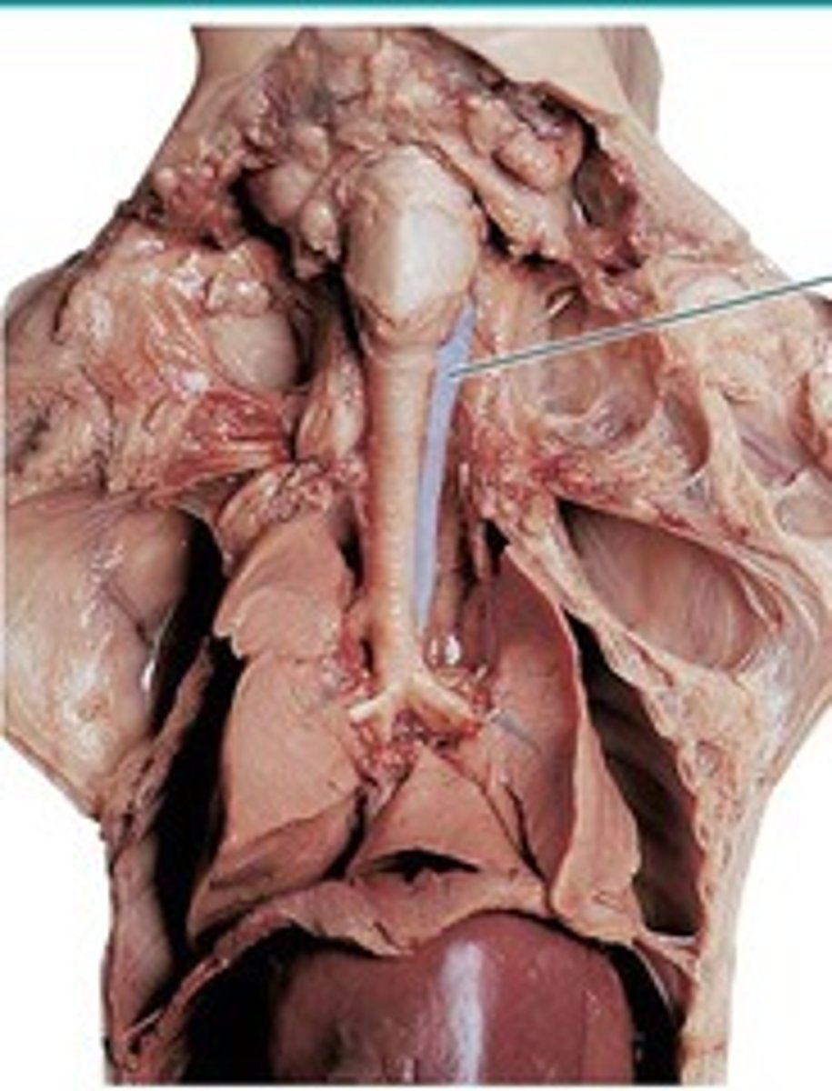
esophagus
path: pierces diaphragm at the esophageal hiatus, left of the median plane --> enters the stomach at the level of T10
retroperitoneal structure - only anterior and lateral parts are covered by peritoneum
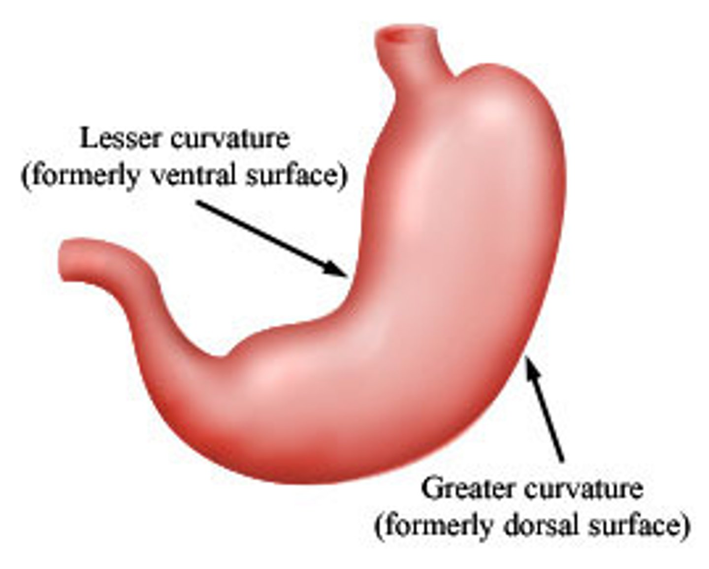
curvatures of the stomach
greater curvature - inferior, attachment point for greater omentum
lesser curvature - superior, attachment point for lesser omentum
angular notch - point on lesser curvature that separates body from pyloric region
sections of the stomach
cardia - stomach/esophagus connection
fundus - most superior part of stomach, deviates left, expands superiorly into the gastrophrenic recess when there is a high volume of food
body - majority of the stomach, contains greater and lesser curvatures
pylorus - funnel to duodenum (contains antrum, canal, sphincter, and orifice)
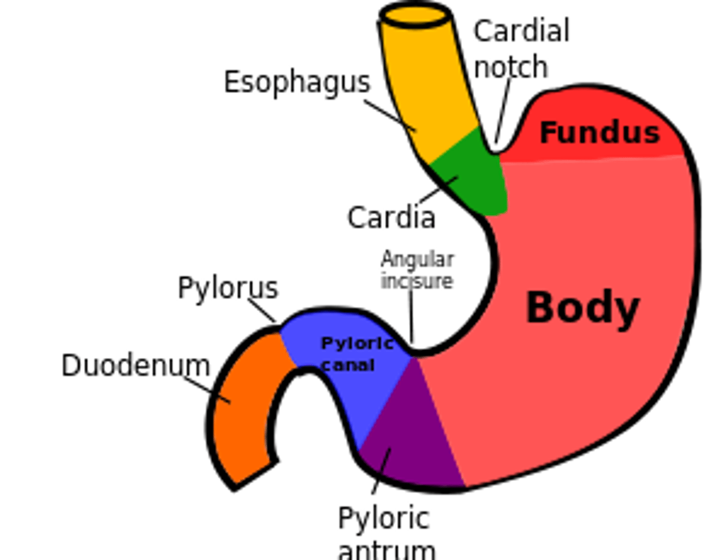
parts of pylorus of stomach
pyloric antrum - vestibule, packs large volume of food into a smaller volume
pyloric canal - stomach-duodenum junction
pyloric sphincter - controls opening/closing of orifice to turn digestion on/off
pyloric orifice - point of passage of chyme from stomach to duodenum
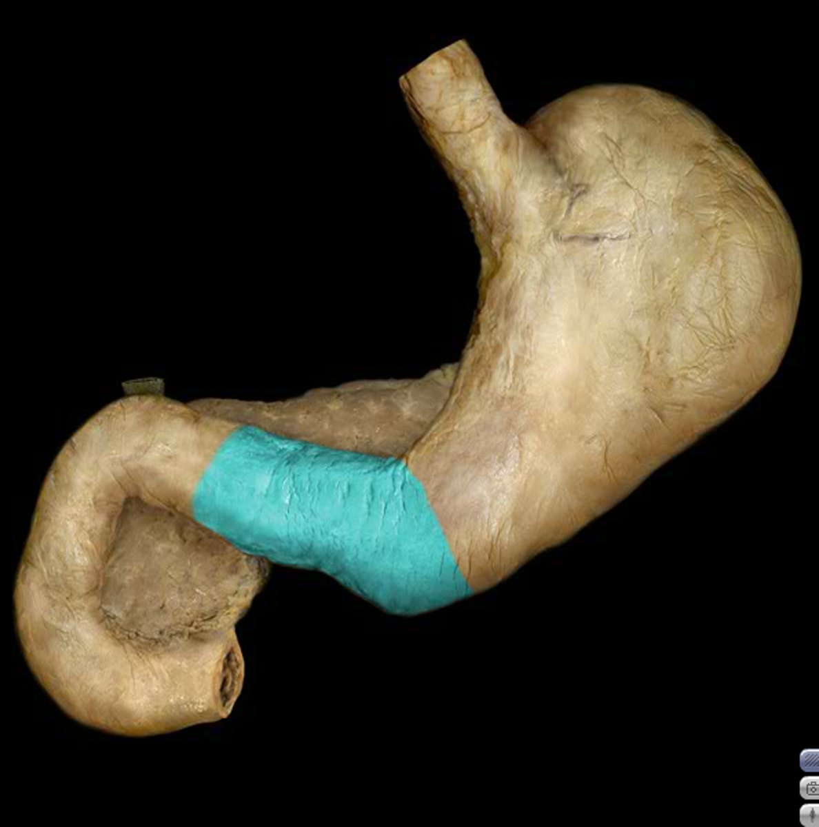
functions of the stomach (4)
- convert food to chyme
- enzymatic digestion
- release HCl (pH ranges 1-4)
- produce gastrin (hormone that stimulates the production of stomach acids)
blood supply to stomach
celiac trunk
branches: right and left gastric a. to the superior portion and lesser omentum; right and left gastroepiploic (gastroomental) a. to inferior portion and greater omentum
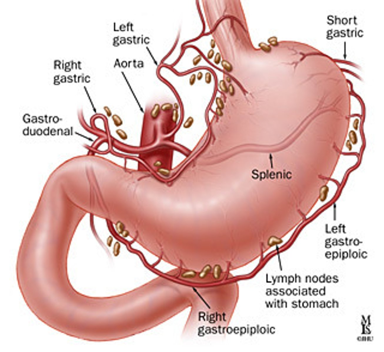
small intestine
digestive organ that extends from the pylorus of the stomach to the ileocecal junction
parasympathetics stimulate secretion and increase motility; sympathetics reduce motility of processing, reduce secretion, and induce vasoconstriction
3 parts: duodenum, jejunum, ileum
4 parts of the duodenum
superior - short, hooks up w/ pyloric canal, lines anterolateral to L1
descending - descends to the right of L1-L3 (retroperitoneal)
horizontal - crosses L3 (right to left) and loops around the head of the pancreas (retroperitoneal)
ascending - short, left of L3 (retroperitoneal)
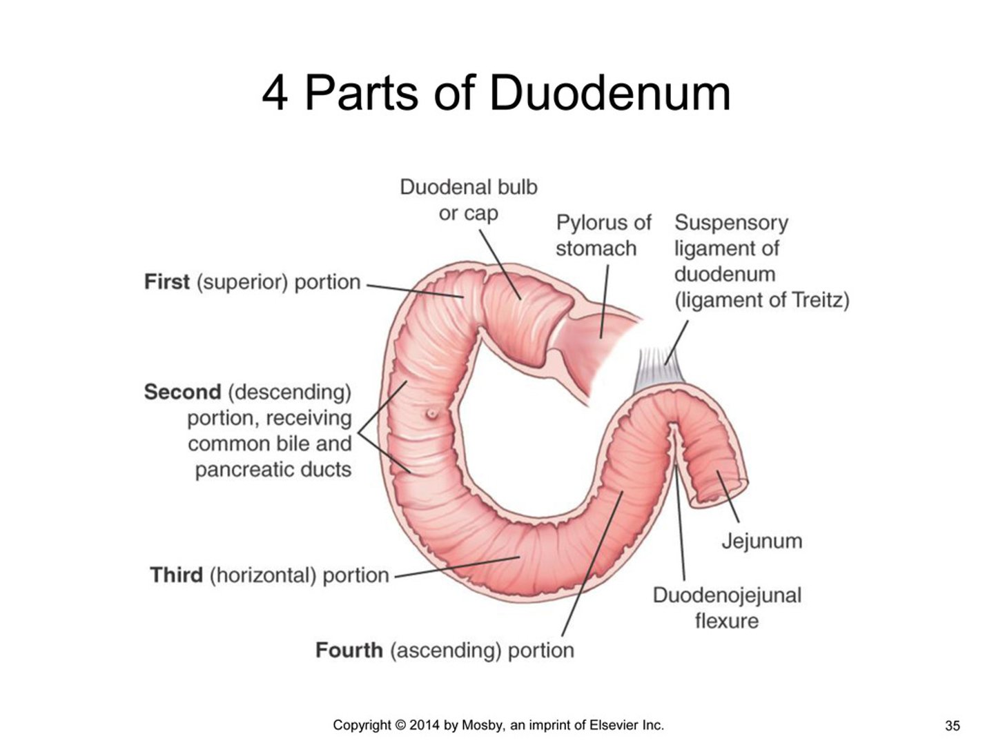
duodenum
first part of the small intestine, shortest but widest, has a mesentery, only superior part is intraperitoneal
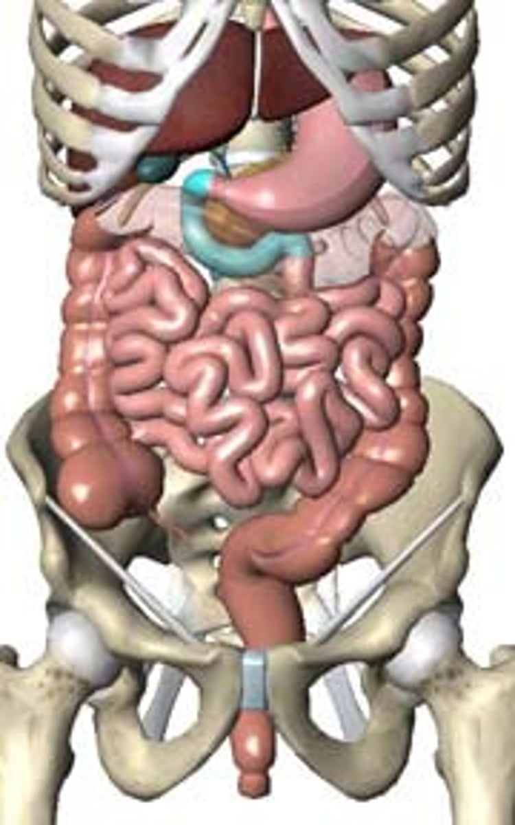
4 parts create a C-shaped course around the pancreas --> joins jejunum at the duodenaljejunal flexure (held in place by suspensory m. of duodenum/ l. of Treitz)
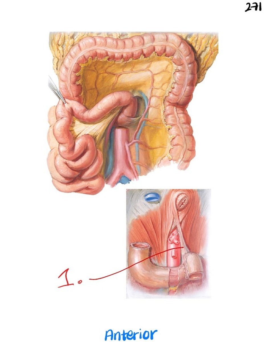
suspensory m. of duodenum (ligament of Treitz)
ligament connecting the ascending part of the duodenum to the right crus of the diaphragm
contracts/loosens based upon sympathetic/parasympathetic activation - parasympathetic allows for chyme to flow into the duodenum, sympathetic halts digestion
functions of the duodenum
- further digestion of chyme (pH = 6)
- production of mucus (by Brunner's glands)
- release bile to emulsify fat (liver --> common bile duct --> major duodenal papilla)
- release pancreatic juices (pancreas --> main pancreatic duct --> major duodenal papilla OR pancreas --> accessory pancreatic duct --> minor duodenal papilla)
blood supply of the duodenum
celiac trunk --> duodenal a. (proximally) and superior mesenteric a. (distal to bile duct), portal venous system
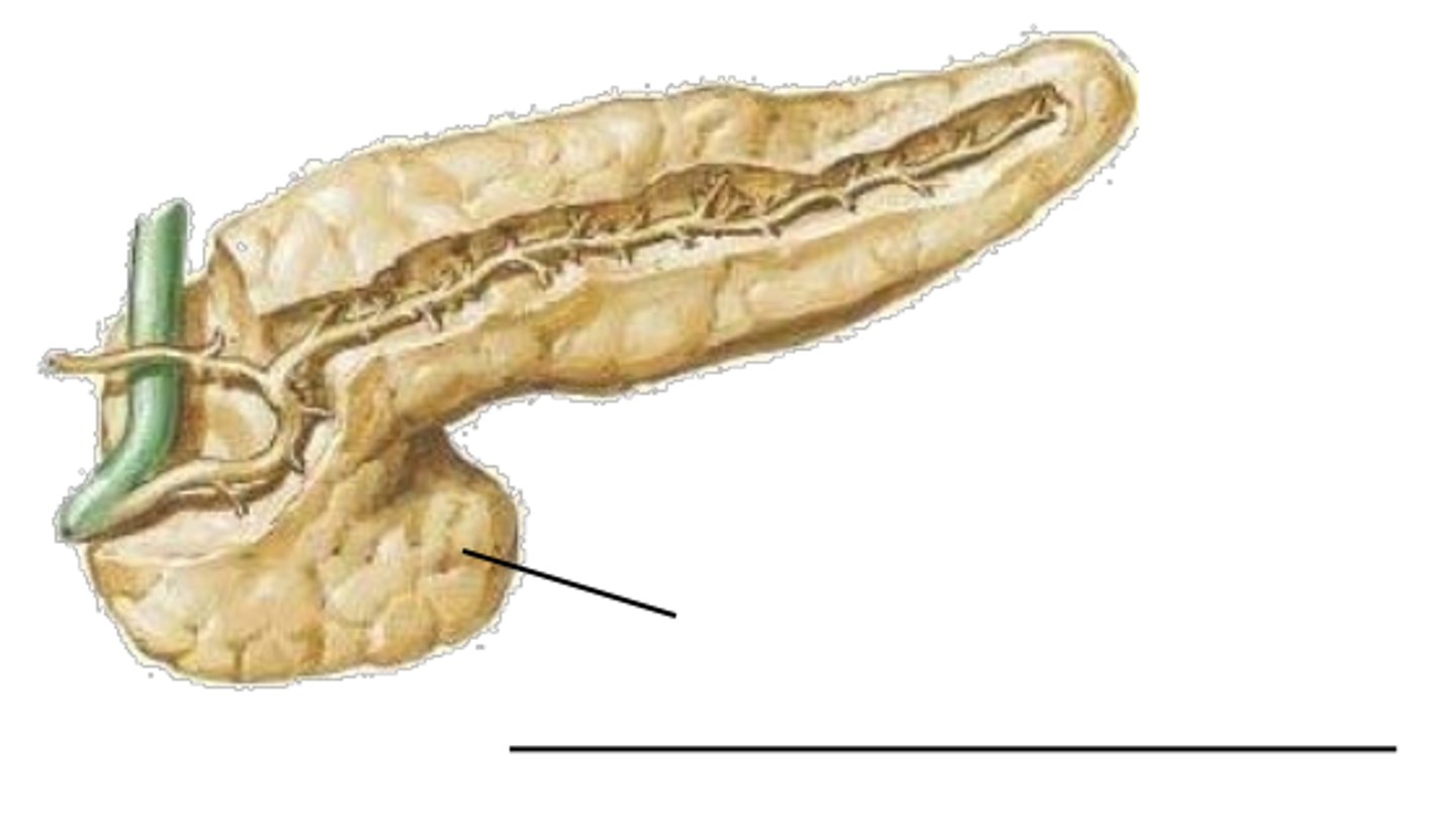
uncinate process of the pancreas
small projection from the lower part of head of pancreas - portal v. found directly posterior as it travels to the liver
very common place for pancreatic tumors --> puts pressure on portal v. and shuts down digestion
jejunum
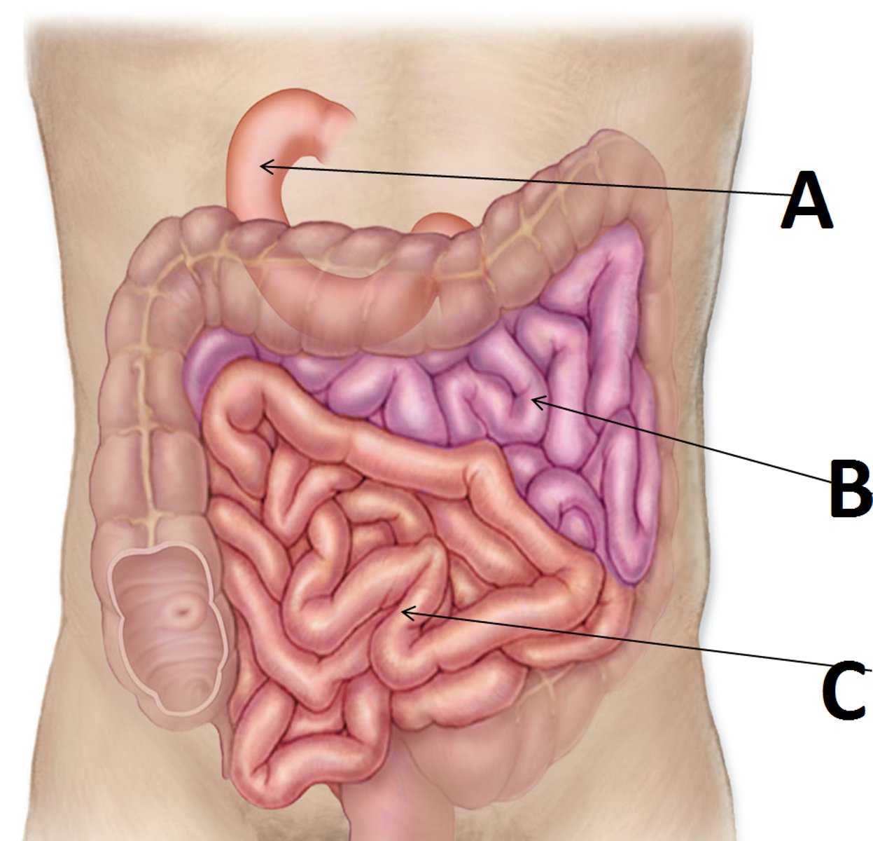
second part of the small intestine, found in the umbilical region, flow is mediated by the suspensory m. of the duodenum (l. of Treitz)
further processes chyme (pH = 7-8) and absorbs water and electrolytes
(B in image)
differences between jejunum and ileum
jejunum has less encroaching fat in mesentery, less complex arcades (arterial structures) --> larger "clear windows", longer vasa recta, and more plicae circulares inside
ileum has more complex arcades --> smaller "clear windows", shorter vasa recta, and more Peyer's patches (secondary lymph structures) inside
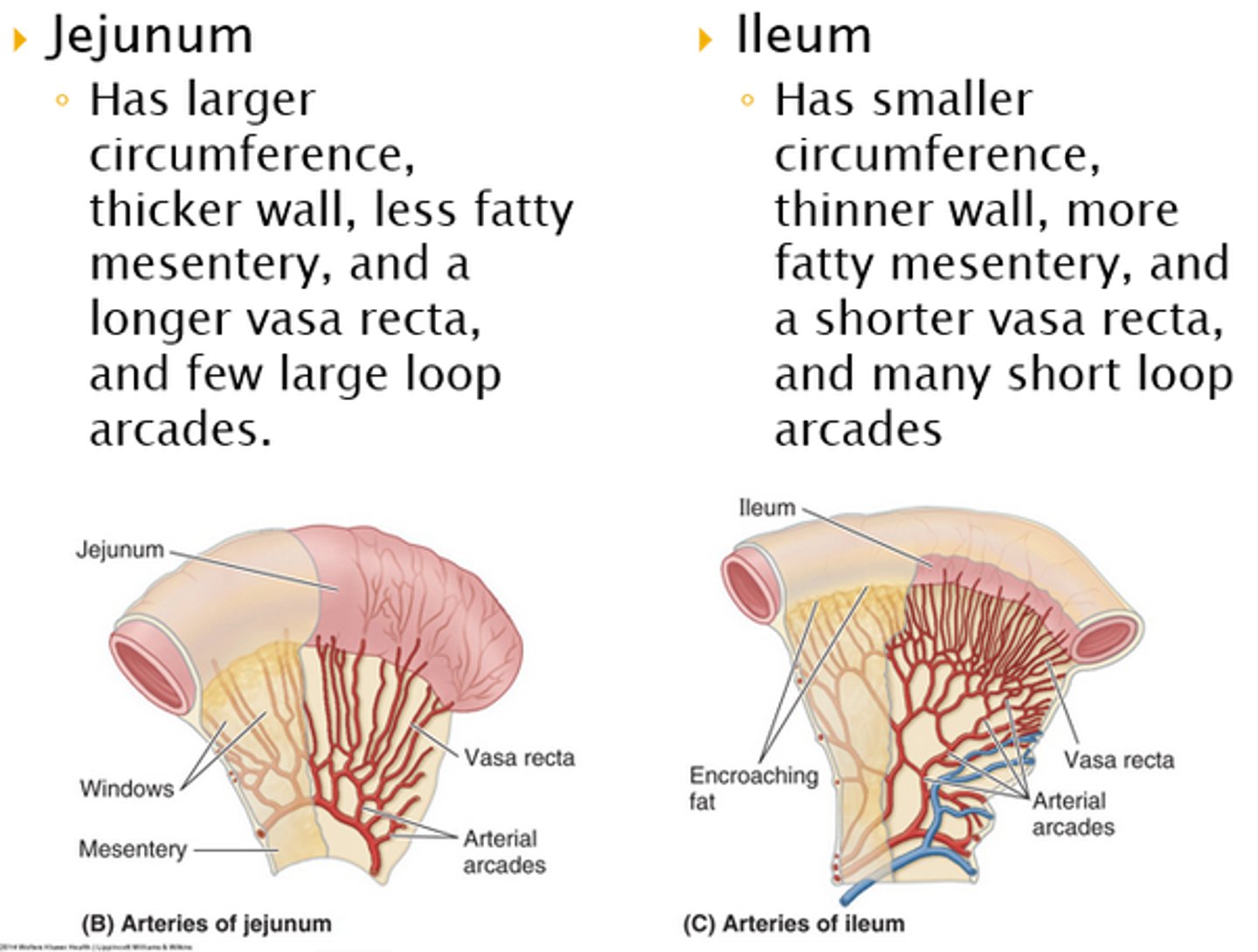
similarities between jejunum and ileum
collectively 6-7 meters long (jejunum is 2/5ths, ileum is 3/5ths), pH = 7-8, blood supply by colic branches of SMA
blood supply of the jejunum and ileum
colic branches of superior mesenteric a., portal venous system
functions of the liver (3)
- detox blood
- store glycogen
- secrete bile
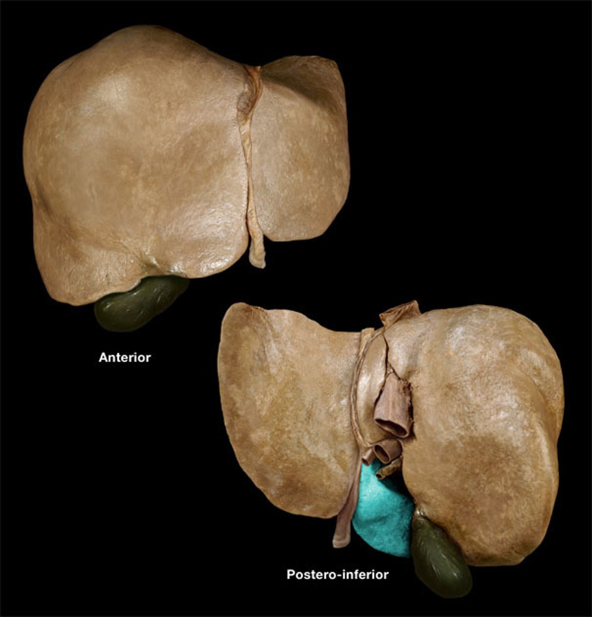
surfaces of the liver
- diaphragmatic - smooth posterior surface, covered w/ peritoneum except "bare area" where the diaphragm attaches (movement of diaphragm = movement of liver)
- visceral - anterior, sharper, angular margins, functional areas where filtration occurs, covered w/ peritoneum except where porta hepatis and gallbladder attach
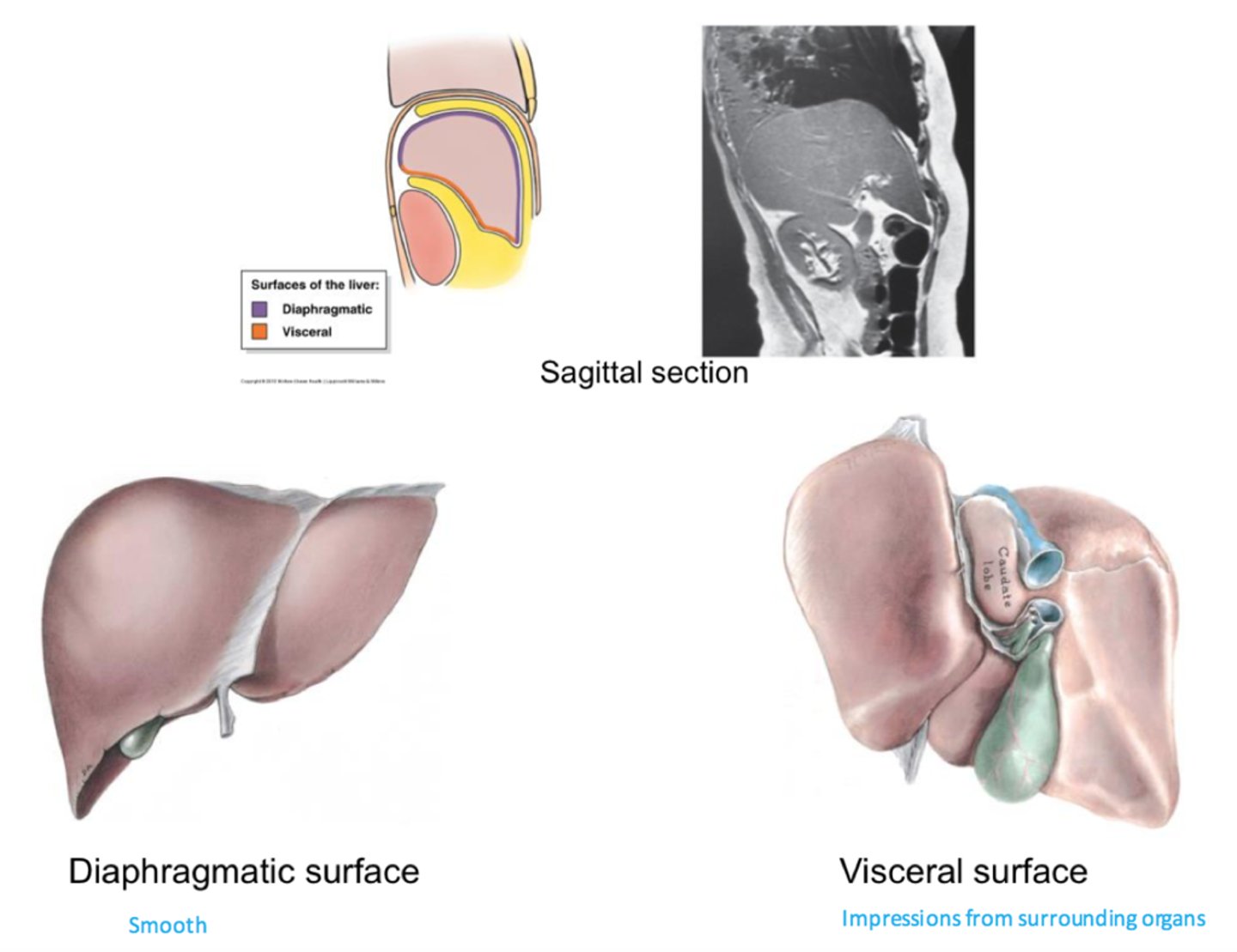
porta hepatis (portal triad)
central area of the liver where the hepatic a. and portal v. enter and the bile duct exits
(tributaries to hepatic a. and portal v. run to independent functional areas and do NOT anastomose)
enveloped by the lesser omentum, has connection to the lesser curvature of the stomach (by hepatogastric l.) and the superior duodenum (by hepatoduodenal l.)
lobes of the liver
- right lobe - bigger, from gallbladder to IVC
- left lobe - situated over stomach
- caudate lobe - superior lobe housed w/in left
- quadrate lobe - inferior lobe housed w/in left, situated right next to gallbladder
the lobes act independently from each other
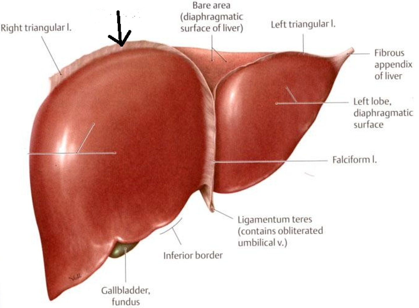
ligaments of the liver (4)
- coronary l. - connects diaphragm to liver, has anterior and posterior layers, defines bare area, connects to falciform l.
- falciform l. - connects liver to posterior aspect of rectus sheath and separates right and left lobes
- round l. (teres l.) - remnant of fetal umbilical v. - attaches to falciform l. and posterior aspect of belly button
- ligamentum venosum - remnant of ductus venosus (fetal connection to the placenta), located b/w caudate lobe and left lobe
blood blow to the liver (venous)
splenic v., superior mesenteric v., inferior mesenteric v., etc --> portal v. --> through porta hepatis
blood is moderately oxygenated and nutrient filled, represents 70-80% of oxygenated blood used by the liver
blood flow to the liver (arterial)
celiac trunk --> hepatic a. --> through porta hepatis
blood is highly oxygenated, represents only 20-30% of oxygenated blood used by the liver
**hepatic a. is much smaller than hepatic v. - used as a supplement, not main feed
blood flow FROM the liver
in the liver, blood is filtered and bile is produced from the waste
filtered blood - central (hepatic) v. --> IVC --> central circulation
bile -> biliary system (to the duodenum)
function of the gallbladder
storage and concentration of bile
portions of the gallbladder
cystic duct, neck, infundibulum (superior, gallstones form here), body, fundus (inferior, rounded)
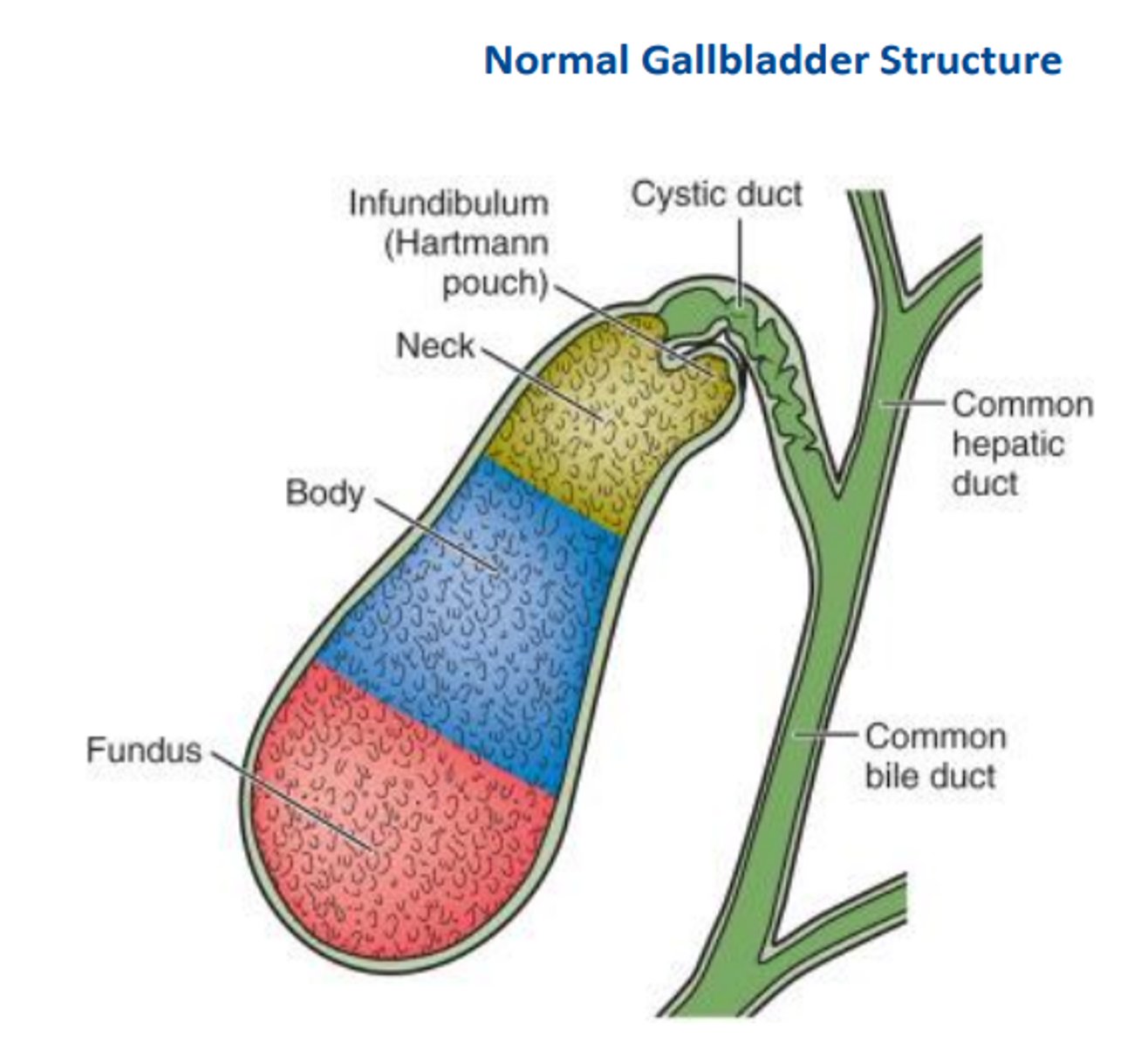
blood supply of the gallbladder
cystic a. (from hepatic a.)
portal hypertension
Pressure gradient (IVC is 2-6mmHg, portal v. is 9mmHg) normally present to help push blood through the liver to be filtered
cirrhosis (liver disease), tumors, etc --> increase pressure in the portal vein (10+ mmHg) --> backflow, pushes blood to other venous structures (ex. IMV, SMV, celiac v. and their tributaries) --> dilation of vessels --> varicose veins --> can lead to esophageal varices (-->ulcers), rectal varices (-->hemorrhoids), and gastric varices (-->ulcers)
function of the spleen
lymphatics - hemopoietic until 5th month and then aids in the process of natural RBC death in adulthood
features of the spleen
- diaphragmatic margin (convexity to fit diaphragm)
- anterior margins (sharp turns)
- held in place by gastrosplenic l. (stomach to spleen), splenorenal l. (kidney to spleen), and phrenicolic l. (left colic flexure to thoracic diaphragm - anchors spleen superiorly)
blood supply to the spleen
splenic a. (from celiac trunk)
drains from tributaries --> hilum --> splenic v. --> inferior mesenteric v. --> portal venous system
location of right v. left kidneys
right - about 2.5 cm lower (due to liver), partially covered by 12th rib, superior pole at 11th rib
left - mostly covered by 11th and 12th ribs, can project into 10th intercostal space
both surrounded by renal fascia and perirenal fat - protection from injury
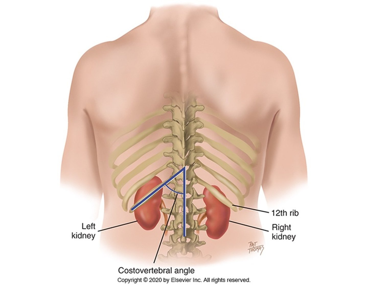
functions of kidneys (2)
fluid/electrolyte balance and urine production
internal structure of the kidney
cortex (outside) --> medulla (inside)
medulla is comprised of 8-20 renal pyramids --> nephrons come through to secrete urine into a papilla at the tip of each pyramid--> minor calix at the end of each papilla --> minor calices join to form ~3 major calices --> major calices dump into renal pelvis (urine collection center, connection for renal vasculature) --> connects to ureter --> urine to the bladder
