BIO 201 Digestive System
 Call Kai
 Learn
Learn Practice Test
 Spaced Repetition
 Match
Match Flashcards
 Knowt Play
1/30

There's no tags or description
Looks like no tags are added yet.
Name | Mastery | Learn | Test | Matching | Spaced | Call with Kai | Chat |
|---|
No analytics yet
Send a link to your students to track their progress
31 Terms
Digestive System
Set of organs that breaks down food items into nutrients. Two types of organs include:
Organs of alimentary canal
aka gastrointestinal tract
Accessory organs
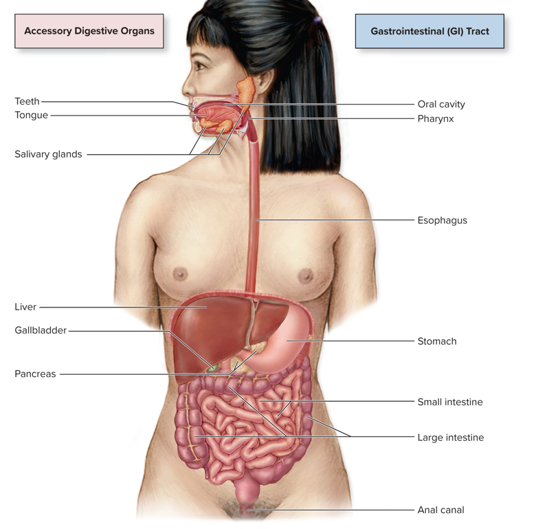
Accessory Organs of the Digestive System
Teeth
Tongue
Salivary Glands
Liver
Gallbladder
Pancreas
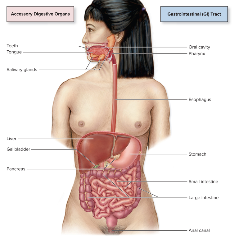
Organs of the Alimentary Canal (GI Tract)
Oral Cavity
Pharynx
Esophagus
Stomach
Small Intestine
Large Intestine
Anal Canal
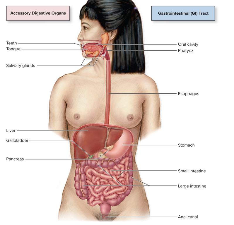
General Functions of the Digestive System
Take food into the body and break it down into its component nutrients (Most Fundamental Function)
Taking in water and electrolytes via diet & delivering them to the blood. (Fluid, electrolyte, and acid-base homeostasis)
Ingesting vitamins & minerals
Producing hormones
Excreting metabolic wastes
Six Basic Processes of the Digestive System
Ingestion
Secretion
Propulsion
Digestion
Absorption
Defecation
Ingestion
Intake of food and water via the mouth.
Basic process of the digestive system
Secretion
Release of mucus, enzymes, acid, and hormones by endocrine and exocrine glands to aid other digestive processes.
A basic process of the digestive system
Propulsion
Movement of ingested food and liquids from one digestive organ to the next.
Basic process of the digestive system
Accomplished largely by peristalsis
Aided by mucus secreted by multiple organs
Peristalsis
Rhythmic contractions of smooth muscle of the alimentary canal that helps with the propulsion of food and liquids.
Digestion
Food Breakdown
Basic process of the digestive system
Two Kinds:
Mechanical Digestion
Chemical Digestion / Enzymatic Hydrolysis
Mechanical Digestion
Physical breakdown of food into smaller pieces
Examples: chewing & mixing by muscles
Chemical Digestion / Enzymatic Hydrolysis
Enzymes catalyze reactions that break chemical bonds
Absorption
Nutrients move through the wall into blood or the lymphatic vessels.
Basic process of the digestive system
Water, electrolytes & vitamins also move into the bloodstream
Defecation
Materials that are not digestable or usable by the body continue their transit through the alimentary canal until they exit the body as feces.
Basic process of the digestive system
A specialized form of propulsion
Motility/Movement
A key process in every region of the alimentary canal. Includes:
Swallowing
Churning
Peristalsis
Defectation
Regulated by the nervous system
Autonomic Nervous System (ANS)
Enteric Nervous System (ENS)
Also regulatied by the endocrine system
Hormones
Motility Using Skeletal Muscle
Oral Cavity
Pharynx
Superior Portion of the Esophagus
Last Portion of the Large Intestine
Motility Using Smooth Muscle
All of the alimentary canal except for:
Oral Cavity
Pharynx
Superior Portion of the Esophagus
Last Portion of the Large Intestine
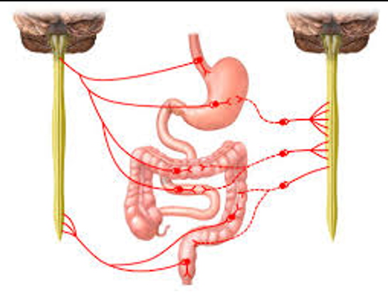
ANS Regulation of the Digestive System
Known as long reflexes.
Must travel outside of the local digestive neurons to the CNS
Sympathetic Nervous System
Inhibits digestive processes
Parasympathetic Nervous System
Activity stimulates digestive processes
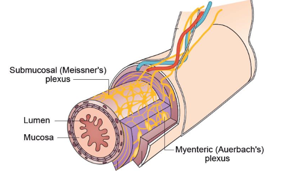
ENS Regulation of the Digestive System
Known as short reflexes.
Reflex pathways are confined to local neurons
Self-contained branch of the ANS
Extends from the esophagus to the anus
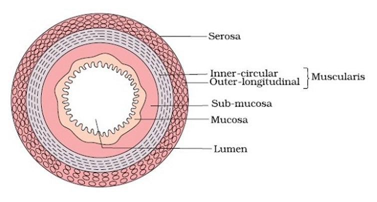
Histology of the Alimentary Canal
Organs follow the same general tissue patterns of other hollow organs:
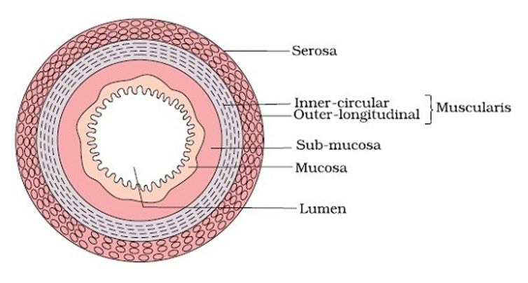
Concentric layers of tissue surround a space called the lumen
Most regions contain four layers (tunics):
Mucosa: Inner Epithelium
Submucosa: Connective Tissue
Muscularis: Smooth Muscle
Serosa: Outer Layer of Connective Tissue
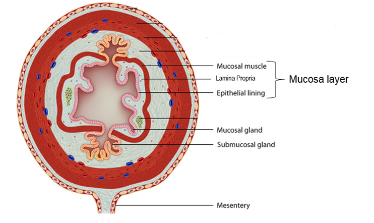
Mucosa
Inner epithelial layer of the alimentary canal.
Contains three components:
Layer of epithelium faces the lumen
Thin layer of loose connective tissue called lamina propria
Thin layers of smooth muscle known collectively as the muscularis mucosae
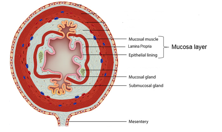
Lamina Propria
Middle layer of mucosa between epithelium and mucosal muscle.
Houses blood and lymphatic vessels, glands, and mucosa-associated lymphoid tissue (MALT)
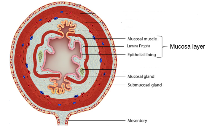
Muscularis Mucosae
Outermost layer of the mucosa between the lamina propria and the submucosa.
Contractions facilitate release of secretions into the lumen
Increases contact of materials with mucosa
Submucosa
A layer of connective tissue between the mucosa and muscularis layers of the alimentary canal.
Composed of dense irregular connective tissue with blood and lymphatic vessels as well as submucosal glands
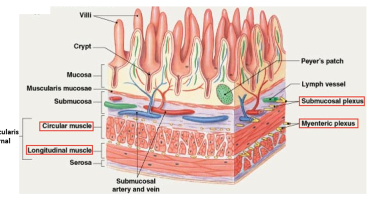
Houses submucosal plexus and MALT Tissue
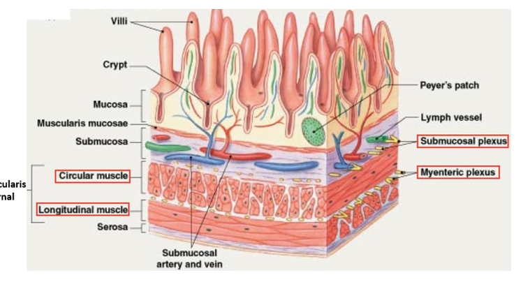
Submucosal Plexus or Meissner’s Plexus
Nerve clusters in the submucosa that are a part of the ENS.
Regulate secretion and blood flow
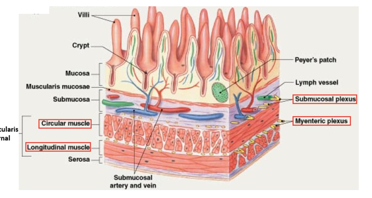
MALT Tissue of Submucosa
Prevents ingested microbes from crossing the GI tract wall
Peyer’s Patches in distal small intestine
Muscularis Externa
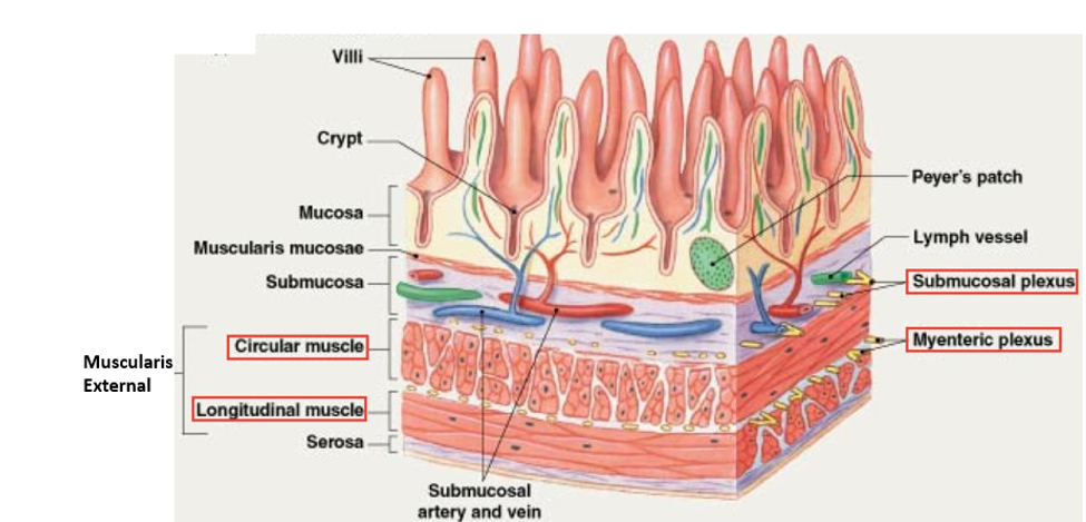
A thick, smooth muscle layer found in most of the alimentary canal. Composed of two layers:
Inner circular layer of smooth muscle
Outer longitudinal layer of smooth muscle
Also houses the myenteric plexus (Auerbach’s plexus)
Myenteric (Auerbach’s) Plexus
Nerve clusters of the ENS are found in the muscularis externa layer of most of the alimentary canal.
Regulate the motility of the muscularis externa
Serosa (Visceral Peritoneum)
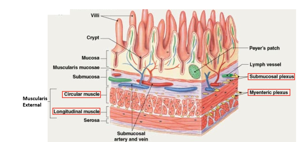
The outer layer of connective tissue found in the organs of the alimentary canal that are surrounded by the peritoneal cavity.
Composed of simple squamous epithelial tissue and loose connective tissue
Supports the digestive organs and anchors them to surrounding structures
Adventita
The outer connective tissue layer found in the organs located outside the peritoneal cavity within the alimentary canal.
Composed of dense irregular connective tissue
Supports digestive organs and anchor them to surrounding structures
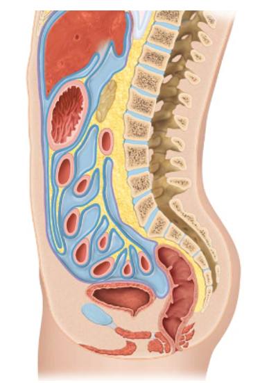
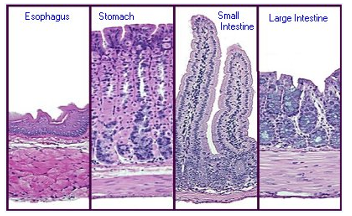
Epithelium of Mucosa (Stomach → Large Intesine)
Simple columnar epithelium with copious goblet cells that secrete an alkaline mucus.
Mucus coats the epithelium and protects surrounding tissues from ingested food and chemicals secreted by digestive organs.
Regenerative epithelial cells that have a high rate of mitosis allow the mucosa to replace epithelial cells as they undergo apoptosis and are sloughed off in the alimentary canal