C1: Cell Structure
 Call Kai
 Learn
Learn Practice Test
 Spaced Repetition
 Match
Match Flashcards
 Knowt Play
1/44

Earn XP
Description and Tags
Vocabulary flashcards reviewing cellular biology organelles, structures, microscopy measurements, and comparisons based on the lecture notes.
Name | Mastery | Learn | Test | Matching | Spaced | Call with Kai | Chat |
|---|
No analytics yet
Send a link to your students to track their progress
45 Terms
Unit conversions (cm → mm → ųm → nm)
1cm=10mm=10000μm=10000000nm

What is the diameter of a typical plant cell?
100 ųm
What is the diameter of a typical animal cell?
40 ųm
Name the structures that plant and animal cells have in common. (CB pg 5)
nucleus with nucleolus and chromatin
cytoplasm containing mitochondria, Golgi apparatus and other small structures
cell surface membrane
Name the structures that are found in plant cells only. (CB pg 5)
chloroplast
large permanent central vacuole
cell wall with middle lamella and plasmodesmata
Name the structure that is only found in animal cells. (CB pg 5)
Centriole
What is the meaning of resolution in microscopy?
The ability to distinguish between two separate points
The higher the resolution, the greater the detail that can be seen
Greater in electron microscopes than light microscopes because the wavelength of an electron is shorter than the wavelength of light.
Nucleus
Structure:
has double-membrane,
has chromatin, nucleolus, and surrounded by nuclear envelope
Function
Contain genetic materials
Controls cell activities

Nucleolus
Structure:
made of loops of DNA from several chromosomes
deeply staining material in the nucleus
rounded structure
Function:
Make ribosomes (combine mRNA with proteins)
Chromatin
Structure:
deeply staining material in the nucleus
mass of coiled threads
Function:
chromatin threads coil together to form chromosomes
Nuclear envelope
Structure:
continuous with the endoplasmic reticulum
double membrane
has nuclear pore
Function:
Allow exchange of materials between the nucleus and the cytoplasm (mRNA and ribosomes)
Substance leaving the nucleus: mRNA, tRNA, ribosomes
Substance entering nucleus: proteins (to make ribosomes), nucleotides, ATP, hormones such as thyroid hormone T3
Cell surface membrane
a selectively permeable boundary enclosing the cytoplasm
regulates the movement of substances in and out of the cell
contains molecules essential for cell signalling and recognition.
Rough Endoplasmic Reticulum (RER)
Structure:
Has ribosomes on surface
Connected cisternae is connected to nuclear envelope
Flattened shape of cisternae/membranous sacs
Vesicles contain protein
Function:
make, modify and transport proteins from transport vesicles to Golgi apparatus
Smooth Endoplasmic Reticulum (SER)
Structure:
Tubular/looped
Connected cisternae
No ribosomes
Vesicles contain lipids
Function:
Make lipids and steroids such as cholesterol
Detoxify the body from metabolic wastes and drugs / drug metabolism
Makes reproductive hormones such as oestrogen and progesterone
Major storage site for calcium ions
Abundant in muscle cells where calcium ions are involved in muscle contraction
Mitochondria
Structure:
double membrane
inner membrane folded to form cristae, which projects into the interior called matrix
Has circular DNA and 70S ribosomes
can move, change shape and divide
Functions:
Synthesise ATP via cellular respiration
Synthesise lipids

ATP
Adenosine triphosphate
Made of a ribose, adenosine and three phosphate groyps
Golgi apparatus (Golgi body or Golgi complex)
Structure:
flattened/bent/curved
Form secretory vesicles that contain protein
Some vesicles mature to become lysosome inside the cell
Function:
processes/modifies proteins
Lysosomes
Contain digestive enzymes
Break down waste substances and old organelles
Work fastest in acidic pH
Contain enzymes such as protease, lipase and nuclease which break down protein, lipids and nucleic acids respectively
Functions:
Engulf and destroy unwanted cell components such as molecules or organelles
Endocytosis
Exocytosis
Self digestion (autolysis)
80S Ribosomes
larger type of ribosome found in eukaryotic cells
located freely in the cytoplasm and attached to the rough endoplasmic reticulum
70S Ribosomes
smaller type of ribosome found in prokaryotic cells
Found in chloroplasts and mitochondria
Ribosome
makes protein
70S: chloroplast and mitochondria
80S: cytoplasm and RER
Centrioles
Structure:
exist as a pair
Arrange in right angle to each other
Contain a ring of 9 triplets microtubule
Function:
make cilia and flagella
Microtubules
Structure:
Long, rigid, hollow tubes
Combine with actin filaments and intermediate filaments to from cytoskeleton
Made of protein called tubulin (alpha and beta tubulin) that combine together to form dimers
Dimers join together end to end to form protofilaments
13 protofilaments line up in a ring to form a cylinder with a hollow centre (microtubule)
Function:
determine/maintain the shape of cell
Centrosome
act as microtubule organising centres (MTOC)
form spindle fibre during cell division
Cilium/cilia
A hair-like projection extending from a cell surface
Short and many cilia per cell
Motion: back and forth motion, moving ecf past cells
Flagellum/flagella
Long and one or few per cell
Motion: wave-like movement/snake-like motion, propel through ecf
Function of flagella and cilia
Aid in the movement of cell
Microvilli
folding on cell surface membrane
Increase the surface area for absorption or exchange of materials
Chloroplasts
Structure:
double-membrane
contain stroma, thylakoid stacks called grana connected by lamella
has circular DNA (contain genetic code to make protein for photosynthesis) and 70S ribosomes.
contain starch granules (product of photosynthesis)
Function:
Absorb light energy during photosynthesis

Cell wall
Gives the cell a definite shape
Prevents the cell from bursting when water enters through osmosis
Porous to allow the movement of substances to the cell surface

Plasmodesma/plasmodesmata
Linked to neighbouring cells by means of fine strands of cytoplasm
Allow the movement of substances (organic substances, ions, water, amino acids, sugars) between cells
Large central/permanent vacuole and tonoplast
help regulate osmotic pressure of the cells
Maintain cell turgidity
Prokaryotic Cells
Small (1−5μm)
typically unicellular organisms
lacking a nucleus or membrane-bound organelles
Eukaryotic Cells
Larger (up to 40μm)
typically multicellular cells containing a true nucleus, membrane-bound organelles, linear chromosomes with histones, and 80S ribosomes.
Compare prokaryotes and eukaryotes

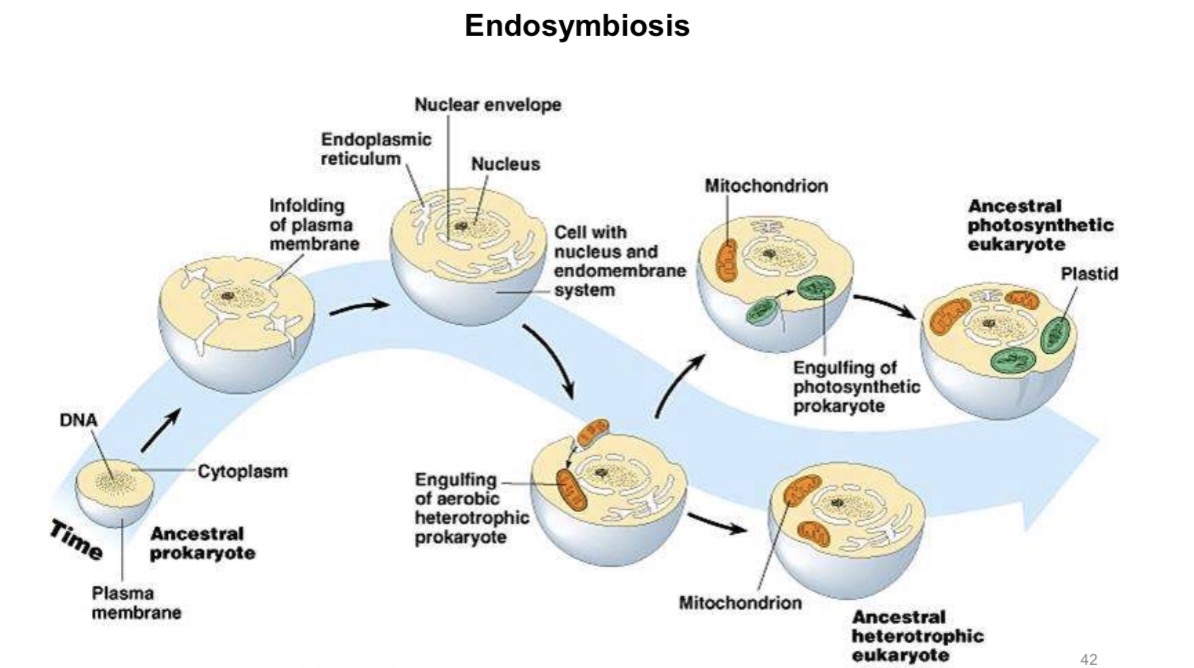
Endosymbiont theory
Mitochondrion and chloroplast have 70S ribosomes and circular DNA like bacterium.
Mitochondrion and chloroplast are in fact ancient bacteria which now live inside larger cells of animals and plants.
This is known as endosymbiont theory.
‘Endo’ means inside and a ‘symbiont’ is an organism which lives in a mutually beneficial relationship with another organism.
Viruses
acellular/not made of cells
has a protein coat/capsid which is made from capsomere
Size: 15 nm to 1000 nm
Capsid
The outer protein coat of a virus that encloses its genetic material and may feature attachment proteins for host cell entry.
Types of microscopes
Light microscope
Electron microscope
Transmission electron microscope (TEM)
Scanning electron microscope (SER)
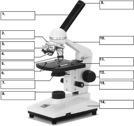
Label the parts of a light microscope and state their functions
Why organelles such as mitochondrion look different?
Have different shape
Have different orientation
Cut at different sections
Mitochondrion → divides
Transmission electron micrograph (TEM)
Shows internal structures/details of organelles (e.g. double membrane)
Show very thin sections through the cell
Show 2D / flat views of cell structure
Image is in one plane
Scanning electron micrograph (SEM)
Show outer/external view of cell structure/organelles
Good focus with different depth
Show surface, contour, 3D views
Compare light microscope and electron microscope

What are the advantages of using an electron microscope rather than a light microscope?
EM has higher resolution because of shorter wavelength
The solution is equal to half the wavelength
Resolution of light microscope is 0.2 ųm / 200 nm
Resolution of electron microscope is 0.0005 ųm / 0.5 nm
More detail can be seen/much clearer at the same magnification