Chapter 9 - Vision
1/104
There's no tags or description
Looks like no tags are added yet.
Name | Mastery | Learn | Test | Matching | Spaced |
|---|
No study sessions yet.
105 Terms
What are Sensations?
The registration of physical stimuli from the environment by the sensory organs.
What are important factors for Reception and transduction?
Conversion to neural activity:
electromagnetic (vision) → ch9
air pressure, mechanical (audition) → ch10
thermal, mechanical, electrical (somatosensory) → ch11
chemical (taste and olfaction) → ch12
What is Encoding? What are important terms for it?
Differentiation between sensations.
Activity (action potentials) → frequency, modulation, rhythm.
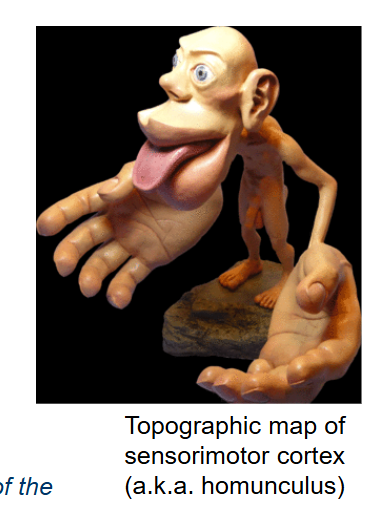
Spatial (topographic map) → neural-spatial representation of the body or of the areas of the sensory world perceived by a sensory organ.
What is a Topographic map?
A neural-spatial representation of the body or of the areas of the sensory world perceived by a sensory organ.
What is Perception? What is it influenced by?
Subjective experience of sensation.
influenced by context, emotional state, past experiences.
What does an organism’s perception of the world depend on?
On its nervous system’s complexity and organization.
Does the same sensory stimulation result in the same perceptions?
No, the same sensory stimulation may result in different perceptions → Perception is an idiosyncratic representation of reality.

What does it mean when we say that Perception is an idiosyncratic representation of reality?
Perception depends on the individual and culture.

What is the primary sensory experience in humans?
Visual
How may visual information alter perception in other senses?
Visual-auditory (e.g. speech-in-noise, McGurk effect)
Visual-tactile (e.g. thermal perception, rubber hand illusion)
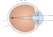
What does the structure of the eye look like?
Pupil = aperture.
Iris = determines size of aperture and hence the amount of light.
Lens = focuses light to retina/film
Retina = film/sensor, transduces light
The eye works like a camera (actually: reverse)
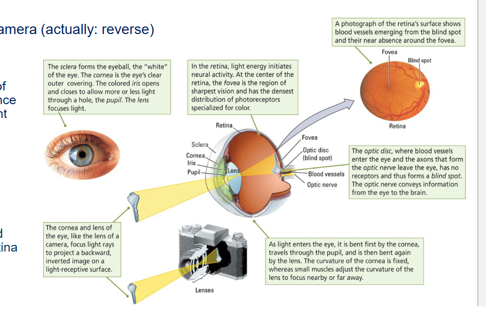
What does it mean when we say that the eye works like a reverse camera?
Visual information is processed ‘upside-down’ and left/right reversed on the retina → corrected by brain.
What is the Sclera?
It forms the eyeball, the “white” of the eye.
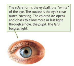
What is the Cornea?
The eye’s clear outer covering.

What does the colored Iris do?
It opens and closes to allow more or less light through a hole, the pupil.

What does the Pupil do?
Light passes through the pupil.

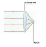
What does the Lens do?
It focuses light.

What do the Cornea and the Lens of the eye do?
They focus light rays to project a backward, inverted image on a light-receptive surface (like a camera).
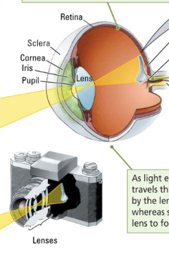
What happens in the Retina?
Light energy initiates neural activity.

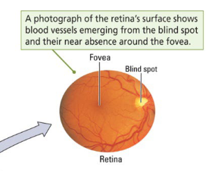
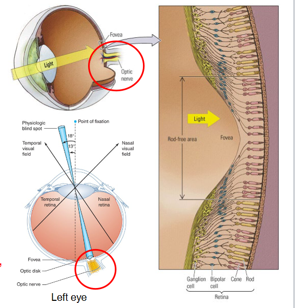
Where is the Fovea located and what does it do?
At the center of the retina, the fovea is the region of sharpest vision and has the densest distribution of photoreceptors specialized for color.

What is in the Optic disc?
Here blood vessels enter the eye and the axons that form the Optic nerve leave the eye, has no receptors and thus forms a blind spot.

What is the function of the Optic nerve?
It conveys information from the eye to the brain.
The place where it leaves the eye is called a blind spot.
How does light travel through the eye?
Light enters the eye
Light is bent by the cornea
The curvature of the cornea is fixed
Light travels through the pupil
Light is bent again by the lens
Small muscles adjust the curvature of the lens to focus nearby or far away
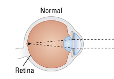
What does a Normal eye structure look like?
The lens focuses incoming light directly on the retina.
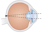
What happens in Myopia? What is it caused by?
aka. Nearsightedness
People cannot bring distant objects into clear focus, because the focal point of light falls short of the retina.
Most commonly caused by the normally round eyeball being elongated → eyeball is too long. Can also be caused by excessive curvature of the front of the cornea.
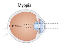
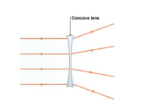
Is this Myopia or Hyperopia? How can it be corrected?
Myopia, can be corrected with a diverging/concave corrective lens (-).
What happens in Hyperopia? What is it caused by?
aka. Farsightedness.
People cannot focus on nearby objects, because the focal point of light falls beyond the retina.
The eyeball is too short. Can also be caused by the lens being too flat to refract adequately.
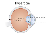
Is this Myopia or Hyperopia? How can it be corrected?
Hyperopia, can be corrected with a converging/convex corrective lens (+).
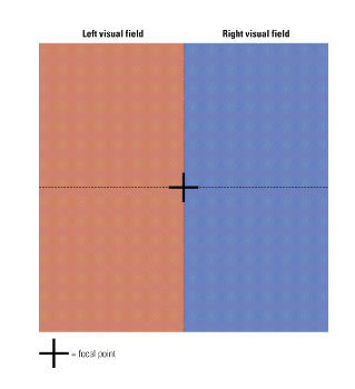
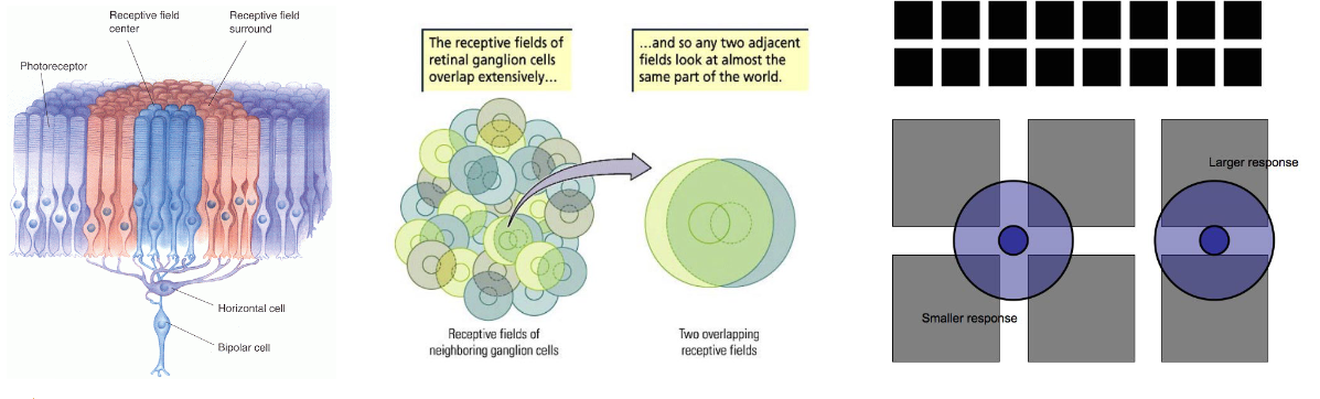
What is the difference between a Visual field and a Receptive field?
Visual field
Part of the visual space seen by the eyes = what a person sees.
Receptive field
Part of the visual space that activates a certain cell = what a cell “sees”.
What does Light consist of? What kind of lights are there?
Electromagnetic waves.
Direct (lamp)
Indirect (reflected of objects)
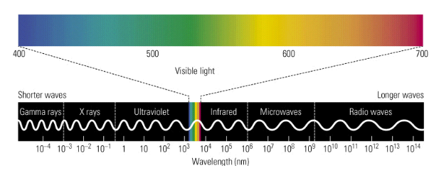
The electromagnetic energy visible to humans varies in wavelength from which nm to which nm?
~ 400 nm (deep purple) to 700 nm (red).

What limits the color we can see?
These constraints follow from the properties of our ~120 million photoreceptor cells in the retina.
What is the Retina?
A light-sensitive layer (‘film’) of the eye where the transduction of light (photons) into neural activity takes place by photoreceptor cells?
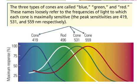
What are the 3 types of photoreceptors?
Rods
Cones
Photosensitive retinal ganglion cells (see ch13).
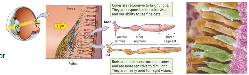
What are the qualities of Rods?
Sensitive to dim light
Black/white and night vision, not color
What are the qualities of Cones?
Blue, green, red
Sensitive to bright light
Color vision and fine detail (visual acuity)
What are the qualities of Photosensitive retinal ganglion cells (see ch13)?
Synchronize circadian rhythms, regulate pupil size
Regulate melatonin release by pineal gland
Where is the Fovea located and what does it contain?
In the center of the retina (macula), it is densely packed with cones, no rods.
Fine detail / sharp, color vision
Where is the Peripheral visual field located and what does it contain?
Around the fovea, less densely packed, predominantly rods.
See less sharp.
Where is the Blind spot / Optic disc located and what does it contain?
Medial to fovea, nasal side of retina.
Axons from retinal ganglion neurons exit the eye here to form the optic nerve to the brain.
Blood vessels also enter and exit the eye here.
No photoreceptors here, so we cannot see with this spot.
Because the two eyes’ visual fields overlap, the right eye can see the left eye’s blind spot and vice versa.
Why can we not see in the Blind spot / Optic disc?
There are no photoreceptors here.
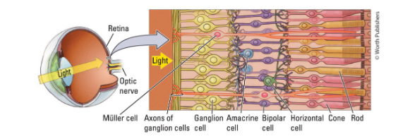
What are Photoreceptors connected by?
By two layers of retinal neurons:
Layer 1
Horizontal cells
Bipolar cells
Amacerine cells
Layer 2
Retinal ganglion cells (RGCs), two types:
Parvocellular
Magnocellular
What are the two types of Retinal ganglion cells (RGCs)? What are their qualities?
Parvocellular
small, mostly in fovea, input from cones → color, fine detail.
Magnocellular
large, throughout retina, input from rods → light, movement, not color.
What forms the Optic nerve?
The axons of the retinal ganglion cells.
How does light go to the back of the eye?
Light goes through the axons to the back of the eye, where it stimulates the photoreceptors (rods, cones).

What is the Optic chiasm?
The part where the optic nerves partly cross, just before entering the brain.
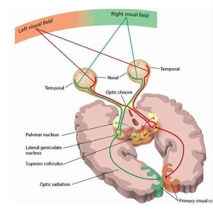
Where do the Optic nerves cross in the Optic chiasm?
Medial (nasal) parts of the optic nerves cross to the other side (contralateral).
Lateral (temporal) parts of the optic nerves travel back to the same side (ipsilateral).
In other words:
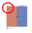
Input from the Right visual field goes to the Left hemisphere.
Input from the Left visual field goes to the Right hemisphere.

Before the optic chiasm (PNS) it is called the Optic nerve. What is the optic nerve called after the optic chiasm (CNS)?
Optic tract
What is the Optic tract?
The optic nerve after the optic chiasm (CNS).
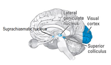
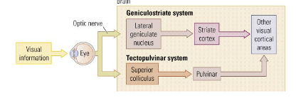
What are the 3 routes to the Visual brain?
Geniculostriate system
Tectopulvinar system (the “where” system)
Retinohypothalamic tract (see ch13)
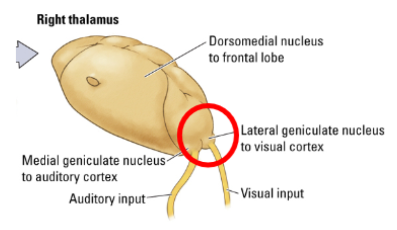
What is the route for the Geniculostriate system? What does the system consist of?
From retina to lateral geniculate nucleus (LGN, in thalamus), to layer IV of the primary visual cortex (V1, striate cortex), and then to other visual areas.
All parvocellular retinal ganglion cells (RGC) axons _ some magnocellular
When you describe what you see, this is processed here.
Info processed by the fovea & RGC is processed here.
What is the route for the Tectopulvinar system? What does the system consist of?
aka. the “where” system.
From retina to superior colliculi (in tectum, midbrain), to pulvinar nuclei in the thalamus, and then to other visual areas (i.e. bypasses the primary visual cortex).
Only magnocellular RGC axons)
notes
doesn’t use the visual system.
knows there are objects around us without being consciously aware.

What is the route for the Retinohypothalamic system? What does the system consist of?
From photosensitive RGC axons to suprachiasmatic nucleus (SCN) in hypothalamus.
Circadian rhythms and pupillary reflex.
notes
sensitive to blue lights
transmits information about what time of the day it is.
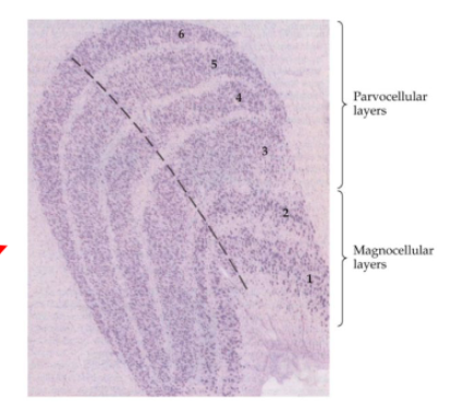
Of how many layers does the Lateral geniculate nucleus (LGN) consist of?
6 layers.
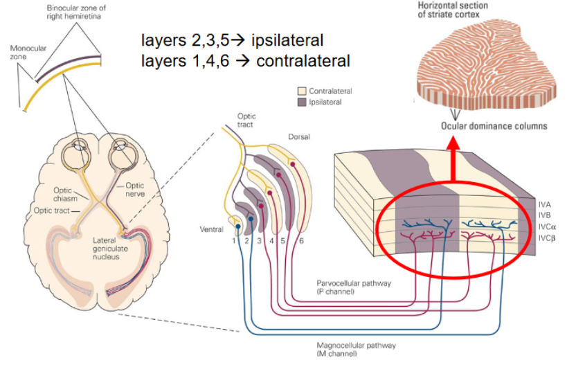
layers 2, 3, 5 → ipsilateral
layers 1, 4, 6 → contralateral
(you don’t need to know the exact layers for the exam).
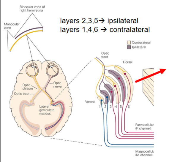
How are the 6 layers distributed in the Lateral geniculate nucleus (LGN)?
Layers 3-6: Parvolellular → color and form (cones)
Layers 1 and 2: Magnocellular → movement (rods)
Input from the magno- and parvocellular retinal ganglion cells and from the two eyes arrives in different LGN layers.
NB: do not confuse with the 6 cortical cell layers (e.g. in V1)
Where does the segregation of visual input remain?
In layer IV of the primary visual cortex (V1).
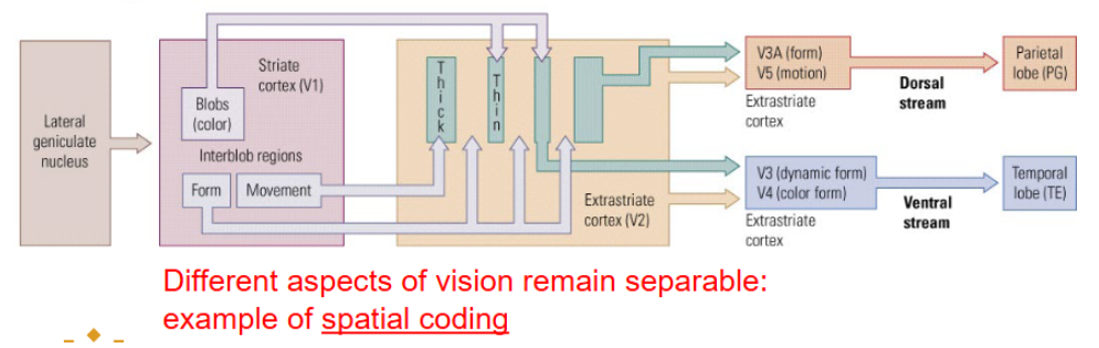
Is the Geniculostriate system an example of spatial coding?
Yes, different aspects of vision remain separable.
Of how many different areas does the Occipital cortex consist of?
6 different areas (not just layers!)
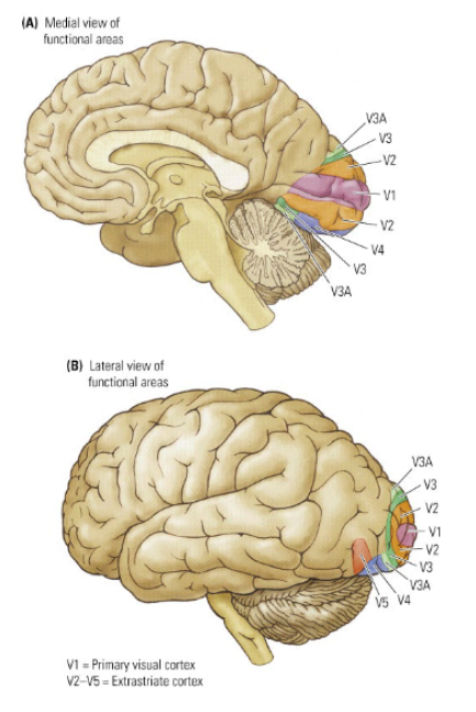
What is the primary projection area (receiving input from LGN) called? What are the other areas (V2, V3, V3A, V4 and V5) called?
V1 (animals)
Brodmann area 17 (humans)
Striate cortex
The other areas are called extrastriate cortex.
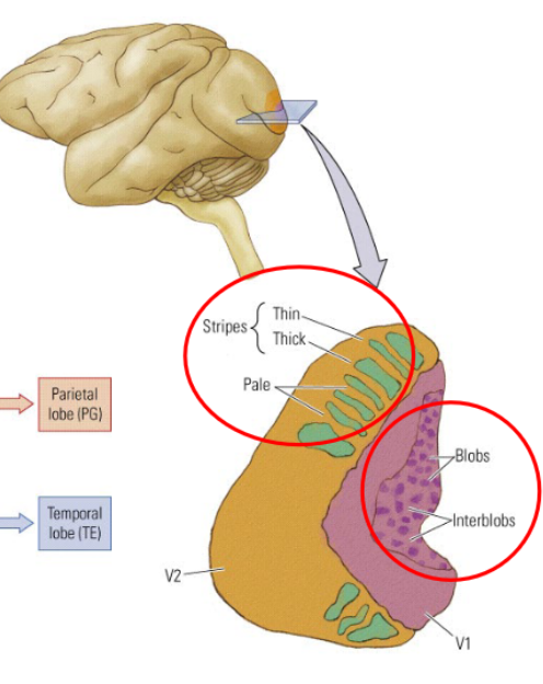
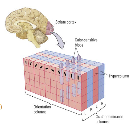
The input from the LGN gives the occipital cortex distinctive patterns, what are they?
in V1 → blob pattern
blobs = color, interblobs = form, motion
(you don’t need to know exactly what the blobs are).
In V2 → striped pattern
thick stripes = motion, thin stripes = color, interstripes = form
(you don’t need to know exactly what the stripes are).
How is the Occipital cortex an example of spatial coding?
Different aspects of vision remain separable.
What does it mean when a neuron has a large receptive field?
It means that the cell “sees” (i.e. responds to) a large part of the visual field.
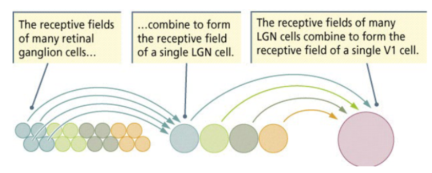
What happens to the receptive fields of neurons as the cell is located further in the processing stream?
The receptive fields of neurons become larger.
The receptive fields of many retinal ganglion cells combined to form the receptive field of a single LGN cell.
The receptive fields of many LGN cells combine to form the receptive field of a single V1 cell.
What happens to Sensory areas that have more cortical representation?
They can provide a more detailed construct of the external world (e.g. fovea).
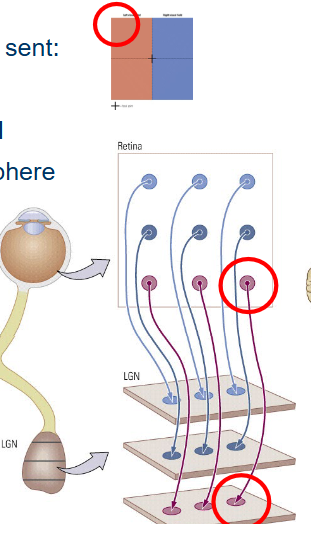
What happens to Information from a receptive field in the retina when sent to the LGN and V1?
It retains its spatial relation.
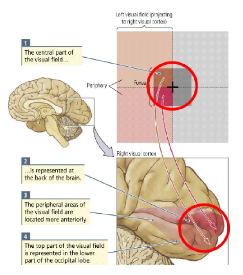
Where is the input from the top of the left side of the visual field sent to? There are 3 locations.
to the bottom-right receptive fields of the retina.
to the bottom-right receptive field of the right LGN.
to the lower-anterior part of V1 in the right hemisphere
Where does spatial coding in V1 take place?
By cortical areas (not by layers, like in LGN).
Other regions of the visual cortex (V3, V4, V5) have similar topographical maps as V1.
How do we perceive shapes on a Neural level?
Neurons at each level of the visual system are selectively sensitive to characteristics of the visual world.
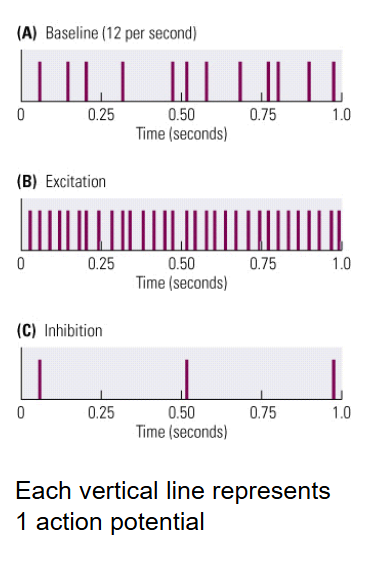
What are the 3 possible responses of a cell to a specific stimulus?
No reaction at all (baseline activity)
Excitation
Inhibition
What do Retinal ganglion cells (RGCs) respond to?
They respond to luminance contrast (not shape).
RGCs respond to the presence or absence of light.
What is Luminance?
Amount of visible light reflected to the eye from a surface.
note: difference in contrast in visual fields.
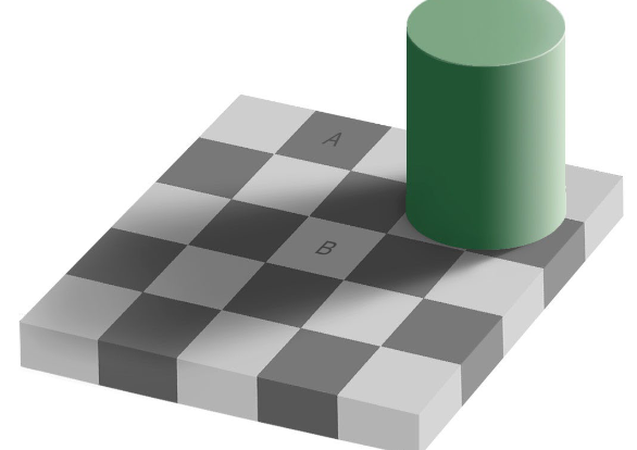
What is Contrast?
Difference in luminance between adjacent parts of that surface.

Why do Retinal ganglion cells (RGCs) respond to luminance contrast → to the presence or absence of light?
Because their receptive fields are miniscule dots (circular).
What are the 2 types of RGCs?
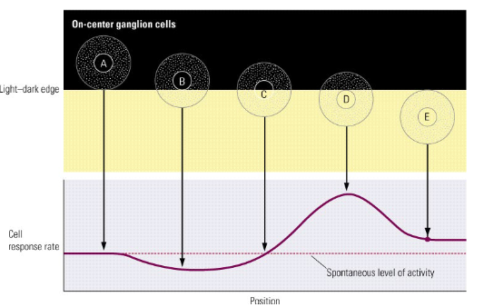
ON-center, OFF-surround
Increase firing rate when light falls on its center
decrease firing rate when light falls in periphery
OFF-center, ON-surround
Decrease firing rate when light falls on its center
increase firing rate when light falls in periphery
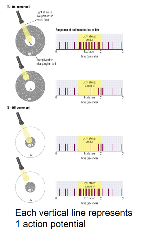
When does a Response of an ON-center cell happen?
If light from the edge of a stimulus falls on it.
The ganglion cell smost affected by the stimulus are those lying around on the edge (B and D).
Regions containing differences in luminance )=areas along the edges) are emphasized.
Information transmitted from RGCs to the visual areas in the brain does not give equal weight to all visual field regions. Is this true or false?
True.
What do RGCs send signals about?
About edges and edges form shapes.
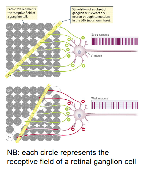
Where does a single V1 cell get its input from?
From multiple retinal ganglion cells (via the LGN).
V1 cells hence have a larger receptive field.
V1 cells are maximally excited by bars of light oriented in a particular direction → orientation detectors.
What do Cells in V1 respond to?
To line segments (not to dots).
What are the qualities of Simple V1 cells?
have an on-off receptive field
respond to line segments (rectangular)
with a specific orientation
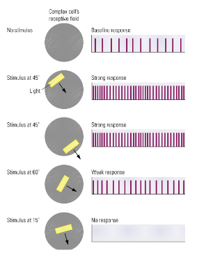
What do Complex V1 cells respond to?
To moving line segments.
with a specific orientation.
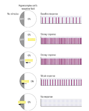
What do Hyper complex V1 cells respond to?
To moving line segments (like complex cells)
Also have a strong inhibitory area at the end of their receptive field (on-off receptive).
What are the 2 different Cell organization in Processing shapes in V1?
Orientation columns
host neurons that respond to the same line orientation.
Ocular dominance columns
receiveinput from the left or right eye.
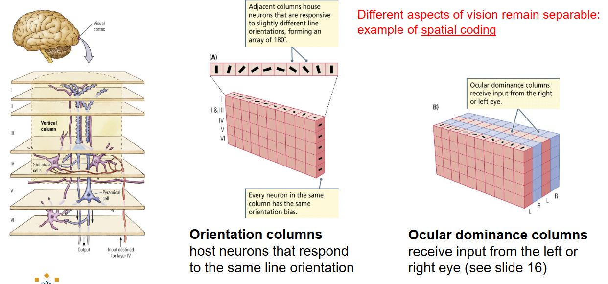
Is the cell organization of Processing shapes in V1 an example of spatial coding.
Yes, different aspects of vision remain separable.
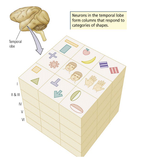
What part does the Temporal lobe have in processing shapes?
Object recognition.
Neurons in the temporal lobe form columns that respond to categories of shapes.
What type of organization does the Temporal lobe have?
Columnar organization:
In Area TE of the temporal lobe, columns host neurons that respond to categories of shapes (e.g. faces, fruit).
What is an object represented by in the Temporal lobe?
By the activity of many neurons with slightly varying stimulus specificity that are grouped in the same column.
What is Stimulus equivalence?
Recognizing an object as remaining the same despite being viewed from different orientations.
Why and How do we see color?
Why → Competitive evolutionary advantage → detect ripe fruit.
How → By cones in the retina.
What are the 2 theories for Seeing color?
Trichromatic Theory
Explains color vision by photoreceptors.
Opponent Processes Theory
Explains color vision at neural level.
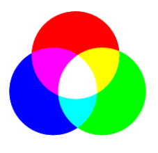
What is the Trichromatic theory by Helmholtz?
We see 3 primary colors because we have 3 types of cones (red, green, blue).
If all primary colors are equally active, we see white.
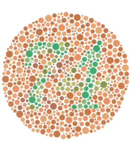
How does the Trichromatic theory explain Color blindness?
If we lack one cone receptor type, we cannot process as many colors as we could with all three (e.g. dichromacy, red-green color deficiency).
What is one disadvantage of the Trichromatic theory?
It does not describe everything: colors seem to occur in pairs, e.g. red-green, blue-yellow, black-white → color-after-effect.
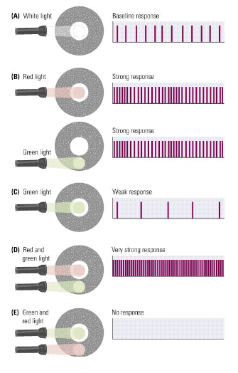
What is the Opponent processes theory by Hering?
ON/OFF, center/surround organization in retinal ganglion cells (RCGs) creates color-opponent cells.
there are three opposing color pairs:
black-white
green-red
blue-yellow (red+green)
What does the Red-green color-opponent RGC look like?
ON-center excitation for red
OFF-center excitation for green
What happens in V1 for seeing color?
The color-sensitive blobs are integrated with the cortical columns and respond in a similar color-opponent way.
V1 contains several thousand ‘modules’, each analyzing color and contour for a particular region of the visual field.
What is perception of color influenced by?
Color constancy (V4).
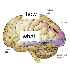
Vision beyond the primary visual cortex (V1); What are the 2 visual streams after the occipital lobe?
Ventral stream to the Temporal lobe.
What functions
Dorsal stream to the Parietal lobe.
How functions
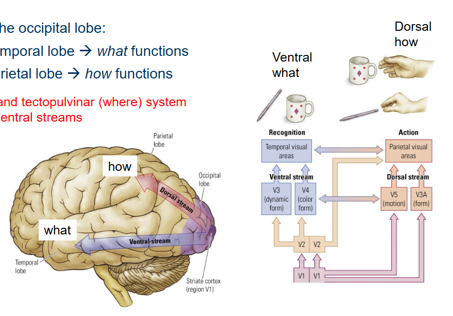
What 2 systems contribute to both the dorsal and ventral streams?
Both the geniculostriate and tectopulvinar (where) system contribute to the dorsal and ventral streams.
Where is the Ventral stream located and what areas does it have?
(what) system.
In the Temporal visual cortex.
Parahippocampal place area (analyzing landmarks, recognizing scenes).
Fusiform face area (face recognition)