What are the two articulation between the costal ribs and the vertebra?
costovertebral
costotransverse
costochondral
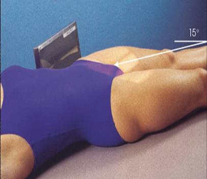
When the AP axial projection is performed for the os calcis:
The leg should be fully extended 2.The plantar surface of the foot should be parallel to the cassette 3.The central ray is directed 40 degrees cephalad to the long axis of the foot
The central ray enters the foot at the head of the fifth metatarsal
A cylinder cone may be used for this projection
For the lateral projection of the scapula:
Patient should be upright to reduce pain
Patient is positioned obliquely with unaffected scapula centered to the cassette 3.Body is adjusted by palpating axillary and vertebral borders of the scapula so that the scapula is lateral 4.Scapula must be projected free of the rib cage
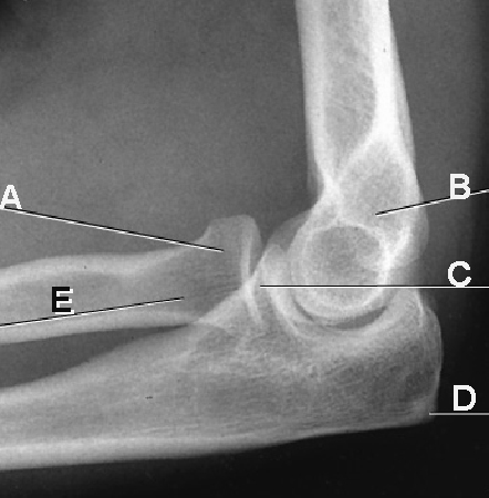
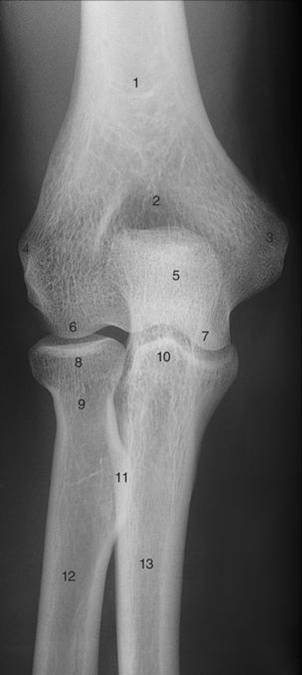
For the AP projection of the elbow:
The forearm and humerus should be at right angles
The central ray is directed perpendicular to the joint
The forearm and humerus should be parallel to the table
The hand must be pronated
The patient may have to lean laterally to ensure AP alignment
For the lateral projection of the forearm:
The ulnar surface must be in contact with the image receptor
The thumb should be in a relaxed position
The humerus and forearm should be in contact with the table 4.The elbow should be flexed 45 degrees 5.The central ray is directed toward the injured joint
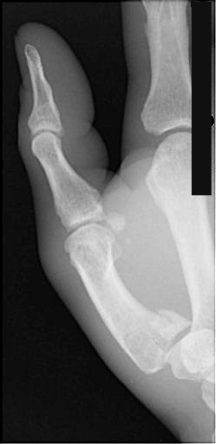
A patient unable to extend his or her arm is seated at the end of the x-ray table, elbow flexed 90 degrees. The CR is directed 45 degrees medially. Which of the following structures will be demonstrated best?
Radial head
Capitulum
Coronoid process
Ulnar flexion/ deviation will best demonstrate which carpal(s)?
Medial carpals
Lateral carpals
Scaphoid
A lateral projection of the hand in extension is often recommended to evaluate
a fracture.
a foreign body.
soft tissue.
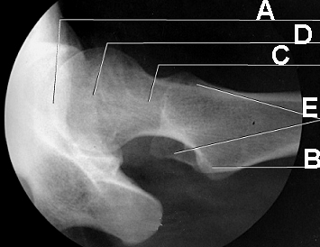
Which of the following will demonstrate the femoral neck free of superimposition
modified cleaves
Lauenstein
Hickey