Equine Unit3 Aos2 - Biomechanics
1/124
There's no tags or description
Looks like no tags are added yet.
Name | Mastery | Learn | Test | Matching | Spaced |
|---|
No study sessions yet.
125 Terms
4 functions of the skeletal system
Movement - Bones attach to muscles, when muscles contract this enables movement.
Support & Structure - Frame work to hold correct shape, place of attachment for muscles, support soft tissue.
Protection - Create protected areas for delicate organs. Cranial, thoracic, spinal.
Production of red blood cells - In the bone marrow, skeleton is a mineral store for calcium.
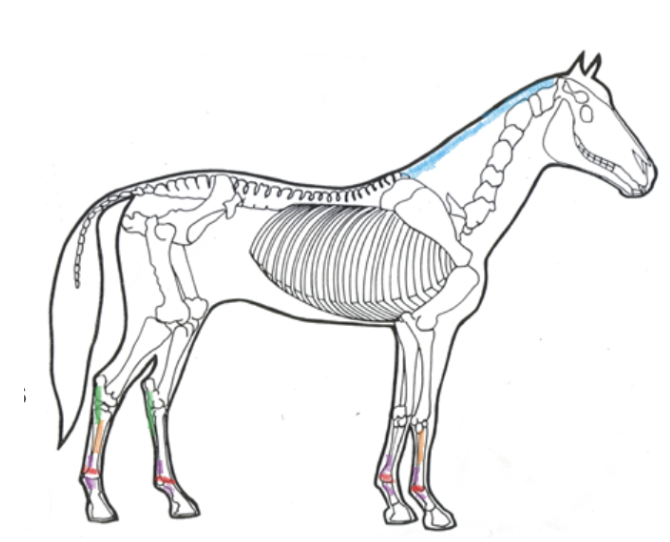
The stay Apparatus
system of muscles, tendons and ligaments that hold a horse in the standing position while its muscles relax. They also use their stay apparatus while they're awake to minimise fatigue due to standing.
F: In the front leg, the stay apparatus is always in place and a horse needs only to relax to take advantage of it.
B: Hind leg, a horse must rotate its hip and literally hook one bone up over a knob on another bone. Complex system based on suspensory ligament, flexor tendons and check ligament
Bone
A living tissue with nerves and blood vessels.
Contains proteins and minerals, particularly calcium and phosphorus(supplement).
Bone is not just a hard, inactive structure. It is a dynamic organ, undergoing constant change and remodelling.
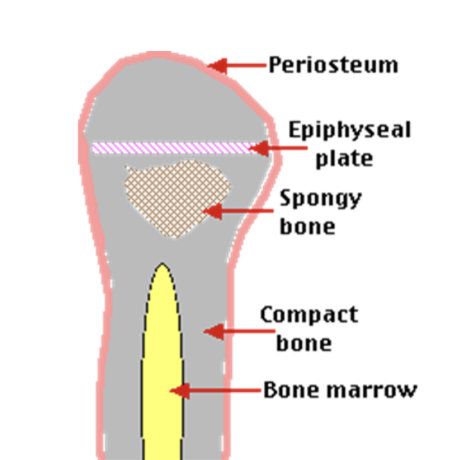
Bone structure PSMBE
Periosteum - entire surface of the bone is covered by a thin protective membrane.
Compact bone - of varying thickness, hard outer cortex, it encases an inner area of: spongy bone.
Medullary Cavity - Spongy bone, Within this cavity is bone marrow.
Bone Marrow - Create new blood cells.
Epiphyseal Plate - (growth plate), a line of cartilage at the end of the long bones.
Types of bones
Long bones – the limbs
Short bones - the knee and hock bones
Flat bones - the scapula, skull, ribs
Irregular bones - the vertebrae
Bone components
Bone has two main components:
- Protein approximately 46%
this is the organic part which forms the framework.
- Minerals approximately 54%
This is the inorganic part which gives bone its hardness. Mainly Calcium (Ca) & Phosphorus (P)
No. of bones
Skeleton - 205 bones
Axial skeleton - 125 bones
Appendicular skeleton - 80 bones (appendages)
Axial skeleton
(Protect vital organs)
- Made up of the skull, spine and ribs
- Acts to protect the horse’s vital organs
Skull - 34 bones
Spinal column - 54 bones
Ribs - 36 bones
Breastbone - 1 bone
Appendicular skeleton
Made up of the appendages - front and back legs
Supports the horse and allows movement
Forelimbs - 40 bones
Hindlimbs - 40 bones
Skeletal system ailments - 5
DOD
Athritis
Laminits
Fractures
Splints
(DALFS)
Developmental orthopedic disease (DOD)
Desc, Cause, treatment, Prevention.
D: A range of problems with the development of the bone.
C: Nutrition, rapid growth, genetics, or trauma to the metaphyseal growth plate/ articular cartilage including poor conformation, lameness, lack of exercise, or excessive exercise.
T: Dependant on the problem, not all forms have a treatment
P: Good feeding management during growth, including during pregnancy and good management practices to minimise the likelihood of injury
Fractures
Desc, Cause, treatment, Prevention, first aid, long term.
D: Damage to the bone resulting in the cracks or breaks
C: Severe trauma or repetitive stress
T:
First Aid – Keep the horse from moving, splinting the injury and calling the vet
Long Term – If treatable stabilising the bones and rest. Not always treatable
Prevention: Good Management
Splints
Desc, Cause, treatment, Prevention
D: Ossification of the ligament that connects the splint bone to the cannon bone. Inflammation of the splint bones which lie on either side of the cannon bone. Splint bones are remnants of toes. May be due to knock to area or overwork. Generally horses >6yo affected.
C: Stress or Trauma to the leg, can be caused by a blow to the area, poor conformation, excessive work.
T: Rest
P: Reducing stress on legs, protecting legs during work, good management. Protective bandages provide support, decreases risk
Arthritis
Desc, Cause, treatment, Prevention.
D: Degenerative Joint Disease – the wear and tear on joints. Usually leads to changes within the bone or cartilage in the joint.
C: Trauma to a joint in the form of stress over time or a wound or infection.
T: No cures, but it can be managed through good management, feed supplements and injections to aid the function of the joints
P: Good management to minimise wear and tear of the joints
Laminitis
Desc, Cause, treatment, Prevention, first aid and long term.
D: Inflammation of the laminae of the hoof
C: Toxicity of the blood – can be caused by too much sugar, infection retained placenta, reaction to drugs, extra pressure on the hooves or excessive concussion.
T: Call the vet for advice
First Aid - Remove the source of the toxicity, stand the horse on soft ground such as deep sand, icing of feet is good but has to be constant and avoid temperature changes
Long term – Prescribed anti - inflammatory, rest, corrective shoeing if P3 has rotated
P: Monitoring and managing susceptible horses off grass, good management to prevent horses over indulging, careful checks of mare postpartum.
Joints
are where two bones meet (Joints bear weight and determine movement).
1) synovial joints
2) cartilaginous or partially movable joints
3) fibrous or immovable jointsS
Synovial Joints
freely moveable joint e.g hip joint, fetlock.
Ligaments attach the two bones together.
The joint is enclosed by a capsule, joint surface is covered with smooth articular cartilage.
•The joint contains a small amount of lubricating fluid, called synovial fluid, This fluid is produced by the synovial membrane lining the joint.
Problems with Synovial joints
If joint is damaged or stressed, excess joint fluid is produced, causing puffy, soft swelling
- Synovial sheaths and bursae may become inflamed and produce excess fluid.Puncture wounds over joints are very dangerous – WHY?
Cartilaginous joints
Partially moveable joints (e.g joints between vertebrae in spinal column).
bones held by cartilage, with no joint cavity.
The articulating bones are held together tightly, allowing little movement.
Fibrous joints
Immovable joints e.g skull
•bones held by fibrous connective tissue, with no joint cavity.
•Only the adult horses have immovable joints. The joints are not fused in foals to allow for their birth and growing.
•These joints are in the pelvis and the skull.
Muscular system purpose and function
Movement – Muscles made up of overlapping filaments that pull together to shorten muscle. One end is attached via a tendon to part of the skeleton to maintain stability, other end attaches to a bone that moves when muscle contracts and relaxes.
Strength– contracting action with other muscles, tendons and ligaments gives the horse strength.
Joint Support – muscles, tendons and ligaments hold each joint together ensuring it moves in the correct directions. Ligaments held absorb concussion and act as a spring in lifting legs in strides.
Muscle composition
Composed of myosin and actin filaments and have the ability to contract, generating a force transferred to the bones via tendons.
Tendons have spring like qualities that link muscle to bone
Require blood supply to keep working
Easily damaged and slow to repair
Factors like conditioning, gender and feed can affect the size of muscles
How muscles work
Muscles work by contracting and relaxing
One end is attached via a tendon to a part of the Skeleton which remains stable
The other end is attached to a bone that moves when the muscle contracts
Muscles work in groups to enable movement
Deep and superficial muscles
Overlay the skeleton and internal structures in layers
Nearest surface – superficial
Primary involved in movement and more prone to external trauma
Closest to internal structures – deep
Deep stabilize the skeleton and maintain the location of internal organs.
Muscles of the neck and head + purpose
Muscles of the head and neck: Trapezius, splenius, Masseter, Brachiocephalicus.
Purpose - Responsible for facial expression, eyelids, lips, large and bulky to mobility and carriage of the head enabling horse to alter its centre of gravity.
Muscles of neck/shoulders + purpose
Muscles of neck and shoulders: Rhomboideus, sternocephalicus, deltoid, pectoral muscles.
Purpose - Horse forehand carries two thirds of the body weight, Movement and head position change the centre of gravity.
Tendons
Fibrous cord of connective tissue attaching muscle to bone, cartilage or other muscle.
•Tendons are more elastic than ligaments, limited extension, have a crimped structure
•Blood supply is poor, collagen is required for effective life
•Where a tendon is positioned to rub on bone or hard surface it is enclosed in a sheath.
Primary tendons of the fore limb
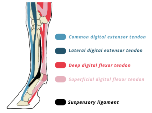
Flexor tendons
On back side and allow the leg to move forward and absorb force of leg, Attached to muscle.
Deep Digital Flexor Tendon runs down back of cannon bone and attaches to coffin bone, Superficial Flexor Tendon runs parallel to Deep Flexor Tendon and attaches to pastern.
Extensor tendons
On the front surface and allow the leg to extend forward and bear weight.
Five extensor tendons main one is the common digital extensor tendon and runs the entire length of leg and attaches into the coffin bone.
Ligaments + common issues
Bands of white (inelastic) and yellow (elastic) fibrous tissue.
Somewhat inflexible, tough, limiting the movement of a joint according to function, the movement required the more white bands. Prevents the leg from dropping too far when bearing weight.
Poor blood supply, lots of sensory nerves (damaged ligaments are difficult to heal due to poor blood flow).
Do not withstand prolonged stretching
Severe stress likely to break the bone
Ligaments ensure bones stay supported together and aligned during movement.
5 ligaments
Nuchal Ligament - Attaches top of skull to withers.
Annular ligaments - Bands around the fetlock to keep flexor tendons stable.
Check Ligament - Attaches the deep flexor tendon to the cannon bone, the only ligament that attaches tendons to bone.
Plantar Ligament - Connects cannon and splint bone to hock bones.
Suspensory Ligaments - Supports the fetlock joint.
Filament movement within muscles
Filaments: Actin (thin) & Myosin (thick)
Relaxed; is a space between the thin filaments(red). Contracted; the thick filaments(blue) pull the thin filaments closer together.
•Myosin filaments release the Actin filaments and allow them to slide back to the relaxed state.
Filament movement responsibility
As muscles can only exert force through contraction, muscles will work in opposite pairs:
The muscle responsible for the intended movement is called the agonist. The muscle responsible for the opposite movement is called the antagonist. Extensor and flexor muscles work in agonist/antagonist pairs.
To extend the leg the extensor muscle contracts while the flexor relaxes. To flex the leg the flexor muscle contracts while the extensor muscle relaxes.
Strength
The contraction of muscle for movement also creates strength
The joints work with the muscle to act as a lever system which increases strength
Muscle fibre can be built up through use and exercise to increase strength
Joints also can increase in strength with exercise
Unconditioned muscles and joints are susceptible to injury
Joint support
Ligaments join bone to bone and hold the joint together
The annular ligament of the lower leg supports the fetlock and pastern
Muscles surround joints for stability
Muscles, ligaments and tendons help absorb concussion as it travels up the leg.
Muscular system injuries and ailments
Soft tissue
Over use or stretching of ligaments, muscles and tendons
Location and severity of the stretch or separation of fibres and type of tissue determines the type of injury
Sprains, strains, tears, ruptures, bowed tendons, suspensory ligament desmitis, curb, azoturia, bruising.
SSTRBTSDCAB
soft tissue injuries
Desc, Cause, treatment, Prevention, impacts.
D: Over stretching of ligaments, muscles or tendons.
C:Stress on tissues, repetitive strain, single misstep, overwork.
T: rest, icing, anti-inflammatory, ultrasound, controlled excursive.
P: good mgmt, appropriate workload, avoiding deep footing.
I: long healing time (L+T) due to blood supply, can become chronic.
Sprain
Stretching or tearing of structures within a joint including ligaments. Joint forces beyond normal range.
Strain
overuse or stretching of muscle or tendon fibres, resulting in damage to tissue.
tears
Tendons stretched to fibre separations resulting in a gap. Identified on ultrasound requires extended time off work and recovery.
ruptures
Complete or partial breakage of fibres in a tendon or ligament. Worst type of injury requiring extensive treatment and rehabilitation.
Bowed tendon
Thickening/inflammation of superficial flexor tendons due to injury usually in front legs. Often lameness and painful bulge along the back of the lower leg.
Suspensory ligament desmitis
Inflammation of the suspensory ligament, chronic when in hind limbs, acute when in front limbs. Can lead to “dropped pastern” or fractures and further tears of ruptures.
Curb
Thickening of the plantar ligament due to strain.
Azoturia - Tying up
Desc, Cause, treatment, Prevention.
D; Muscle Spasms, cramping and stiffness of the muscles of the hind quarters during exercise.Pain often causes sweating and laboured breathing and reluctance to move. Overall stiffness, soreness, muscle tremors.
C: Diet imbalances / Sudden increase in exercise. Hormones / Weather / Can be inherited - Breed.
T: Rest, anti-inflammatories, careful observation for kidney damage
RER – low starch diet, avoid long periods in box, reduce stress
PSSM – Daily exercise to reduce glycogen, low starch, sufficient protein.
P: Ensure balanced diet, carbohydrates(sugar) electrolytes, minerals (esp selenium) & vitamins. Look for behaviour change. Electrolyte intake.
Types of Azoturia
Sporadic Exertional rhabdomyolysis
Any type of breed age, sex as a result of stress and trauma, including over exertions and or dietary or electrolyte imbalance
Chronic Recurrent exertional rhabdomyolysis (RER)
Affects Thoroughbred, Standardbred and Arabian dependent breeds, linked to bodies ability to regulate calcium and is triggered by stress of excitement
Polysaccharide storage myopathy Types 1 and 2 (PSSM)
Affects drafts and quarter horses. Storing large amounts of glycogen leading to spasms.
Malignant Hyperthermia
Genetic mutation found in 1% of quarter horses that can be triggered under general anesthesia.
Bruising
Desc, Cause, treatment, Prevention.
D: Diffuse bleeding under the skin due to a trauma. Hematomas are collection of blood under skin when vein or artery is damaged.
C: A trauma including a blow, a tear, or pressure that damages underlying blood vessels
T: Generally resorb, Ice immediately and Rest, Heat therapy after 36 hours.
P: Good management, safe environment
I: Pain and horse need to rested. Hematomas often need surgical procedure to release blood from skin.
Lameness and leg problems
Unevenness of horse’s stride when moving
Mostly in forelegs ~ 90% are the knee and below. Hind Legs ~ 80% involve hock or stifle
May be acute (sudden onset) or chronic (comes and goes)
Prevention is better than cure.
First Aid – rest & confinement to prevent further damage and initiate healing
If heat & swelling – cold therapy
Diagnosis – avoid further pain and harm
Vet – seek out veterinary advice
Diagnosis
Signs of Injury – blood, heat, swelling
Objects in Hoof? – stone, wire, nail etc
Weight Bearing
Trot Out on hard surface
Raising head to relieve sore leg when trotted out
X –ray
Nerve block
Preventing lameness
Good hoof care – frequent attention from farrier & regular cleaning, oiling & inspection
Purchase horses with good conformation – poor conformation predisposes to lameness
Ride horses according to their level of fitness – do not over-exercise unfit horses
Take care with working surfaces
- Too deep can lead to tendon injuries
- Too hard can lead to concussion injuries
- Avoid stony or pot-holed ground
Make sure the horse has a healthy balanced diet with adequate vitamins and minerals.
- Calcium for bones
- Biotin for hooves
Muscle soreness
Horses need to be warmed up, worked according to fitness level, & cooled down appropriately
Overuse of muscles can make the horse sore. This may be seen –
- stiffness in the stride when lunged or ridden
- horse may dip its back when groomed
Check saddle fit and work schedule.
May need attention from massage therapist or physiotherapist to assist healing.
Common leg problems
Sore shins
Suspensory ligament strain
curb/bowed tendon
Bursal enlargements:
Wind galls
Bog spavin
Thorough pin
Bursal enlargements
Capped elbow/hock
Boney enlargements:
Splints
Ringbone
Bone spavin
Side bone
Ring bone
Term refers to bony enlargement (new growth) below the fetlock, more common in forelegs.
High Ringbone involves the pastern joint
Low Ringbone involves the coffin joint
In severe cases the bone growth can encircle the bones giving “ringbone” its name. Treatment is usually pain management.
Sore shins
Common in 2 & 3yo race horses - Inflammation of the front of the cannon bone.
Due to incomplete bone growth and working too hard or on hard surfaces.
Cold therapy, poultices and rest will fix the problem.
Bone spavin
Bony growth inside the hock below the joint.
Generally the result of a blow
Horses will show shortness of stride and tend to drag the hind toes
Side bone
Often called Ringbone’s “cousin”
Calcification of the collateral cartilages of the pedal bone These are found on either side of the foot protruding above the level of the coronary band. The lateral cartilages support the hoof wall and provide an important role in the support and cushioning provided to the heel.
The front feet are most commonly affected. Caused by – concussion, narrow feet, poor conformation draft breeds predisposed
Suspensory ligament strain
Inflammation of the suspensory ligament. Causes pain, swelling and heat over the ligament. Often requires 6 months rest
Cold therapy alleviates condition
Wing galls
Soft swelling above & behind the fetlock joint
Increased fluid production of joint fluid in the bursae, Horse generally not lame.
Swellings can be reduced by cold therapy, Sometimes corrective shoeing is required, Common in horses who have jumped.
Bog spavin
Swollen hock joint due to soft, spongy Bursal enlargement of the hock joint capsule. Located towards the front inside of the hock joint. Often associated with lameness.
Caused by bang or blow to the joint, wear & tear from work on hard surfaces. Poor conformation & possibly vitamin & mineral imbalance while horse is young.
Thorough pin
Bursal enlargement of the deep digital flexor tendon sheath in hollow area between the back of the hock joint & the point of the hock.
Caused by stress to the tendon from over exertion, over extension and trauma to the tendons and joint. Poor conformation of the hock joint.
Capped elbow/hock
Often caused by trauma to area
Cold, soft swelling on point of hock or elbow
Usually painless
Prevention:
- Hock boots when travelling
- plenty of bedding to protect hock & elbow
Fractures
Outcome depends on the bone affected and the type of fracture
Bone chip injuries can be treated by removal of chip
Small fractures may be treated by removing bone fragment and use of pins
Severe fractures are more difficult, especially as horse's have large weight bearing requirements
Treatment is expensive – usually for breeding horses worth money
Nervous system functions
Control of all the functions the brain and the nerves send and receive electrical impulses that inhibit or activate parts of the body. Neurons (nerves) communicate with each other, cells in the body such as muscle and secretory cells via electrical signals.
Movement neurons send message from brain to specific muscles to signal them to contract and relax and carry back messages about the movement.
Sensation, Sensory organs collect and detect stimuli information about the external environment and transmit it via electrical signal to the brain to process to ensure the horse reacts appropriately.
Central nervous system CNS
Central Nervous system – lies within protective bones – brain +spinal cord.
Brain - centre for processing and storing info.
Cerebrum, the centre of conscious decision-making Cerebellum, involved in movement and motor control. Brain stem, control basic functions.
Spinal cord - bundle of nerve fibres carry messages to body. divided into cervical, thoracic, lumbar, sacral, and caudal segments.
Peripheral nervous system PNS
Peripheral System comprises the nerves that carry the signals to and from the brain.
Sensory Organs
Receives messages from the outside world
Motor
Sends messages from the brain to the body to coordinate movement of the body
Autonomic nervous system ANS
controls the involuntary systems - respiratory and circulatory.
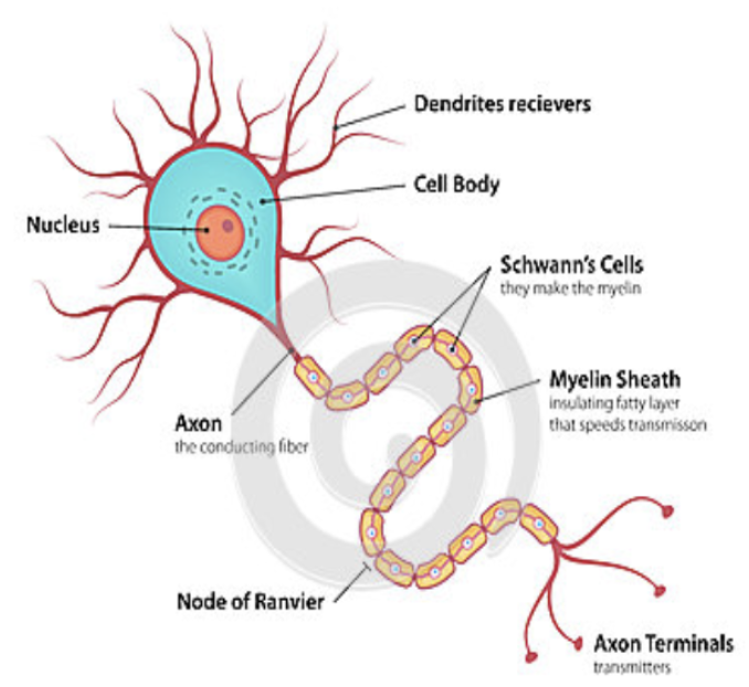
Neurons
Central and peripheral nervous systems contain billions of cells known as neurons.
Sensory – transmit pulses from receptors
Motor- carry impulses to muscles and glands
Intermediate –connect motor and Sensory
Neurons connect with each other to form neurological circuits. Information travels along these circuits via electrical signals.
Center portion is called a cell body and 2 extensions called dendrites and axons.
Dendrites receive signals from other neurons and transmit electrical charges to the cell body.
Axons transmit the electrical charges away from the cell body. When the current reaches the end of the axon, the axon releases chemicals called neurotransmitters.
Neurotransmitters pass the signal to the dendrites of other neurons, or to muscles or glands.
Peripheral system nuerons form and pairs
The spinal nerves extend axons outward into the front and hind legs and to the chest, abdomen, and tail.
Nerves subdivide into smaller nerves to cover the entire surface and interior of the body.
The cranial nerves include sensory and motor neurons that connect the head and face to the brain.
Sympathetic nervous system
Responds to stress or danger
Triggers the fight or flight response (flight stronger in horses)
Makes changes in the body to allow the horse to react to danger
Releases Adrenaline
Slows down digestion
Increases heart and breathing rate
Releases glucose from the liver
Parasympathetic
Responsible for maintaining daily functions
Stimulates digestive system
Relaxes lungs and heart
Allows production of urine
sensation
Skin – Whiskers Sensory Receptors
Eyes - Retina
Ears - Ear Drums
Nostrils - Olfactory Receptors
Tongue - Taste buds
carry signals to the brain for it to be processed
Detection of stimuli
Nervous systems detect stimuli from the outside environment so the horse can react to ensure survival.
First
- Sensation of the stimuli and transmission of signal
- Each sense has specialised organs to convert the stimulus into an electrical signal
Second
- Electrical signal is received and processed in the brain and turned into a thought
Smell
Allows horses to detect objects in its environment including when breathing in
Location of water and food
Predators
Recognise familiar and unfamiliar horses
Sensory receptors that respond to various chemicals that float in the air (smells)
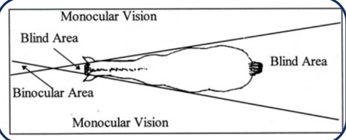
sight
Used to see objects and detect light and dark
Can see in bright and dim light
Have a large field of vision
Very sensitive to variation in their field of vision (movement)
Hearing
Ears used for hearing and balanced
Ears used to pick up sounds from the environment
Moveable ears to locate direction of and focus on different sounds
Horses hear a larger range of sounds than people
Touch
The skin can sense:
Touch
Pressure
Vibration
Temperature – Hot and Cold
Pain
Sensory receptors distributed around the skin
Areas with the most receptors include
Lips
Nose
Eyes
Taste sensation of tongue
Sensation of tongue
Important for the horse to allow them to
Avoid inedible food
Avoid tainted food or water
Select the best feed
illnesses and abnormalities of the nervous system
Wobbler syndrome, tetanus, string halt.
WTS
Wobbler syndrome
Desc, cause, prevention, treatment, impacts.
D: Equine Unco-ordination, wobbly or uncoordinated gait.
C: Compression of spinal cord in the cervical vertebrae which can be caused by malformed vertebrae, arthritis, hypertrophy or inflammation of the spinal cord which causes swelling of the cord.
P: Monitor growth rate in young horses, ensure sufficient Vitamin E in diet, be aware of neck conformation in breeds with higher susceptibility.
T: Neurological test to confirm wobblers: x-rays to find the narrowing, Change feed to slow growth rate, joint injection, and rest. Surgery can be done, Some stablise with rest, diet management and corticosteroids.
I: Loss of coordination which can put the horse and handlers at risk
55% of surgery successful
Tetanus
Desc, cause, prevention, treatment, impacts.
D: Systematic spasms of the muscles due to a neurotoxin.
C: Bacteria Clostridium Tetani from the soil growing in a oxygen free pocket in a wound and releasing a neurotoxin that binds to the nerves contracting the muscles.
P: Vaccination, good wound management.
T: Clean the infection from the wound site, antibiotics, dark quiet stable. If treated early anti-toxin can be administered.
I: If identified early horse can be treated,
50% will die or be euthanized.
String Halt
Desc, cause, prevention, treatment, impacts.
D: Neurological disease, exaggerated and uncontrolled movement of the digital extensor muscle.
C: Toxicity due to the ingestion of flatweed (Hypochaeris radicata) most commonly in late summer on poor quality pasture
P: Good Pasture management, removal of weeds.
T: Remove from pasture, Call vet, Horse may recover over 6-9 months, once removed from the pasture.
I: Some make full recovery, Some may return to work but have weakness. Potentially become a roarer. Poor to no improvement - euthanised.
Respiratory system function
Responsible for supplying oxygen to the bloodstream and removing carbon dioxide from the bloodstream. Body needs oxygen to enable a chemical reaction that produces the energy of life.
The respiratory system of the horse is well adapted to athletic exercise, with unrestricted upper airway diameters, and a large lung capacity afforded by 18 ribs. The lungs can expand 4 times their size when galloping and enable air intakes of up to 1800 litres per minute.
Organs in respiratory system
Nostrils and nasal cavity
Pharynx, larynx,
Airways- Trachea, Bronchioles and Bronchioles
Lungs
Alveolus
Pleura
diaphragm
Respiration process
Breathing is the process of moving air into the lungs (inspiration) and out of lungs (expiration).
This requires movement of the diaphragm and intercostal muscles (between ribs). Respiration rate is determined by stride frequency.
Normal respiration rate is 8-14 breaths/min -> 100
The breathing rate can be counted by watching the horses flanks.
Oxygen as energy
The body needs energy, to grow, repair, reproduce.Energy comes from food eaten passing through the process of digestion.
The main product from digestion is glucose.
Glucose then needs to be combined with oxygen to produce energy plus by products carbon dioxide and water.
Respiratory process - oxygen intake
The diaphragm contracts, pulling caudally on the lungs. This creates a vacuum within the lungs that sucks the air in.
Air then travels - In through the nostrils and nasal cavities, Through the pharynx and larynx, Down the trachea, Into the left and right bronchus.
Through bronchioles in the lungs to the alveoli, Oxygen gets transported into the blood stream, Carbondioxide gets transported from the blood stream and exhaled.
nostrils and nasal cavity
The first structures seen are the nasal turbinates.
Their function is to warm, clean, moisten, and channel the air as it is inhaled into the lower respiratory system (larynx).
Pharynx, larynx and Trachea
The Larynx produces sound by vibrating the vocal cords. The pharynx is a common chamber for the openings to both the lungs via the trachea and the stomach via the oesophagus. Epiglottis on the floor of the pharynx and above are the arytenoids cartilages, which form an upside down V.
These structures form the opening to the trachea. When a horse swallows food or water, the epiglottis covers the opening of the trachea
Trachea - windpipe
Is a rigid tube supported by 50-60 cartilaginous rings
The Trachea divides into the left and right bronchi which further divide into the bronchioles.
Smooth muscle fibres of the tracheal muscle join the inner surface of the free ends of the cartilaginous rings
Extra cartilaginous plates at the foot of the trachea fill in the gaps between the free ends of the main rings.
Lungs
Two large elastic organs which completely fill the chest cavity (apart from the cavity occupied by the heart) between the ribcage and the diaphragm. Surrounded by the pleura, a smooth. slippery membrane that prevents friction whilst the lungs expand and contract.
The basic function is to absorb oxygen from inhaled air and excrete carbon dioxide in exhaled air.
Alveoli - gas exchange
Once in the lung air travels to the alveoli where gas exchange occurs
Oxygen O2 and Carbon Dioxide CO2 move across the membrane between the alveolus and the pulmonary capillary
For every O2 molecules moved to the capillary a CO2 molecule gets taken out of the capillary.
Exhalation
Once the lungs are full of air the carbon dioxide needs to be expelled
The diaphragm relaxes, the intercostal muscles contract and the lungs are squeezed which pushes the air out.
Respiratory diseases
Pneumonia, cold (E HPV), Influenza, strangles, roaring, excerise induced pulmonary haemorrhage, inflammatory airway disease.
PICSREI
Pneumonia
Desc, cause, prevention, treatment, signs.
D: Inflammation of the lungs
C: Pleuropneumonia – secondary bacterial infection of the lungs and pleura caused by weakened defence system. long distance travel, general anaesthetic, strenuous exercise. Viral pneumonia – caused by a virus in immuno-deficient foals. often Arabian breeding.
P: Breaks during long trips, good vaccine protocols and stable hygiene to minimise viral infections of Airways, ensure horse is fit for work required.
T: Call Vet immediately. Drain and monitor fluid in the lungs, antibiotics, anti-inflammatories, pain relief and supportive care.
S: Fever, rapid, shallow breaths, coughing, depression, loss of appetite, symptoms of pain around the chest area.
Cold - Equine Herpes virus
Desc, cause, prevention, treatment, signs.
D: An upper respiratory infection
C: Viral infection caused by the Equine Herpesvirus (EHV)
P: Good stable hygiene, quarantining new arrivals, vaccination of pregnant mares, keeping pregnant mares and young stock separate from competition horses.
T: Isolation to prevent spread, rest, and supportive care to assist recovery.
S: Runny nose, cough, fever, lethargy. Some strains can cause late term abortion in pregnant mares or paralysis of the limbs in young horses.
Influenza
Desc, cause, prevention, treatment, signs.
D: Equine Influenza is an acute, highly contagious viral respiratory disease, Considered to be exotic to Australia.
C: Airborne virus that can be transmitted by the air or carried on people, animals or objects.
P: Vaccination, Quarantine, disinfecting cars, floats, people, equipment if in contact with a sick horse.
T: Supportive care until the virus passes
S: Fever, deep dry hacking persistent cough which may last 2-3 weeks, often accompanied by depression, loss of appetite, laboured breathing and muscle stiffness.
Strangles
Desc, cause, prevention, treatment, signs.
D: Bacterial Infection in the upper respiratory tract and the lymph nodes of the throat and head.
C: Streptococcus equi equi, transmitted through the air or by contact.
P: Vaccination, Quarantine new animals, good stable hygiene practices.
T: Call the vet, isolate to prevent spread, supportive care, warm dry dust free stable, rupture and flush abscesses, antibiotics can be used, but can also put the horse at a risk of forming ‘bastard strangles’ or prevent the horse from forming sufficient antibodies.
S: Thick yellow mucus discharge, fever, depression, enlargement of submandibular lymph nodes which can later form abscesses, difficulty swallowing, noisy breathing.
Roaring
Desc, cause, prevention, treatment, signs.
D: Laryngeal Hemiplegia - Paralysis of the (left) side of the larynx due to neuron atrophy resulting in a collapsed arytenoid cartilage.
C: Genetics and size (more common in males with long necks) Trauma, Toxicity from plants.
P: None
T: Tie Back surgery which sutures open the affected cartilage. Can be done by laser to reduce the noise but does not improve.
S: Excessive noise on inhalation and exercise intolerance, often asymptomatic at rest, but can have a odd sounding whinny.
Exercise induced pulmonary haemorrhage.
Desc, cause, prevention, treatment, signs.
D: Bleeding from the upper and/or lower airways.
C: Unknown, but thought to be related to the high vascular pressure in the pulmonary blood vessels during exercise.
P: None - Zero tolerance in Australian racing
T: Drug Furosemide can reduce incidence and severity, Nasal dilator bands can help.
S: Bleeding from the nose, best diagnosed 30 -90 minutes after exercise (endoscopy).