LAB EXAM BISC 303
 Call Kai
 Learn
Learn Practice Test
 Spaced Repetition
 Match
Match Flashcards
 Knowt Play
1/61

Earn XP
Description and Tags
Completed Exercises: 1, 2, 3, 4, 5, 6, 7, 8, 9
Name | Mastery | Learn | Test | Matching | Spaced | Call with Kai | Chat |
|---|
No analytics yet
Send a link to your students to track their progress
62 Terms
magnification of the oculars on the microscope
10X
Steps to establish Kohler
Fully wrack condenser up, diaphragm half open and adjusted for glare.
focus on specimen with 10X objective
close field diaphragm fully
slightly lower condenser until optimum focus
centre hexagon
open field diaphgram until lighted edge of hexagon just clears field of view.
how to establish phase contrast
establish kohler
switch to phase contrast objective
focus on specimen
push angular stop into position
increase light as needed
fine focus
inoculum
number of cells to be inoculated to form a bacterial culture
inoculated
cells were transfered to some medium to form a culture
3 main functions of a streak plate culture
1.Single colonies can be isolated and purified from an uncharacterized primary source (eg. soil, water, sputum etc.).
2. Colonies may be described on a morphological basis; morphological traits generally are expressed stably when environmental conditions stay the same.
3. Isolated colonies provide a source of pure inoculum for sub-culturing and starting further experiments.
size of each small line on stage micrometer
10um apart
chemoheterotrophs
use organic molecules as a source of carbon and energy
anaerobic or aerobic → oxygen is used as the terminal electron acceptor
the organism is aerobic
anaerobic or aerobic → If the organism is using an inorganic electron acceptor in an electron transport chain, that is not O2,
anaerobic respiration.
microaerophilic organisms
require free oxygen but at a very limited concentration
facultative anaerobes
can adapt to aerobic or anearobic conditions
role of sodium thioglycolate in media
promotes anaerobic growth. This compound reacts with oxygen and maintains enzymes in the cell in a reduced condition. Methylene blue is also added to indicate the presence of oxygen in the medium
drawing magnification
actual size of the drawing you made / actual size of the organism (measured via eyepiece)
purpose of Azide blood agar base
used to isolate and differentiate enterococci, which are resistant to sodium azide. supplemented with blood is also used to determine hemolytic properties of enterococci
role of agar deep tubes
test tubes filled with solid media that has been allowed to solidify vertically. Agar deeps are used to culture microorganisms that require less oxygen, such as anaerobic bacteria.
how a brewer anaerobic jar works (ones used in this lab)
several means. We use modern catalysts, that generate carbon dioxide as well as the hydrogen required to react with oxygen and create a strongly reducing environment.
Alternatively, air can be replaced with an inert gas.
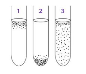
what kind of bacteria is growing in thioglycolate broth tube 2
anaerobic bacteria
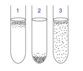
what kind of bacteria is growing in thioglycolate broth tube 1
aerobic bacteria
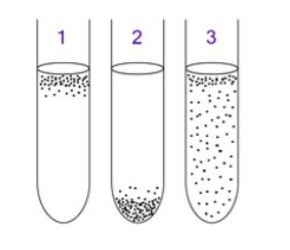
what kind of bacteria is growing in thioglycolate broth tube 3
a facultative anaerobe
Germicides
chemical agents that kill microorganisms
microstatic agents
inhibit the growth of microorganisms but do not kill them. Upon removal of the microstatic agent the microorganisms will resume their growth.
Disinfectants
may be either germicides or microstatic agents but are not considered safe for medical use and are applied only to inanimate objects. Household cleaning agents often contain disinfectants, such as ammonia and bleach (hypochlorite) to control the growth of microorganisms
Antiseptics
are similar to disinfectants but may be applied safely to biological tissues. These substances
are used for topical (surface) applications and are not necessarily safe for consumption. Examples
include alcohol, iodine and silver nitrate
The zone of inhibition technique and how it works
A filter disk is soaked in the disinfectant of choice and placed on the surface of a plate evenly spread with the bacteria of interest. If the disinfectant is effective, it will prevent the growth of the organism in a ring around the filter disk, referred to as the zone of inhibition.
In general, the larger the zone of inhibition, the greater the antimicrobial activity of the substance
The basic dye used in a Gram stain:
Crystal violet
the mordant used in Gram stain
Gram;s iodine
A mordant
a substance—typically a metallic salt—used to set and bind dyes to fabrics
decolourizing agent used in Gram stain
ethyl alcohol (95% Alcohol used in lab)
or an alcohol-acetone mixture
role of the decolourizing agent in a Gram stain
…decolourizes
used to wash unbound stain out of the sample
what happens to Gram Positive bacteria after decolourizing in Gram stain, why?
Gram-positive bacteria bind the dye strongly after treatment with the mordant (iodine) and retain the initial colour throughout the decolourizing procedure.
what happens to Gram Negative bacteria after decolourizing in Gram stain, why?
Gram-negative cells bind less strong to the original dye, so are decolourized and can be restrained with a counterstain of contrasting colour
result of the KOH test on Gram Negative cells and why
Gram negative cells are more permeable and can be lysed by 3% KOH. When the Gram negative cells are lysed, the DNA forms long sticky strands resulting in a viscous mixture
result of the KOH test on Gram Positive cells and why
Gram positive cells are less permeable and are not lysed by 3% KOH. because cells are not lysed, no sticky strands are formed.
How to prep a slide for a gram stain, if using a broth culture.
heat slide by passing through flame
label and draw a dime sized circle with a Grease pencil AKA the well
using aseptic technique, obtain a loopful of organisms, if not turbid try a drop from a pasteur pipette.
air dry smear
ONCE DRY, fix the smear by passing through the flame 2-3 times, should have a warm not hot slide.
How to prep a slide for a gram stain, if using a solid agar culture.
heat slide by passing through flame
label and draw a dime sized circle with a Grease pencil AKA the well
place a loopful of tap water on the slide
emulsify a small amount of culture into the drop, if the colonies are large, use cells from the outer edge of the colony.
air dry smear
ONCE DRY, fix the smear by passing through the flame 2-3 times, should have a warm not hot slide.
How to perform a Gram Stain
Fill well with Gram’s Crystal violet for 60 seconds, then tip off stain and rinse gently with water. remove excess water with wicking or shaking DO NOT WIPE!!
Flood Well with Gram’s Iodine for 60 seconds, then rinse with water, shake and wick.
Decolour with 95% alcohol for Approximately 10 seconds or until colour stops leaching from the slide. IMMEDIATELY rinse with water, gently blot away excess
counter stain with safranin for 10-20 seconds, rinse and blot off excess water
air dry
Endospores
tough, dormant, cell structures produced by certain bacteria like [Bacillus and Clostridium]. Resistant to Desiccation, UV light, hear and chemicals. can remain dormant for extended periods of time
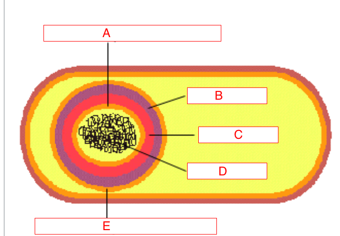
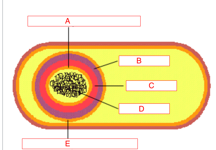
what is A
Inner forespore membrane of an endospore
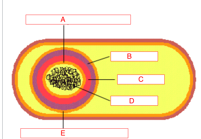
what is B and its role
Spore coat of an endospore (made of several protein layers). responsible for chemical resistance
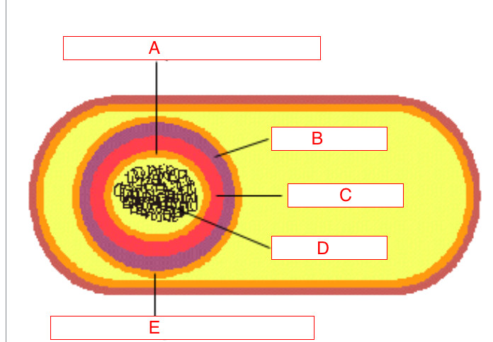
what is C
Cortex (soft peptidoglycan)
what is D
Nucleoid
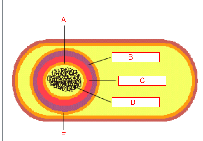
what is E
outer fore spore membrane
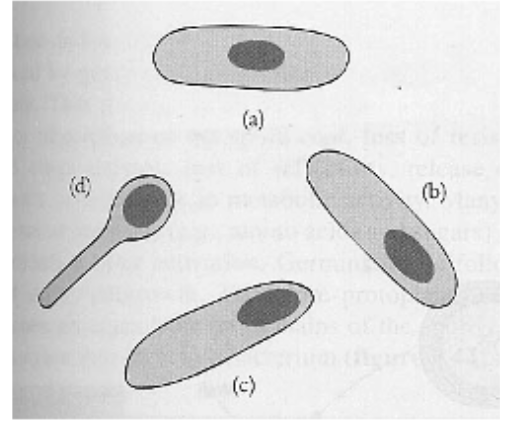
what is the position of endospore a
Central
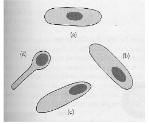
what is the position of endospore b
subterminal
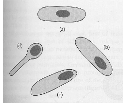
what is the position of endospore c
terminal
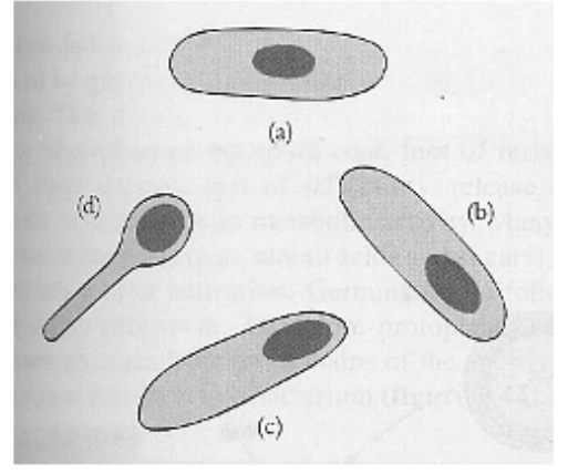
what is the position of endospore d
terminal with swollen sporangium
what cells produce endospores
vegetative cells
Sporangium
vegetative cell containing an endospore
what is used to stain endospores
Malachite green
what colour do endospores appear after staining technique using malachite green
Green
what colour do vegetative cells and the sporangium (not including endospore) appear after staining technique using malachite green
light red
how to perform an Endospore Stain
prepare a thin smear of culture on a clean slide, allow to dry, then fix with heat
In the fume hood
place a piece of bibulous paper (AKA filter paper) on the slide, just covering the film of the bacteria
SATURATE the paper with malachite green
heat slide over boiling water on a hot plate for five minutes, adding more stain if it starts to dry.
dispose of the paper in the waste container, cool the slide and gently rinse with water.
at the lab sink
counterstain with safranin for 30 seconds
rinse with water and blot dry
Selective media
contains an inhibitor which slows/prevents growth of some organisms without affecting the growth of the organisms you want to s
what does Maconkey’s agar do
inhibits Gram positives, is used to pick up Enterobacteriaceae (E. coli and cousins).
also, lactose fermenters will have pink colonies.
manitol salt media
inhibits many bacteria which are not salt tolerant, allowing certain Gram positives to grow unimpeded, especially Staphylococcus.
alsoe S. aureus (an important pathogen) ferments mannitol and produces a yellow colour.
differential media
contains ingredient(s) that make one type of organism appear different from others.
what happens to lactose fermenters on macConkey’s agar
form pink colonies
what happens to S. aureus on mannitol salt agar
ferments mannitol and produces a yellow colour
Blood agar,
made with fresh blood containing whole cells, is used to differentiate between different types of Streptococcus, based on their ability to form a zone of clearing by lysing the red blood cells (called hemolysis).
Enriched media .
contain special ingredients that allow extremely fastidious organisms to grow
chocolate agar
made with autoclaved blood the media preferred by Neiserria gonorrhea and Neiserria meningiditis, two important pathogens that look like Branhamella in a Gram stain.