7.5; Radiography of the Foal - sorry, had to separate this for my own brain
 Call Kai
 Learn
Learn Practice Test
 Spaced Repetition
 Match
Match Flashcards
 Knowt Play
1/43

There's no tags or description
Looks like no tags are added yet.
Name | Mastery | Learn | Test | Matching | Spaced | Call with Kai | Chat |
|---|
No analytics yet
Send a link to your students to track their progress
44 Terms
premature
_________________: a preterm birth (under 320 days)
dysmature
____________: a full term foal who was born with premature signs. Most commonly this is secondary to placental insufficiency
thin body condition, resp distress, floppy ears, domed forehead, thin silky coat, difficulty standing, incomplete ossificationof cuboidal bones, periarticular laxity, weak suckling response, secondary bacterial infection
What are the clinical signs of a premature or dysmature foal? (theres a lot so just throw out a couple)
crush, weight bearing movements
If a foal has failure or delay of ossification, the incompletely ossified bones do not provide normal support for the limb and begin to _________ during __________
lateral, laterally, carpus valgus
In horses with failure/delay of ossification, the _________ aspect of the carpus will collapse and the limb will angle [which direction], causing "__________ __________"
dorsal, curved
In horses with failure/delay of ossification, the _________ aspect of the tarsus will collapse, leading to an abnormally ________ appearance of the tarsus
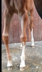
valgus
Which term describes this foals legs?

varus
Which term describes this foals legs?
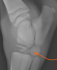
failure/delay of ossification, thin and wedge shaped
What is happening here in these foal bones? How can you tell?
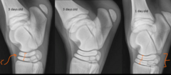
cool
He said not to worry about grading, but here are some tarsal bones, with the least mature on the left side and most mature on the right
angular limb deformities, pathologic fractures, osteoarthritis
non ossified limbs will lead to what three things?
varus
medial deviation of the limb is called
valgus
lateral deviation of the limb is called
true
true/false: angular limb deformities are named by indicating the joint or location where the deviation stars
cool
This is a lot, but I dunno how to make it a question and it DOES seem important:
Using Straight DP projections - Include a significant length of the bone proximal and distal to the joint where the angulation begins
•To determine the cause of the angulation and the degree of angulation that is present, lines are drawn through the central axis of the bones proximal and distal to the affected joint
•The point at which the lines intersect helps determine the cause of the abnormal angulation
•The angle formed at the intersection of the lines is the degree of abnormal angulation

hematogenous
Infectious septic arthritis in foals is usually via the ___________ route of infection
6-8, 10-12
Infectious septic arthritis in foals is usually seen in foals who are _________ weeks but can be seen up to ______ months
false; hematogenous spread means almost always multiple joints
true/false; infectious septic arthritis is, despite it's infectious nature, typically confined to one joint in most infected foals.
soft tissue swelling, aggressive osseous changes
early radiographic signs of infectious arthritis are often only _________ but later signs include __________ as the disease progresses
umbilicus, lungs, GI
What are the most common sources of the bacteremia? (organs of entry)
synovium
Where does type S infectious polyarthritis stem from in neonates?
physes, septic physitis
Where does type P infectious polyarthritis stem from in neonates?
epiphyses, septic epiphysitis
Where does type E infectious polyarthritis stem from in neonates?
tarsal bones
Where is type T infectious polyarthritis limited to in neonates?
carpal bones
Where is type C infectious polyarthritis limited to in neonates?
distension of joint capsule, normal osseous structures
What are radiographic signs of type S infections?
some swelling, widening of joint spaces (i know, i know)
What tips us off that this is type S septic arthritis?

joint capsule distension, lysis and irregular physis
What are radiographic signs of septic physitis?
infection of a synovial structure
septic physitis is generally secondary to or concurrent with ____________________
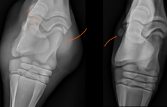
widened joint spaces AND moth eaten physis lines
What tells us this image is of septic physitis?
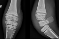
irregular margination, effusion, mineralization
What are the signs of septic arthritis seen here? (hint, they ARE outlined by my crappy orange annotations)
salter harris fracture
_________________: a fracture of the epiphyseal plate in young animals
I
salter harris type ______?

IV
Salter harris type ______?
(notes say: radiolucency surrounding joint)

II
salter harris type ____?
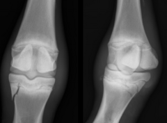
physeal dysplasia
___________: developmental disease cahracterized by disturbance of endochondral ossification at the physis
negative, this is an aseptic physitis
If you cultured a joint that had physeal dysplasia, what is the most likely thing you would find?
active growth phases
When is physeal dysplasia most commonly seen?
distal radial physis, distal MC/MT III physis, distal tibial physis
What three locations did Dr Berry give us that physeal dysplasia appears in most commonly?
rapid cartilage production, mineralization
physeal dysplasia is thought to be secondary to ________________ or defects in the _____________________ within primary spongiosa
warm, painful, firm swelling at distal radius or distal MC/MT III
what are physical exam findings with physeal dysplasia
medial, physeal closure,
Which aspect of the limb is most commonly affected with physeal dysplasia?
And This can lead to premature _______ and to angular limb deformities
exercise restriction, stall rest, NSAIDs, good
What are the treatments for physeal dysplasia?
What is the prognosis?
widening of physis, bony irregularity at pyseal margins, metaphyseal flaring, hook like projection at metaphysis
what are some radiographic signs of physeal dysplasia?