Zoology 153 Quiz 2
 Call Kai
 Learn
Learn Practice Test
 Spaced Repetition
 Match
Match Flashcards
 Knowt Play
1/104

There's no tags or description
Looks like no tags are added yet.
Name | Mastery | Learn | Test | Matching | Spaced | Call with Kai | Chat |
|---|
No analytics yet
Send a link to your students to track their progress
105 Terms
Solute
Dissolved substance
Flow of Concentration Gradient
High concentration to low concentration
Permeable Membrane
Solute can move across a membrane
Impermeable
Solute cannot move across a membrane (water still can)
Osmosis
Movement of water from an area of low solute concentration to high solute concentration
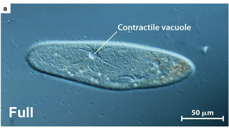
Contractile Vacuole
Absorbs excess water in the cell and expels it to prevent cells from exploding
Hypertonic RBCs
Surroundings have more solutes than RBC cytoplasm so RBC shrink
Isotonic
Surroundings have solutes comparable to the RBC cytoplasm and RBCs maintain their shape
Hypotonic
Surrounds have fewer solutes than the RBC cytoplasm and the RBCs swell
Cell Wall Function
Provides structural support and protection
Turgor Pressure
Pressure within cell when water moves into the cell through osmosis
Causes plants to wilt
Simple Diffusion
Molecules move directly across the membrane
Facilitated Diffusion
Molecules move from high to low concentration through transporters embedded in the membrane (channels)
Uniport
When the transporter or channel allows movement of one solute
Cotransport
Two solutes move through a single channel
Symport
Two solutes move in the same direction
Antiport
Two solutes move in opposite directions
Primary Active Transport
Cells move substances against their concentration gradient and energy must be expended (ATP)
NA+ / K+ + ATPase is an active transporter / pump that exchanves 3 Na+ for 2 K+ in a four-step process
Secondary (indirect) Active Transport
One solute is moved against its concentration gradient by active transport and a second solute’s movement can be coupled to that of the first by cotransport
Second relies on the active transport of the first
Prokaryotic
“before a nucleus”
Cells (bacteria) that lack a nucleus and typically have little or no internal compartmentalization
Eukaryotic
“true nucleus”
Cells that have a nucleus and internal membrane-bound organelles
Endomembrane System
System of internal membranes that allows specific cellular functions to occur within the localized spaces in the cell
Exocytosis
Fusion of vesicle with the cell’s plasma membrane
Endocytosis
Internalizes materials from the surface to create a new vesicle
Nuclear Envelope
Double membrane that surrounds the nucleus
Regulates traffic in and out of the nucleus
Nuclear Pores
Nuclear envelope is perforated with these large multi-subunit channels that regulate inward and outward movement of solutes and macromolecules
Endoplasmic Reticulum (ER)
Continuous with the nuclear envelope and is a site of lipid and protein synthesis
Rough ER
Studded with ribosomes
Many proteins destined for secretion and transmembrane proteins are synthesized here
Smooth ER
Site of lipid and steroid synthesis
Golgi Apparatus
“stack of pancakes” structure
Modifies proteins and lipids produced in the ER
Sorts proteins and lipids as they move to their final destinations
Synthesize carbohydrates
Golgi Apparatus is made up of
A series of flattened sacs called cisternae
Edges bud off as vesicles carrying proteins or modified sugars to the cell membrane of organelles in the cell
Enzymes within the Golgi Appartus ….
Chemically modify proteins and lipids in a sequence of steps in a different region of the organelle
Lysosomes
Derived from the Golgi apparatus
Degrade damaged or unneeded macromolecules
pH of Lysosomes
Maintain a pH of 4-5
Proton pumps help maintain this low pH
Exocytosis
Transport (along microtubules) vesicles migrate to the membrane, fuse with it, and release their contents outside the cell
Production of vesicles, budded from Golgi
Docking of vesicles with the plasma membrane; fusion of vesicles with the plasma membrane & contents are released; recycling of leftover membrane
Endocytosis
Macromolecules are taken into the cell in vesicles
Membrane forms a pocket that deepens and pinches off, forming a vesicle
Receptor-mediated endocytosis
Vesicle formation is triggered by solute binding to receptors
Receptor proteins bound to specific solutes are clustered in coated pits that form coated vesicles. The coats are formed by special proteins that aid membrane bending and pinching
Emptied receptors are recycled to the plasma membrane by the same vesicle
Mitochondria
Extract energy from metabolites
Found in all eukaryotes, double membrane, divide independently and contain their own DNA
Cholorplasts
Harness light energy to create carbon-containing compounds
Found in plants, double membrane, divide independently and contain their own DNA
Endosymbiosis
Biological relationship where one organism lives inside the body or cell of another, usually to the benefit of both
Reason that eukaryotic cells came from prokaryotic cells
Cytosol
The fluid portion of the cytoplasm inside a cellW
Where are mRNAs bound?
Cytosol by ribosomes
Translation
Making of proteins which begins in the cytosol
Sorting Sequence
Specific amino acids incorporated into the polypeptide dictate where it will be transported
Proteins destined for the ________ system are made in the ____
Endomembrane system; rough ER
Squirted into the lumen (center) of the ER
Some are completely released, other remain inserted into a piece of the ER membrane
Proteins that are completely released will …
Remain in the aqueous medium inside a vesicle; typically screted
Proteins that remain inserted into a piece of the ER membrane will …
Be inserted into a membrane (like the plasma membrane)
ER-Targeting Signal Sequences
Specific N-terminal signal sequence with attached ribosome bind to the ER
Signal sequence is bound by the signal recognition particle (SRP)
When SRP binds, translation pauses until the SRP binds the SRP receptor on the ER
Translation begins again
SRP will disassociate from the polypeptide
Polypeptide will be fed into the ER lumen
Hight temperature on membranes
Causes greater molecular motion of the bilayer which can increase permeability to solutes and can eventually destabilize the bilayer
Low temperature on membranes
Causes excessive membrane rigidity and impedes proper membrane transport
Homeoviscous Adaptation
Altering lipid composition to adjust membrane viscocity
Extremophiles
Organisms that have more drastic adaptations (more disulfide bonds, other modifications, etc.)
Cytoskeleton
Provides internal structural support
Enables transport within the cell via motor proteins
Formed from long chains of protein subunits joined together
3 Main Polymers of Cytoskeleton
Microfilament; from actin monomers
Intermediate filament; from protein subunits
Microtubule; from tubulin dimers
Centrosome
Central organizing center where a microtubule has their minus end
Why are Microtubules Important
Important for chromosome segregation during cell division
Allow vesicle transport
Formation of Cilia and flagela
Where does growth occur in a microtubule and microfilament
Growth occurs more quickly at the plus ends while minus ends are less dynamic
What causes a bend in a microtubule
Dyneins are positions to slide adjacent microtubules which are locked togther into an integrated network; this uses energy (ATP)

Kinesin Function
Moves cargo toward the plus end of MTs; requires energy (ATP)
Dynein Function
Moves cargo toward the minus end of MTs; requires energy (ATP)
What are cells organized into?
Tissues
Extracellular Matrix
Associated with sheets of cells
Lies outside of cells
Cell-cell Adhesion
Cells stick togetherC
Cell-ECM Adhesion
Cells need to stick to ECM
Cadherins
Cadherins are cell adhesion proteins that help connect one cell to another cell. They bind to cadherins on neighboring cells and are connected to the cytoskeleton inside the cell.
Integrins
Integrins are cell adhesion proteins that attach to the ECM and connect to the cytoskeleton inside the cell.
Junctions
Adhesion molecules assemble in sheets of cells (epithelia)
Tight Junctions
Permeability barrier, sealing the sheet so solute movement is restricted
Adherens Junctions
Contain cadherins and confer adhesion
Desmosomes
Connect to intermediate filaments to form mechanically strong “spot welds” between cells
Gap Junctions
Allow passage of small molecules between cells
Cisternae
A series of flattened sacs of Golgi
Metabolism
Building and breakdown of carbon sources to harness or release energy
Anabolism
Building of molecules from smaller units, requiring an input of energy (ATP)
Catabolism
Breakdown of molecules into smaller units, producing energy (ATP)
First Law of Thermodynamics
Energy cannot be created or destroyed; it can only be transformed
Potential Energy
Stored energy
Chemical Energy
Considered a form of potential energy
As energy in the cell is transformed …
The energy available to do work in the cell decreases
Entropy (S)
Measure of disorder; cells do work to combat increases in entropy
Gibbs Free Energy
Measure of the energy available to do work
Allows us to predict whether a reaction is favored
G = H - TS
Exergonic
DG is negative and the reaction is favored
Products store less free energy than the reactants and it is spontaneous
Liberate energy
Endergonic
DG is positive and the reaction is not favored
Products store more free energy than the reaction and it is non-spontaneous
Require energy input
Is glucose oxidation exergonic or endergonic?
Exergonic!
How to make an endergonic reaction favorable?
Couple it with an exergonic one like the synthesis of glutamine!
Phosphorylated Intermediate
The P from ATP gets moved around when you couple a reaction to ATP
Minority Shareholder of ATP
GTP
Minority Shareholder of NADH
FADH2
Intermediate Energy
Allows ATP to drive reactions but also be readily replenished
Reduced
Molecules acquire electrons
Oxidized
Molecules lose electrons
Important electron carriers in cellular respiration
NAD+ and FAD
What helps a reaction proceed
Enzymes!
What do enzymes do?
Lower the activation energy; delta G is the same
Enzymes often require a ….
Cofactor: metol ion, etc.
Enzyme process
Enzyme binds to substrate
Complicated interaction occurs between the reactants and the enzyme
Substrate is converted to products
Products are released
the same enzyme is ready to undergo another round
Active Site
Formed within the function protein, depends on tertiary structure
Helps stabilize the transition state
Hexokinase
Enzyme that catalyzes the first step in glycolysis
Induced Fit
Enzyme’s shape changes to fit substrate more tightly
Competitive Inhibition
A substance binds to the active site and blocks it from substrate