Integumentary System Overview for NPTE Exam
 Call Kai
 Learn
Learn Practice Test
 Spaced Repetition
 Match
Match Flashcards
 Knowt Play
1/65

There's no tags or description
Looks like no tags are added yet.
Name | Mastery | Learn | Test | Matching | Spaced | Call with Kai | Chat |
|---|
No analytics yet
Send a link to your students to track their progress
66 Terms
Integumentary System
Largest body organ, 15-20% of body weight.
Primary Function
Protection, insulation, sensory, fluid balance, temperature control.
Epidermis
Outer skin layer containing keratinocytes and melanocytes.
Dermis
Inner skin layer with collagen and blood vessels.
Keratinocytes
Cells producing keratin, a protective protein.
Melanocytes
Cells responsible for skin pigmentation.
Langerhans Cells
Immune cells in the epidermis.
Basal Cells
Cells in the epidermis that regenerate skin.
Venous Insufficiency
Inadequate drainage of venous blood, causing edema.
Arterial Insufficiency
Lack of adequate blood flow to body regions.
Clinical Presentation
Signs and symptoms observed during patient assessment.
Pressure Ulcers
Wounds over bony areas, staged by depth.
Meissner Corpuscles
Sensory receptors for light touch and pressure.
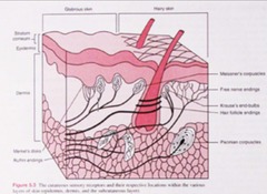
Krause End Bulbs
Receptors for cold sensation in the skin.
Pacinian Corpuscles
Receptors for deep pressure and vibration sensation.
Ruffini Endings
Receptors for skin stretch and sustained pressure.
Hemosiderin Staining
Brown discoloration due to iron deposition.
Granulation Tissue
New connective tissue formed during wound healing.
Pitting Edema
Swelling that leaves an indentation when pressed.
Hyperkeratosis
Thickening of the outer skin layer.
Plan of Care
Strategy for patient treatment and rehabilitation.
Differential Diagnosis
Process of distinguishing between different conditions.
Stage 1 Pressure Injury
Reddened area that does not disappear.
Stage 2 Pressure Injury
Involves first two skin layers, superficial.
Stage 3 Pressure Injury
Subcutaneous fat may be visible.
Stage 4 Pressure Injury
Extends down to and includes bone.
Unstageable Pressure Injury
Wound base obscured by necrotic tissue.
Diabetic Ulcers
Located on weight-bearing surfaces of the foot.
Venous Insufficiency Ulcers
Found proximal to medial malleolus, edematous.
Arterial Ulcers
Located on lateral malleolus or distal toes.
Pressure Ulcers
Result from unrelieved external pressure.
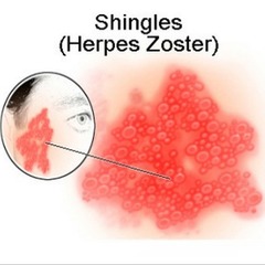
Herpes Zoster Symptoms
Initial pain and paresthesia in affected dermatome.
Herpes Zoster Rash
Presents as unilateral, raised, pink with silvery appearance.
Contact Precautions
Used for herpes simplex virus type 1.
Airborne Precautions
Required for herpes zoster (shingles).
Localized Skin Edema
Irregular areas indicating possible wheals.
Macerated Wound
Identified by white, friable, overhydrated skin.
Causes of Maceration
Uncontrolled drainage, perspiration, or incontinence.
Transparent Films
Used for very mild exudate wounds.
Hydrogel Dressing
Appropriate for minimal exudate wounds.
Hydrocolloid Dressing
Used for minimal to moderate exudate.
Foam Dressings
Suitable for moderate exudate wounds.
Calcium Alginates
Used for heavy exudate wounds.
Hydrofiber Dressings
Max capacity for heavy exudate management.
Debridement
Removal of nonviable tissue from a wound.
Selective Debridement
Only nonviable tissues are removed.
Sharp Debridement
Use of scalpel, scissors, and forceps.
Enzymatic Debridement
Topical application to remove nonviable tissue.
Autolytic Debridement
Body's mechanisms remove nonviable tissue.
Nonselective Debridement
Both viable and nonviable tissues are removed.
Wet to Dry Dressings
Moistened gauze applied to necrotic tissue.
Wound Irrigation
Pressurized fluid removes necrotic tissue.
Hydrotherapy
Whirlpool agitation for wound debridement.
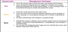
Red Yellow Black System
Color-coded system for wound assessment.
Superficial Burn
Affects epidermis; heals in 5 days.
Superficial Partial Thickness Burn
Involves epidermis and some dermis; heals in 10-14 days.
Deep Partial Thickness Burn
Involves epidermis and dermis; heals in 3 weeks.
Full Thickness Burn
Involves all skin layers; requires surgical intervention.
Subdermal Burn
Involves all skin layers and subcutaneous tissue.
Hypertrophic Scar
Thick fibrous tissue within original wound borders.
Keloid Scar
Excessive scar tissue beyond original wound margins.
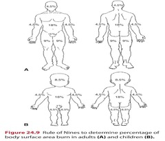
Rule of Nines
Estimates burn surface area based on body regions.
Burn Depth
Classification based on tissue involvement and healing.
Capillary Refill
Time taken for color to return after pressure.
Necrosis
Death of tissue due to lack of blood supply.
Granulation Tissue
New connective tissue and blood vessels in healing.