BIOL 4431 Exam 2 Study Guide
1/131
Earn XP
Name | Mastery | Learn | Test | Matching | Spaced | Call with Kai |
|---|
No analytics yet
Send a link to your students to track their progress
132 Terms
General Senses in Skin, Muscles, and joints.
Touch
Pressure
Proprioception
Temperature
Pain
General Senses in Internal Organs.
Pain
Pressure
Special Senses
Receptors are located within specific organs.
Smell
Sight
Balance
Taste
Hearing
Sensory Transduction
Converts environmental stimulus into a neural signal.
Chemoreceptors
Sense chemicals.
Photoreceptors
Sense light.
Thermoreceptors
Sense temperature.
Mechanoreceptors
Sense pressure.
Nocireceptors
Sense pain.
Generator Potential
An EPSP that is triggered by a stimulus. A sufficient GP triggers an action potential.
Frequency Coding
Increased stimulus intensity is encoded as increased action potential frequency.
Cutaneous Receptors
Free nerve endings located throughout the skin that sense light touch, temperature, and pain.
Meissner’s Corpuscles (encapsulated)
Located in the upper dermis that sense texture and slow vibration. 2 point discrimination.
Merkel’s Disks
Located at the base of the epidermis, sense sustained touch and pressure.
Ruffini Endings
Located deep in the dermis, senses sustained pressure.
Pacinian Corpuscles
Located the deepest in the dermis. They are encapsulated and sense deep pressure and fast vibrations.
How does encapsulation of a receptor affect what is transduced/sensed?
The encapsulation of a receptor increases the dendrites sensitivity.
Visceral Receptors
Located in internal organs; sense pressure & pain. Output transmitted by vagus nerve.
Muscle Spindle Apparatus
Sensory receptor located within skeletal muscles that detects changes in muscle length and helps regulate muscle contraction.
Extrafusal Muscle Fiber
Outside the muscle spindle apparatus. Contracted by alpha motor neurons.
Intrafusal Muscle Fibers
Located inside the muscle spindle apparatus. Maintain stretch receptor length. Gamma motor neuron innervation.
When are muscle spindles activated and what is their action on the muscle?
Activated when a muscle is stretched/lengthened, muscle spindles detect changes in muscle length and trigger a reflex contraction to prevent overstretching.
Golgi Tendon Organs
Sensory receptors (proprioceptors) located at the junction between muscles and tendons that inhibit muscle contraction.
Sensory Unit
A sensory neuron and its associated receptors.
Receptor Field
The skin area served by a sensory unit.
Lateral Inhibition
When a sensory unit receiving most stimulation inhibits adjacent sensory units.
Fine Touch, Pressure, and Proprioception Pathways
Sensory neurons enter dorsal spinal cord. They travel up the dorsal spinal cord on the same side as the stimulus. They synapse with the 2nd neuron in the brainstem. 2nd neuron crosses over to the other side of the body and travels to the thalamus. Synapse with 3rd neuron in thalamus. 3rd neuron → Sensory cortex.
Pain and Temperature Pathways
Synpase immediately with the 2nd neuron which immediately crosses over. Ascends in the anterospinal cord to thalamus. Synapse with 3rd neuron in thalamus. 3rd neuron → Sensory cortex.
Sensory Cortex
Somatotopically organized (specific areas serve specific body regions).
Head → more lateral
Foot → More medial
The greater the sensitivity the greater the cortical area.
Sensory information influences motor output.

Cornea and Sclera
Cornea is clear. Sclera are the whites of the eyes. Outermost layer of eye.
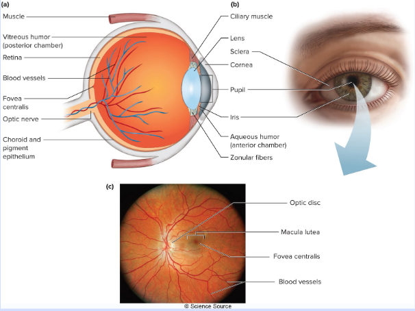
Anterior Chamber
Between the cornea and the lens, contains aqueous humor.
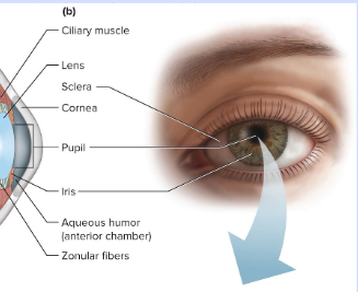
Iris
Regulates light entrance.
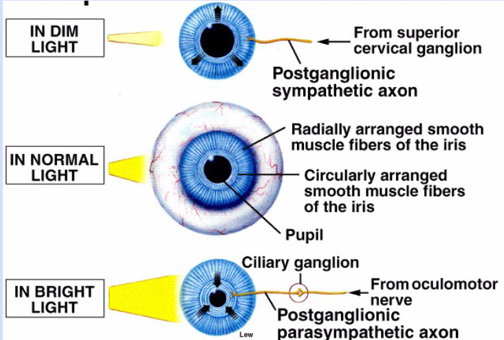
Pupil
Opening in the middle of the iris. Dilated by the sympathetic nervous system and constricted by the parasympathetic.
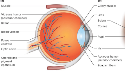
Lens
Refracts light; focuses images.
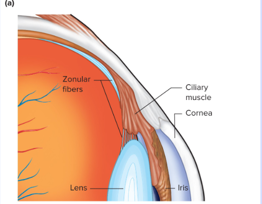
Cilliary Muscles
Attached to the lens by suspensory ligaments, they adjust the refraction of the lens.
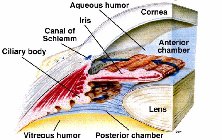
Vitreous Humor
A thick, viscous liquid that fills the eyeball.
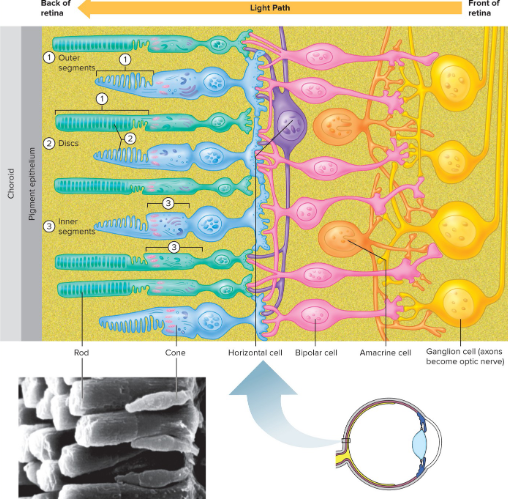
Retina
Photoreceptor layer. Has layers itself; innermost layers are nerve cell axons from the optic nerve, the outermost is a photoreceptor layer.
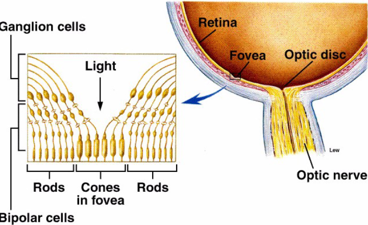
Fovea centralis
The retinal area with the greatest visual acuity. Here light falls directly on the cones (focused by lens).
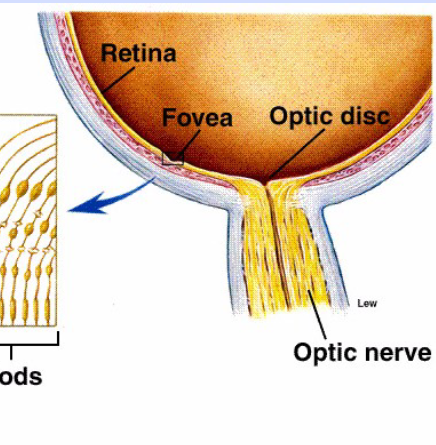
Optic Disk
Where the optic nerve exits the eye; this is a blind spot.
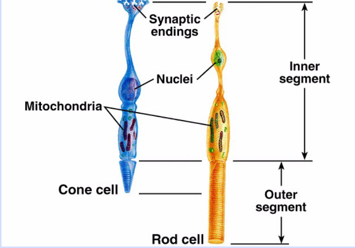
Rods
Sense low levels of light.

Cones
Sense high levels of blue, green, and red light.
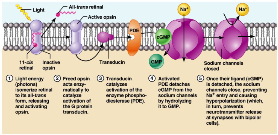
What happens when light strikes photoreceptors? What are the effects on the receptor and on the other cells in the retina?
When light strikes photoreceptors, it triggers a series of chemical reactions leading to hyperpolarization of the receptor cell (activates g-protein cascade which closes Na+ cells and hyperpolarizes the cell). This signal is then transmitted to other cells in the retina, such as bipolar and ganglion cells, eventually reaching the brain for visual processing.
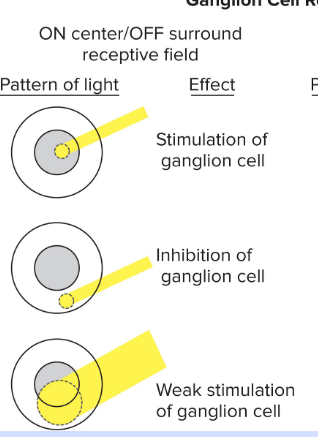
On-Center/Off-Surround Ganglion Cells
Stimulated when light hits the center of the field. Inhibited when light hits the edge of the field.
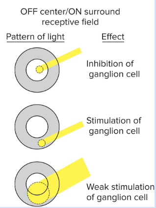
Off-Center/On-Surround Ganglion Cells
Inhibited when light hits the center of the field. Stimulated when light hits the edge of the field.
Rhodopsin Dark Adaptation
When exposed to light, receptors are “bleached” and rhodopsin decreases in rods. First 5 minutes in the dark, rhodopsin increases in cones. For the next 20 minutes rhodopsin increases in rods.
Pathway of Sound
The pathway of sound begins with sound waves entering the outer ear, passing through the ear canal to the eardrum, then vibrating the ossicles in the middle ear, transmitting to the cochlea in the inner ear, where hair cells convert vibrations into electrical signals sent to the brain via the auditory nerve.
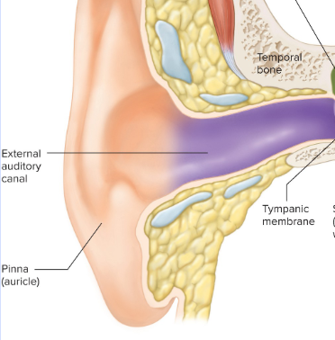
Outer Ear Structures
Auricle (pinna) - Ear
External Auditory Canal - Ear Canal
Ceruminous Glands - Secrete cerumin
Tympanic Membrane - Ear drum
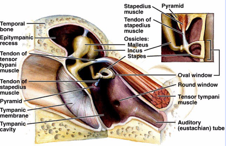
Middle Ear Structures
Bones - Malleus (on TM), Incus, and Stapes (Vibrates cochlear fluid; amplify TM vibrations)
Muscles - Tensor tympani (on malleus), stapedius (on stapes), dampen vibration of TM and stapes.
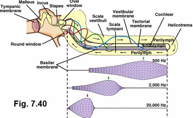
Cochlea (Inner Ear) Structures
Fluid is continuous with vestibular apparatus.
Scala Vestibuli - First fluid (perilymph) vibrated by stapes
Cochlear Duct - Contains the Organ of Corti
Scala Tympani - Final fluid vibration pathway to round window.
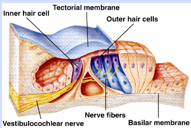
How is sound transduced in the cochlea? Which regions transduce which frequencies?
Hair cells in the organ of Corti respond to fluid vibrations. High frequency pitches happen closer to the stapes and low frequency pitches happen further from the stapes.
How are the hair cells in both the cochlea and in the vestibular apparatus activated (or inhibited)
Stereocilia – most of hairs
• Kinocilium – large hair
• Stereocilia bend toward kinocilium – stimulation
• Stereocilia bend away – inhibition
What are the structures involved in transducing linear and angular movement?
Otolith organs (with utricles and saccules) sense linear movement. Semicircular canals sense angular movement; one in each 3d plane.
Nasal epithelium
In the roof of the nasal cavity, nasal turbinates stir air past.
Bipolar Cells
Receptor & sensory neuron
Dendritic End
: In nasal cavity, covered by mucous layer
Olfactory Hairs
Dendrites of receptor, sites of transduction.
Axons
Profect through cribiform plate. Synpases in the olfactory bulb.
Receptor Sites
In the olfactory hairs, one odor may activate combination of receptors. Humans may detect up to 10,000 different odors.
Papilla
Small, projections found on the surface of the tongue that give tongue its rough appearance.
Largest to smallest: Papilla → Taste Bud → Taste Cell → Taste Receptors → Taste pores → Taste hairs
Salty
Na+ depolarizes taste receptors.
Sour
H+ depolarizes receptor.
Sweet (sugar)
2nd messengers close K+ channels, this depolarizes the receptor.
Bitter (quinine)
2nd messengers release intracellular Ca++, which triggers NT release.
Umami (Amino Acids)
Amino acids trigger 2nd messengers, which allows Ca++ entry which triggers NT release.
Facial Nerve (CN VII)
Anterior 2/3 of tongue.
Glossopharyngeal Nerve (CN IX)
Posterior 1/3 of tongue.
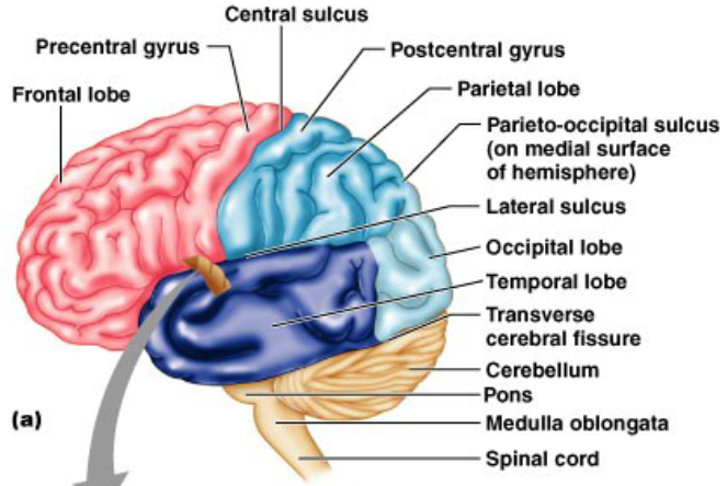
Cerebrum
The majority of the brain, made of gray matter (associated with higher functions) with a left (verbal, analytical) and right (spatial tasks) hemisphere and 5 paired lobes.
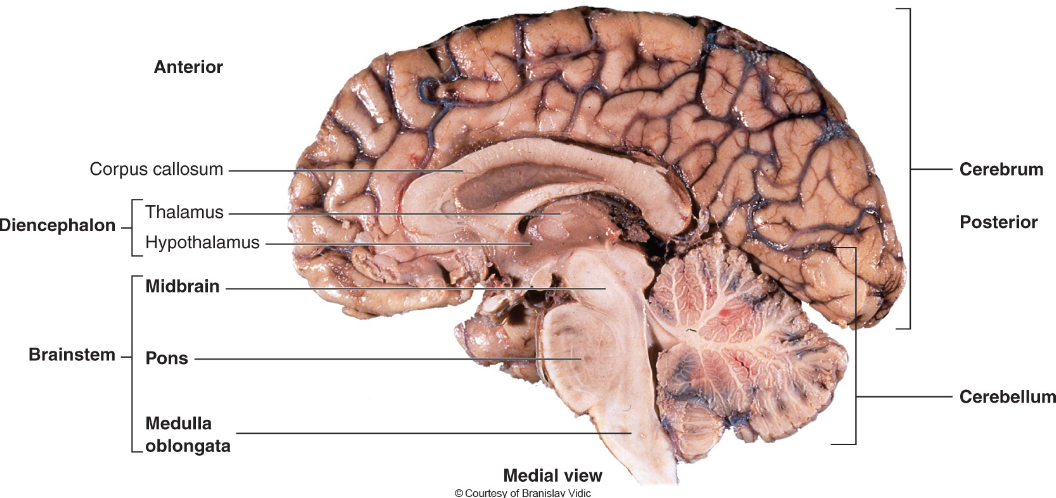
Corpus Callosum
Neural connection between brain hemispheres.
Gyrus
Ridge
Sulcus
Valley
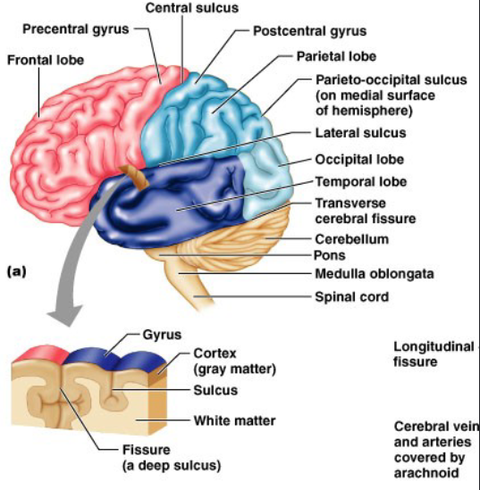
Cerebellum
Smaller, convoluted portion of the brain. For the coordination of movement.
Meninges
Protective covering of the brain.
Ventricles
Circulate cerebrospinal fluid in the brain.

Frontal Lobe
The association area; general mood.
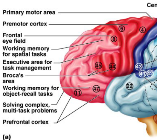
Broca’s Area
Motor/speech.
Lateral Sulcus
Divides the TEMPORAL lobe from the others.
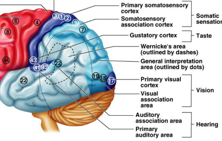
Temporal (lateral) lobe
Auditory signal processing and interpretation area (short term memory, emotion).
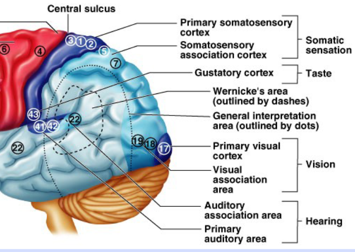
Occipital (back) lobe
The visual cortex.
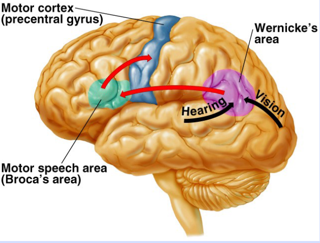
General Interpretive (Wernike’s) Area
Speech interpretation, comprehension.
Electroencephalocardiogram (EEG)
A recording of brain electrical activity. Uses surface electrodes placed on the head.
Waking State
Beta rhythms. Small amplitude waves that indicate the alert state.
Relaxed State
Alpha rhythms.
Non-Rapid Eye Movement Sleep State
Theta and delta rhythms with larger amplitudes. Decreases the ease of arousal, increases the threshold for stimuli, and decreases motor output. Dozing off state.
Sleep spindles/K Complexes
High frequency/large amplitude bursts. Eventually become delta waves (slow wave sleep).
REM Sleep State
Pattern resembles alert state. Intense dreaming occurs.
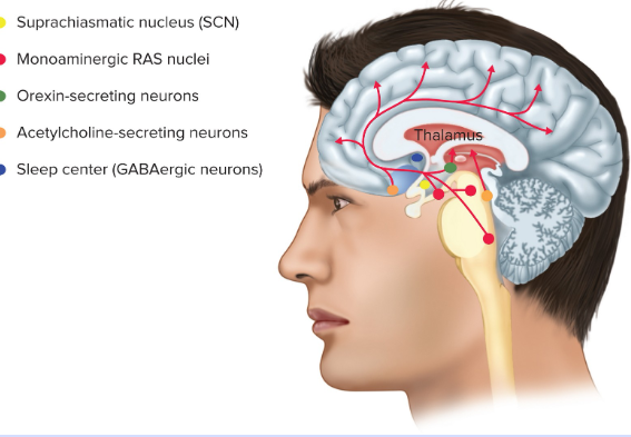
Reticular Activating System (RAS)
Clusters of neurons in the brainstem and hypothalamus with widely distributed axons. Plays a crucial role in regulating wakefulness, attention, and arousal.
Awake State
Neurons release monoamine neurotransmitters which enhance the excitatory synapses.
Orexins & Hypocretins
Peptides released by hypothalamus, these stimulate the firing of RAS neurons. Lack of these is associated with narcolepsy.
Sleep State
Sleep center neurons release GABA (inhibitory) which inhibits the excitatory neurotransmitter/peptide release.
Coma
Extreme decrease in mental function. Still some EEG activity.
Brain Death
No evidence of CNS function above the spinal cord and no spontaneous respiration for 8-10 minutes.
Selective Attention
Avoiding distraction.
Orienting Response
Paying attention to stimulus.
Preattentive Processing
Requires prior experience; helps establish stimulus as meaningful.
Habituation
Due to repeated stimulus and is associated with decreased NT release.
Neural Mechanisms
Receptive field modalities overlap (ex: visual and auditioy). Weak cues can add up.
Excitatory NT release from the locus ceruleus.
ADHD
Associated with a decrease in excitatory NT release and an inability to maintain selective attention.
Primary Drive for Motivated Behavior
The reward pathway (part of the RAS). Neurons originate in the midbrain and release dopamine into the limbic system and prefrontal cortex.