Integument - Exam 1
 Call Kai
 Learn
Learn Practice Test
 Spaced Repetition
 Match
Match Flashcards
 Knowt Play
1/28

Earn XP
Description and Tags
Exam 1
Name | Mastery | Learn | Test | Matching | Spaced | Call with Kai | Chat |
|---|
No analytics yet
Send a link to your students to track their progress
29 Terms
integument
7-10% of BW
skin (epidermis, dermis, hypodermis*)
glands (sweat and sebaceous)
cutaneous scent glands
hair
wound healing
claws (cat and dog)
hooves
feathers
horns and antlers
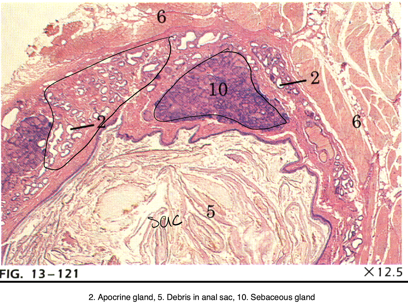
special skin structures (mammary glands, anal sacs)
skin function
protection - dehydration (wounds), infection (physical barrier)
sensory function
secretion/excretion
regulation of body temperature (sweat, blood flow to skin)
absorption of UV (Vit D production)
skin layers
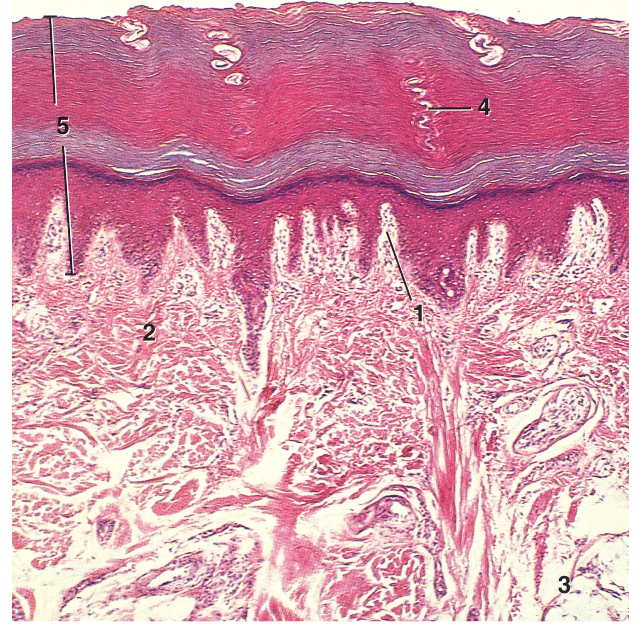
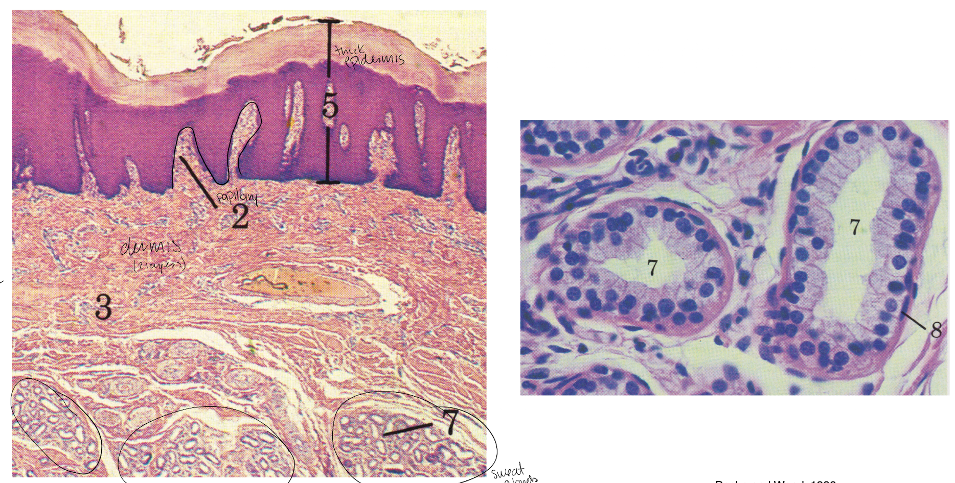
epidermis - epithelium (keratinized, stratified squamous), avascular (5)
dermis - blood supply, relatively few cells, dense irregular CT (type 1 collagen) (1&2)
hypodermis - loose CT, superficial fascia
epidermis
thick skin (glabrous)
no hair follicles
up to 5 identifiable layers
merocrine sweat glands
thin skin (nonglabrous)
has hair follicles
thin stratum corneum
stratum lucidum (absent) and granulosum (hard to define)
apocrine sweat glands

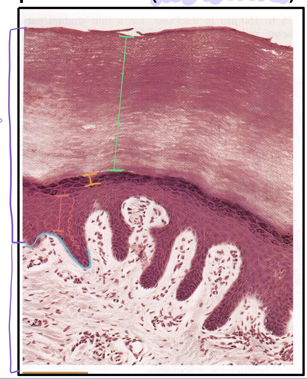
layers of epidermis
stratum basale
stratum spinosum
stratum granulosum
stratum lucidum (not evident on slide)
stratum corneum
straum basale
cell division (stem cells give rise to keratinocytes [cells in epidermis])
sits on basement membrane
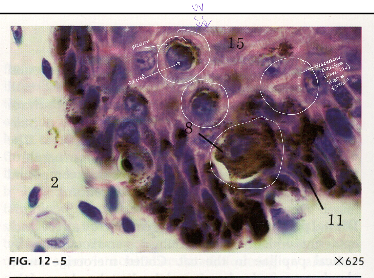
melanocytes → produce pigment
merkel cells → nerve receptor

stratum spinosum
cells attached by desmosomes (cells shrink in staining and desmosomes make it look like they have spines) (all cells in epithelium attached this way)
membrane coating granules
keratinization begins

stratum granulosum
cells still have nuclei (last layer w/ nucleus)
keratohyalin granules

stratum lucidum
packets of keratin from here move up towards surface
only in thick skin
lack organelles and nuclei
keratin filaments
really only in human histo and only in palms/soles, and not all people
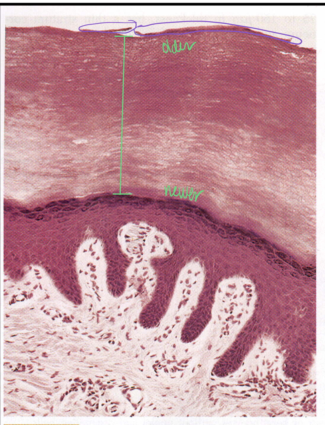
stratum corneum
further from surface - still have desmosomes
closer to surface - desmosomes breakdown, desquamated (sloughed off)
melanocytes
thick and thin skin
large cell, multiple processes coming off
derived from neural crest cells
located in the basal layer
source of melanin for keratinocytes (same # of melanocytes in black and white, differ in melanin production)
also found in the external root sheath and matrix of hair

merkel cells
thick and thin skin
light touch receptors
slow adapting
special stain required to visualize
located in s. basale

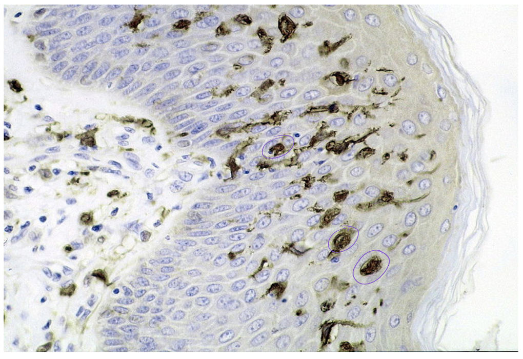
dendritic cells
thick and thin skin
langerhans cells
monocyte derived (patrol for foreign material) (macrophages)
special stain required to visualize
Antigen Presenting Cells (APC) - take pateria to lymph nodes (lymphocyte) to initiate immune response
dermis
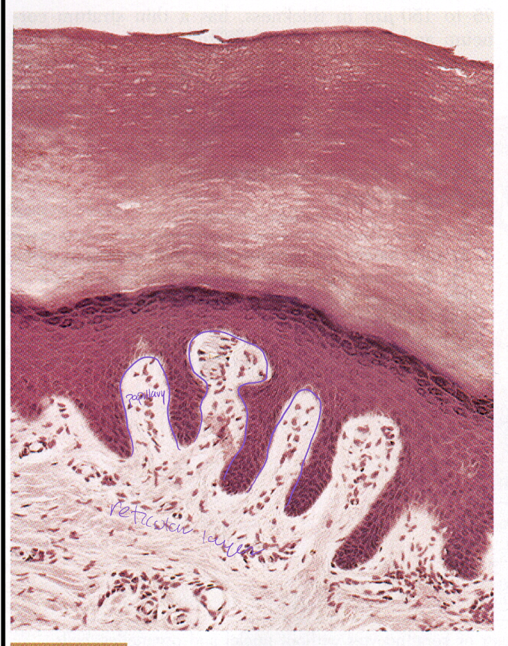
reticular layer (thick and thin)
papillary layer (thick skin only)
reticular layer of dermis
dense irregular CT (type 1 collagen and elastin)
sweat glands
hair follicles, arrector pili muscles
mechanoreceptors
blood and lymph vessels
papillary layer of dermis
dermal ridges
loose CT
anchoring fibrils from basal lamina of epidermis
increase surface area for interaction between epidermis and dermis bc more abrasion in thick skin (foot pads, etc.)
hypodermis
loose CT
anchors dermis to underlying muscle/bone
adipose tissue
fat pads (structural fat) (pads of feet bottoms)
burn classification

sebaceous glands
simple branched acinar (alveolar) glands
release sebum via holocrine secretion
antimicrobial (FAs acidify environment, Fe binding so bacteria can’t grow, antibodies - IgA, IgG)
waterproofing - lipid content
secrete into hair follicle (most frequent)

sweat glands
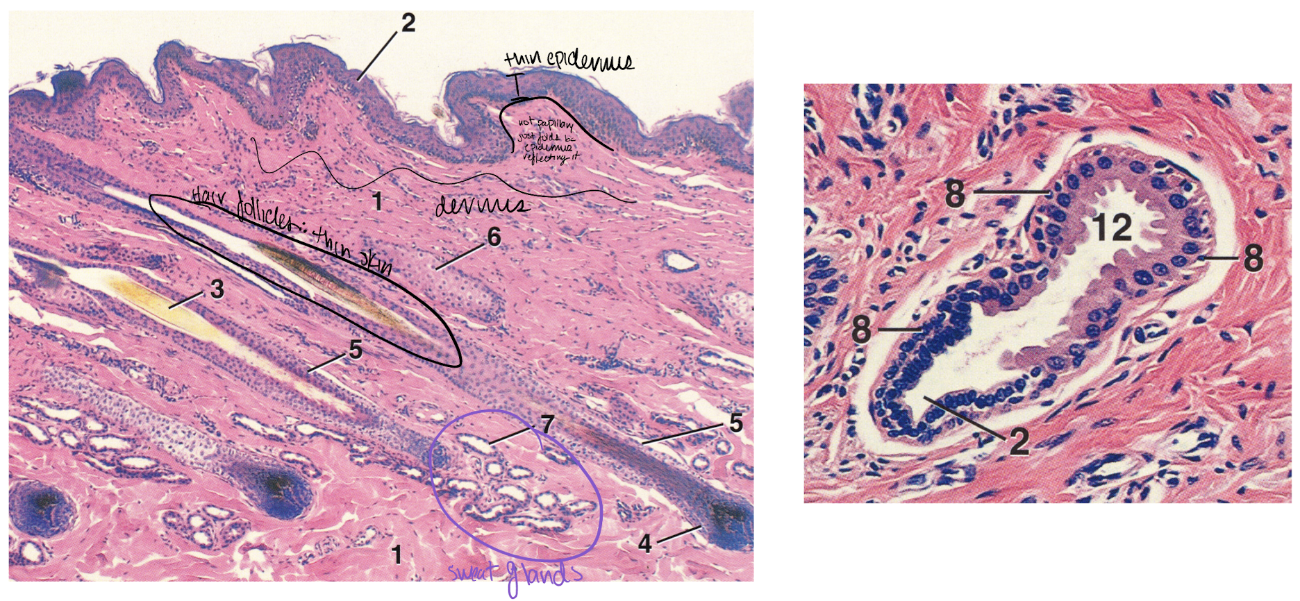
simple coiled tubular glands
apocrine
throughout all haired skin (thin)
viscous secretion that contains scent (pheromone based)
secretes into hair follicle
merocrine (eccrine)
thick skin
limited distribution in animals (very efficient in humans)
foot pads in dogs and cats, nasal planum
watery secretion onto skin surface
function of sweat glands in heat loss varies in importance between sp. (horse>cattle>sheep>dog>cat>pig)
hair follilce
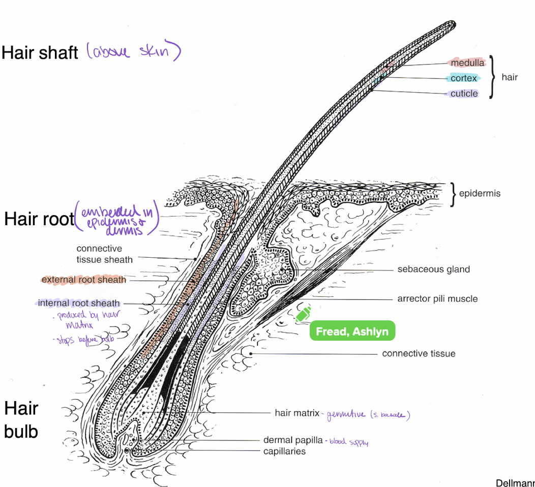
produces hair, a flexible keratinized structure (hard keratin)
embedded in dermis
hair (medulla, cortex, cuticle)
external root sheath - continuous with epidermis, shoots down into dermis
internal root sheath - protect hair from immune system
bulb - at base of follicle
hair matrix - hair matrix cells (analogous to s. basale) produce hard keratin
dermal papilla - blood supply

hair follicle types
follicle includes - hair, sebaceous and apocrine glands, arrector pili muscle (smooth muscle)
Simple (A)
single hair from one external orifice
Compound (B)
one/few primary hair(s), several/many secondary hairs
secondary hairs - thinner (undercoat), reduced/absent medullary
primary hair - outer/ground coat
all hairs emerge from same external orifice
arrangement of compound follicles depends on species/breed/age (newborn animals have simple and become compound with age; Lanugo - organized as simple and no medulla)

sinus/tactile hair follicles
whiskers
blood filled cavity between inner and outer root sheath
innervated by sensory nerve bundles (in external root sheath
movement induced pressure waves through blood filled sinus

hair growth cycle
anagen - active growth phase (hair matrix and dermal papilla)
catagen - growth slows and stops (dermal papilla reduced), entire bulb (matrix) becomes keratinized and begins to move superficially towards the sebaceous glands
telogen - follicle is quiescent (resting), club hair (internal root sheath breaks down, becomes frayed at bulb)
renewed anagen - new hair pushes out old one
under control of hormones, day-length, temperature, and nutritional status

claw
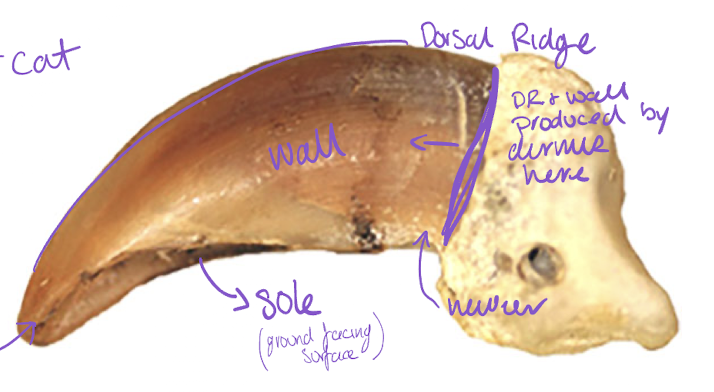
derivative of skin (epidermis and dermis)
covers distal phalanges (cat and dog)
epidermis (dorsal ridge, wall, sole)
underlying dermis (corium) - rich in blood supply, ‘quick’
DR grows faster than wall (curved appearance)
sole produces along entire plane of blood supply

hollow horns
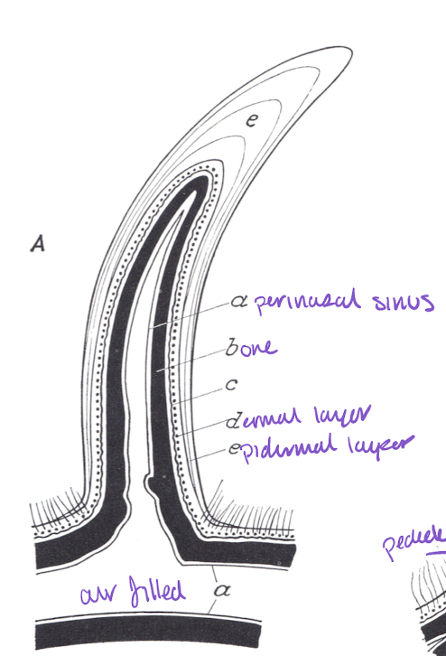
domestic ruminants (cattle, sheep, goats); male and female (male larger)
Basic bony core is the cornual process from the frontal bone
dermal core covering the cornual process (dermis)
covered with keratinized epithelium (epidermis)
permanent
antlers
cervids (generally only males, reindeer and caribou females have small)
bony outgrowth from frontal bone (pedicle) - endochondral ossification
covered with velvet (dermis and epidermis)
antlers shed seasonally
fastest bone production
elk - 1in/d
deer - 0.25-0.5 in/d

mammary gland
compound acinar gland (tubuloacinar in some sp.)
highly modified sweat glands
alveoli, ducts, and interstitial tissue
merocrine (protein and carbohydrates) and apocrine (lipids) secretion
active vs inactive
involution: inactive → active
first 3 lactations, add more mammary tissue
after 3, add more CT and adipose during involution


anal sacs
carnivores (skunk, ferret)
paired diverticula opening into the anal canal at anocutaneous junction
glandular tissue is in wall, sac is innately hollow
dogs - apocrine glands in the wall, sebaceous glands newat the duct (holocrine, viscous potentially harder to express)
cat - sebaceous and apocrine glands in the wall (equal distribution)
do not fuse with perianal (circumanal) glands