Midterm exam
 Call Kai
 Learn
Learn Practice Test
 Spaced Repetition
 Match
Match Flashcards
 Knowt Play
1/71

There's no tags or description
Looks like no tags are added yet.
Name | Mastery | Learn | Test | Matching | Spaced | Call with Kai | Chat |
|---|
No analytics yet
Send a link to your students to track their progress
72 Terms
What types of PPE are used in the lab?
Aprons, splash- and impact-resistant safety goggles (Z87), and gloves. If possible, also add a lab coat for safety.
What are fume hoods
They provide an enclosed air barrier between you and the hazardous materials used. They direct airflow away from you and carry those harmful contaminants out through the exhaust duct.
Where does trash go
Trash canister
Where do points go
Sharps container including razors and scapples
Where does glass go
Glass disposal
How is biological waste handled?
It goes in the biohazard’s container
How are chemicals handled
Go in hazardous waste containers
What is BSL?
Biosafety Level is the classification of microorganisms based on their:
Potential to cause disease
Conditions under which they should be handled
What does the CDC take into account when grouping a microorganism into a level
Virulence
Pathogenicity
Antibiotic resistance
Treatment availability
What level of BSL organisms are used in the lab?
BSL-1 and BSL-2
Why is the level of contamination measured as the number of colonies rather than the size of colonies
Their ability to contaminate is measured by their ability to multiply, as colonies originate from the division of a single cell.
The size of the colony may only help with species identification.
Should one be concerned to find bacteria on the skin
No, because that is our normal microbiota that helps train our immune system, inform, modify, maintain organs, and digest food
How can microbial levels be controlled on the skin, surface, and air
Aseptic techniques are used to control microbial levels
Skin= Washing hands
Surface= Using disinfectants
Air= Using adequate ventilation and airborne precautions
Why choose broth and gar plates
Agar plates are best for separating or isolating bacteria
Broth is convenient for growing large quantities of bacteria quickly
Why are agar plates incubated ‘upside down’ / inverted?
They are inverted so that condensed water does not drip into the agar, interfering with the growth of the colony
What is a colony?
A visible mass of cells resulting from the division of a single cell that can exceed one billion.
Review colony morphology / characteristics (shape, margin, and elevation).
Morphology is the form of organisms and their structures
Bacteria are described by their Configurations (round with scalloped margins, wrinkled, concentric, L-form, or Filamentous), Margins, and Elevation
Know basic use and care of a microscope.
Use both hands, one on the arm and one on the base of the microscope.
Limit of resolution of a typical light microscope VS unaided human eye
The limit of resolution of a microscope is 0.2 uhms, meaning it can distinguish 2 objects as distinct within this range. The unaided eye can only do this at 0.1 mm.
What 2 adjustments can be made to the condenser and how do they affect the image seen
The condenser can be moved up or down to change the focus of the light on the specimen. Up increases the light on the specimen, while moving it down decreases it.
What is a parfocal microscope?
The image remains both centered and in focus when changing from a low-power to a high-power lens
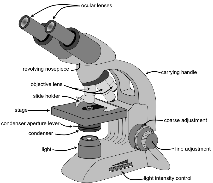
Parts of a light microscope
Remember!
The condenser has a diaphragm that conducts AND regulates the amount of light to the specimen
Objective lenses are:
Low-power, high dry, and oil immersion
What can be done to improve contrast and resolution when using a microscope?
Resolution: Ability to distinguish fine detail
Contrast: Difference in brightness between features
Resolution can be improved by high NA, oil immersion, opening the diaphragm wider for higher resolving power, adjusting the condenser, and being aware of working distance
For contrast, adjust the iris of the diaphragm (close down diaphragm) and use specialized optical techniques like phase contrast, dark field, or differential interference contrast
How to achieve the maximum resolution using oil immersion
Place immersion oil on the slide before using the lens
Open the iris diaphragm wider for higher resolving power
Adjust the condenser when changing objectives
Beware of working at a distance to avoid damage to the lens
How does oil work for oil immersion lens and not others
Oil immersion has the same refractive index as glass, so it creates a continuous lens system that prevents the loss of light when passing from glass to air
List 4 student errors that can result in a dirty or broken microscope.
DO NOT use the coarse adjustment knob w/oil immersion lens
Clean oil off the slide and oil immersion lens before switching lens
Place the dust cover before storing it away
DO NOT use only ONE hand to handle/transfer the microscope
What are aseptic techniques?
They are practices that ensure that
No microorganisms are introduced into culture materials
No contamination occurs to the handler
No contamination remains after work is done
List several basic steps one follows in accordance with aseptic techniques.
Use PPE (aprons, gloves, goggles, and a lab coat)
The work area is to be disinfected
Loops and needles are sterilized by a Bunsen burner
The mouth of the culture tube is flamed to be sterilized
3 reasons why aseptic technique is essential when handling microbial cultures
Ensures that only desired organisms are transferred in each inoculation
Kills microorganisms that may already be present in the workspace
Ensure bacteria deposited outside the workspace are killed, protecting us from infections
Provide 2 examples of how heat is used during inoculation of a tube culture
Used first on an inoculating loop to sterilize and remove undesired organisms
Used last on the inoculating loop to destroy remaining organisms on the loop
How is air contamination prevented when an inoculating loop is used to introduce or take a bacterial sample to/from an agar plate
The plate cover is held diagonally over the plate to allow the inoculating loop to gather bacteria w/o exposing the microbes to other bacteria for long
Where should a label be written on an agar plate
On the bottom of the plate or where the agar is present to ensure identification of the culture
How should agar plates be incubated and why?
They are to be incubated upside down (agar on top) to prevent moisture from condensing on the agar surface and spreading on the inoculated organisms
What are disinfectants good for? What organisms may still remain on the bench after disinfection?
They kill vegetative cells and viruses, but not endospores. Yet, they reduce the likelihood of contamination
How different is bacterial growth in broths, slants, and agar plates. Advantages and disadvantages of them?
Broths → Appears as a presence of cloudiness. Allows for a large presence of bacteria
Agar plates → Appears as visible, isolated colonies. Allows identification of different bacterial colonies
Slants→ Appears as visible, isolated colonies. Allow space saving environment for longer storage
How is a streak plate done?
An agar plate is divided into 4 areas, and one area is inoculated with a certain bacterium. The second area is then inoculated with a little bit of the bacteria inoculated in area 1. The same is repeated in areas 3 and 4 until the cells are diluted over the plate and single cells are separated and able to grow as isolated bacterial colonies.
Advantage of streak plate method over pour plate method
It is very quick to do and requires few materials, but it requires a certain level of skill
Advantage of pour plate method
Requires less skill than the streak plate method, but more materials
3 common errors during streak plating that may result in no isolated colonies after incubation
Failure to let the agar cool down if liquefied, to avoid condensation and moisture
Not inverting the plate to prevent condensation and moisture from reaching the agar plate
Forgetting to flame the mouth of the tube to destroy bacteria around it
You observe isolated colonies on quadrant 2 rather than 4. Why did this happen and would the streak plate be considered a failure
Yes, it is a failure bc the dilution process or spread of bacteria happened too soon, and thus proper isolation of pure culture did not occur
Difference in appearance of surface and subsurface colonies in a pour plate. If they are the same bacteria, why do these differences happen?
Surface colonies are larger in size than subsurface colonies. This is because cells were inoculated into the melted agar before the plate was poured
What is the significance of a streak plate?
It is a method used to obtain pure cultures that isolate a single kind of organism. This helps to study the organism’s culture, morphology, and physiological characteristics.
It can also be used to determine the best treatment options
What are the basic steps in making a smear from solid media and from liquid media?
From liquid medium: 2+ loopfuls of the liquid medium are placed directly on the slide
From a solid medium: 2+ loopfuls of WATER are placed directly on the slide. AN inoculating loop then disperses the organisms in the water
What makes a good smear?
A good smear is done when cells are adhered to the slide of the microscope, shrinkage of the cells has not occurred, and a thin smear is done.
Why is it important to limit the quantity of cells used to prepare a smear
Thick smears w/large clumps can obscure details about arrangement and the presence of internal structures. It may cause staining to prevail in the clumps, preventing visualization
What is the purpose of heat fixation? What problems can arise when the slide is heated in a flame before it is completely dried out? What if you forget to heat fix before staining?
Heat is used to both kill and fix organisms on the slide
If done before the slide is dried, boiling and shrinkage of cells may occur, distorting morphology
If not done before staining, the bacterium may wash out during the rinse-out process of staining
What happens if you forget to turn the slide over after making a target circle and you apply the organism to the slide? After staining, the slide is placed right-side up. What problem will I face when trying to view the organisms?
I wouldn’t be able to focus on the microbes because the glass slide acts as a barrier, preventing sharp focus and clear viewing of details. Lenses may become dirty or contaminated as well, causing damage to the microscope and posing a hazard to me.
What is a chromophore?
Thus, they are attracted to bacterial cells with a negative charge on their surface.
What is the difference between a basic and an acidic dye?
Basic dyes have POSITIVELY charged (cationic)chromophores.
Acidic dyes have NEGATIVELY charged (anionic) chromophores.
Since basic dyes are positively charged, they are attracted to bacterial cells w/a negative charged surface and can stain cells
Acidic dyes are negative and repel from bacterial cells; thus, they only stain the background of the slide
Bacteria are usually stained with a basic dye – why?
Basic dyes can stain the bacterial cells as they have an electrostatic attraction between the negatively charged cells and positively charged chromophores of basic dye
What type of dye is crystal violet and what color do cells take on after it is applied?
It is a basic dye that stains deep purple
What is meant by the palisade arrangement of cells
Parallel arrangement of rod-shaped cells AKA “picket-fence”
What is expected to be seen if a Streptococcus pyogenes smear is stained w/crystal violet
Sphere-shaped cells arranged in chains, stained deep purple are to be expected
What does simple staining reveal
Morphology of the cells like their shape and arrangements
If you were working with an unlabeled simple-stained smear, would you be able to identify the bacterial species by observing it under the microscope?
Simple staining can reveal the shape and arrangement, but the specific bacteria won’t be identified because many microorganisms share these characteristics, so further investigation would have to be done.
What is the difference between a simple and a differential stain?
Simple staining → Reveals morphology (shape and arrangement of cells) using 1 dye
Differential staining → Uses multiple dyes to distinguish between different types of cells and their structures
Gram stain
Purpose: To differentiate between Gram-positive and Gram-negative bacteria based on their cell wall structure and composition
Results: Gram-positive takes on a purple color, and gram-negative takes on a red color
Stains: Crystal violet and Safranin
Procedures: After heat-fixing the cells, they are dyed with crystal violet. Gram’s Iodine (mordant) is added to create an insoluble complex in Gram-positive bacteria. Ethyl alcohol is used for decolorization. Safranin is then added as a counterstain.
Why is gram stain considered a differential stain
It uses 2 kinds of dyes to differentiate between 2 kinds of cells based on their different staining reactions that structures have w/different dyes.
How does age of a culture affect gram staining reactions
The optimum age is 16-8 hours, as anything beyond is when cell walls begin to break, causing them to be leaky and lose crystal violet.
What step in gram staining is more prone to error? If done right, how might it affect the end result?
Decolorization is the most critical step bc if it
Negative stain
Purpose: Negative dyes are acidic, so they reveal the morphology of cells, such as external structures like capsules, by staining the background.
Results: Cells appear as clear objects against a dark background
Stains used: India Ink or Nigrosin
Procedure: Organisms are mixed in a drop of nigrosin and spread over the slide by another slide, making a thin-to-thick smear.
Another is the needle method that just spreads nigrosin and the bacteria w/o the need for a slide.
What type of chromophore is associated w/a negative stain
Negative stains are acidic and have negatively charged chromophores that do not penetrate the cell, thus only stain the background or surrounding of the cell
What step normally associated w/staining bacterial cells is omitted when the size of the cell is determined?
Heat-fixation is avoided to prevent the shrinkage of cells and accurately determine the size of bacterial cells without harming the cells’ structure
Acid-fast stain
Spore stain
Review the Disk Diffusion test (Kirby Bauer method) – review the questions on the lab reports.
What are the standard procedures / protocols required for setting up the Kirby Bauer method? List 4.
What is a zone of inhibition?
Know the mode of action of the following antimicrobials: Penicillin, Imipenem, Tetracycline, Ciprofloxacin, Streptomycin, and Sulfisoxazole-Trimethoprim (folic acid inhibitor – a competitive inhibitor), Isoniazid.
Note the difference between antiseptic and disinfectant.
Review questions in lab report (Ex 32)
Review the effects of UV as a control agent – review the questions on the worksheet.