Disease Correlation
1/85
There's no tags or description
Looks like no tags are added yet.
Name | Mastery | Learn | Test | Matching | Spaced |
|---|
No study sessions yet.
86 Terms
most common etiology (cause) of glomerular disease
immunologic (most common)
metabolic causes
hereditary disorders
what changes occur in the glomerular tissue as a result of glomerular disease
cellular proliferation
leukocyte infiltration (WBCs go to inflammation)
glomerular basement membrane thickening (b/c it’s repairing over and over)
hyalinization with sclerosis (tissue is being replaced by scar tissue which hardens)
how does the disease process of glomerular disease occur
antibody-antigen complexs are formed and get stuck and clog the glomerulus so it gets inflammed anf filtration can’t occur
when glomerulus filter is compromised waste begins to build up and end up in the bloodstream
what specific renal disease is most likely to progress to renal failure and require either dialysis or a transplant
acute glomerulaonephritis
common clinical features that all three of the glomerular diseases discussed in class share
hemeturia, proteinuria, oliguria, azotemia, edema, and hypertension
physical/chemical/microscopic findings of acute glomerulonephritis that make it unique
protein: mild (<1 g/day)
increased casts
physical/chemical/microscopic findings of acute glomerulonephritis
protein: mild (<1 g/day)
blood: positive (variable)
increased RBCs, WBCs, renal tubular epi cells
increased casts (RBC/Hgb/granular)
physical/chemical/microscopic findings of chronic glomerulonephritis
protein: heavy (>2.5 g/day)
blood: positive (usually small)
SG: low and fixed (isothenuria)
increased RBCs, WBCs, rnal tubular epi cells
casts (all types but esp grabular/waxy/broad)
physical/chemical/microscopic findings of chronic glomerulonephritis that make it unique
protein: heavy (>2.5 g/day)
SG: low and fixed (isothenuria)
casts (all types but esp. granular/waxy/broad)
physical/chemical/microscopic findings of nephrotic syndrome
protein: severe (>3.5 g/day)
blood: positive (usually small)
oval fat bodies and free fat globules present
increased casts (esp. fatty/waxy/renal cell)
increased RBCs and renal epi cells
physical/chemical/microscopic findings of nephrotic syndrome that make it unique
protein: severe (>3 g/day)
oval fat bodies and free fat globules present
increased casts (esp. fatty/waxy/renal cell)
what glomerular disease is associated with increased lipids present in the urine
nephrotic syndrome
difference between acute and chronic glomerularnephritis
acute: sudden and swelling and blockage inhibits filtration
chronic: occurs over time, glomerular disease has progressed to point of sclerosis (hardening of tissue) and hyalinization (scarring)
difference between ischemic and toxic acute tubular necrosis in terms of the cause of each
ischemic: due to hypovolemic event (decreased BP) where the kidneys have decreaed perfusion (blood flow)
toxic: due to exposure to a toxin (ex: meds/drugs, poison, or heavy metals)
difference between ischemic and toxic acute tubular necrosis in terms of what part of the tubules is affected
ischemic: proximal convoluted, loop of henle, the distal convoluted, and the collecting duct (more likely to affect the basement membrane, and more spread out)
toxic: proximal convoluted, loop of henle, distal convoluted, and collecting duct (more concentrated in the PCT)
acute tubular necrosis clinical features
oliguria, azotemia, uremia, acid-base imbalance, electrolyte imbalace
for acute tubular necorisis, what treatment is there
remove the causitive agent, once ID and removed normally symptoms go away
what is tubular dysfunction
dysfunction of the tubules system either in a small and isolated area or a large and diffuse area that causes a variety of effects (ex: cystinuria, fanconi’s, renal phospaturia)
why is the GFR normal for tubular dysfunction
the filtration system is working, it’s the tubules that aren’t absorbing the filtered substances
examples of tubular dysfunction disorders
cystinuria (PCT can’t absrob cystine)
fanconi’s (PCT doesn’t absorb at all)
renal phosphaturia (PCT doesn’t absorb phosphate)
renal tubular acidosis (tubules can’t secrete enough hydrogen so acid base balance is off)
what region of the urinary tract is affected by urethritis
urethra
what region of the urinary tract is affected by cystitis
bladder
what region of the urinary tract is affected by pyelitis
pelvis
what region of the urinary tract is affected by pyelonephritis
spaces inbetween where filtration occurs (interstition of the kidneys)
why do UTIs affect females more frequenty than males
women due to having a shorter urethra and its close proximity to the anal region, greater hormone fluctuations, and no prostate (prostatic fluids has natural antibacterial effects)
physical/chemical and microscopic findings for urethritis/cystitis
protein: small (<0.5 g/day)
LE: positive
nitrate: usually positive
blood: positive
increased WBCs and RBCs
bacteria
transitional epi cells
physical/chemical and microscopic findings for urethritis/cystitis that make it unique
protein: small (<0.5 g/day)
transitional epi cells
no casts (b/c kidney isn’t involved)
physical/chemical and microscopic findings for pyelonephritis/pyelitis (acute)
protein: mild (<1 g/day)
blood: positive
LE: positive
nitrate: usually positive
SG: normal to low
increased WBCs, bacteria, RBCs, and renal epi cells
increased casts (granular, waxy, broad, WBC, and renal cell)
physical/chemical and microscopic findings for pyelonephritis/pyelitis (acute) that make it unique
protein: mild (<1 g/day)
SG: normal to low
increased renal epi cells
physical/chemical and microscopic findings for pyelonephritis (chronic)
protein: moderate (<2.5 g/day)
LE: may be positive
SG: low (may have isothenuria)
increased WBCs and macrophages
increased worsening casts (granular, waxy, broad, few WBC, and renal cell
physical/chemical and microscopic findings for pyelonephritis (chornic) that make it unique
protein: moderate (<2.5 g/day)
LE: may be positive
SG: low (may have isothenuria)
increased macrophages
increased worsening casts (few WBC ones)
S/S of urethritis/cystitis
pain or burning on urination (dysuria) and occasional lower abdomen pain
S/S of pyelitis/pyelonephritis
flank pain (in back and sides), painful urination (dysuria), nocturia, urgency, fever, nausia, headache, malaise, and polyuria
etiology of chronic pyelonephritis
birth defects
an acute infection that was never treated properly
common causes of interstitial nephritis
exposure to drugs, toxins, and heavy metals (when removed issue fixes)
what three ways can interstitial nephritis affect the interstitial tissues
immediate allergic response
rapid tubules destruction
slowly and progressing failure of the tubules
most common species of yeast associated with yeast infections
Candida species
specific conditions in which a yeast infection is more likely to occur
normal flora being disrupted by a pH change or steroid use (immunocompromised) or antibiotics
introduced via urinary catherization or contamination
conditions that would contribute to low perfusion through the kidney that compromise normal kidney function
chronic heart failure (not enough force to pump blood)
hypotension (decreased BP)
hypovolemia (decreased volume)
hypertension (increased BP)
atherosclerosis (fatty plaque accumulation in BV)
diabetes (sugar coated BV, become hardened)
what consequences occur when the kidneys are deprived of adequate blood flow
kidneys won’t be able to regulate BP and BV which leads to other organs being damaged
kidney then loses mass as it shrinks and hardens (then it becomes nonfunctional)
kidneys require at least _________ of cardiac output going through the flomerulus at one time
25%
prerenal causes of acute renal failure
the kidney is fine but isn’t getting enough oxgenated blood to function
could be caused by cardiac output issue or loss of vascular integrity (meaning kidneys are retaining sodium and then water follows)
25% of cases
renal causes of acute renal failure
failure is due to the kidney itself
could be caused by glomerular disease, tuular disease, or intravascular disease (kidney can’t retain sodium so they aren’t retaining water either)
65% of cases
postrenal causes of acute renal failure
failure is due to the formation of urine normally due to a blockage of urine exiting
10% of cases
difference between prerenal and renal in term of sodium and water
prerenal: kidneys retain sodium so water follows
renal: kidney loses sodium so it loses water
acute renal failure effects
the function of the kidney (prerenal, renal, or postrenal)
which out of the 3 types of acute renal failure is most comonly associated with acute renal failure
acute renal failure (65%)
lab/urinalysis fnding of acute renal failure
sudden decrease in GFR
azotemia
hematuria
proteinuria
electrolyte imbalance
polyuria
oliguria
anuria (depending on cause)
S/S of acute renal failure
polyuria then dehydration
overhydration
edema
heart failure
fatigue
poor appetite
pale skin
why is chronic renal disease sometimes described as “slow and silent”
because it’s not recognizable until about 80-85% of the kidney’s function is lost (because you can function with just one kidney until this point)
approximate percentage of kidney function that must be lost in oder for kidney function to be adversely affected and require treatment in the form of dailysis or a transplant
80-85%
lab findings of chronic renal failure
azotemia
acid base imbalance
electrolyte imbalance
abnormal Ca and P metabolism
anemia
chornic renal failure urinalysis findings
isothenuria (can’t conc urine)
proteinuria
hematuria
increased casts (waxy and broad)
S/S of chronic renal failure
bleeding tendencies
hypertension
weight loss
nausea
vomiting
edema
common locations where renal calculi form
renal calyces (darining tubes)
pelvis
ureter
bladder
most common location of renal calculi formation
renal calyces (draining tubes)
four factors that contribute to the formation of renal calculi
hydration status (allows for super saturation)
urinary solute status (increased solutes → more likely to precipitate out)
changes in urinary pH (acidic/alkaline crystals form)
urinary stasis (slow/no movement)
most common constituent of renal calculi
calcium (also oxalate, phosphate, uric acid, and cystine)
all kidney stones have a _______________ core
protein
lab findings/symptoms of renal calculi
severe pain (kidney region radiating forward and donward)
nausea
vomiting
sweating
frequent urge to urinate (dysuria)
oliguria/anuria
hemeturia (b/c some stones are pointy)
dysuria
treatment options for patients with renal calculi
lithotripsy (the surgucal crushing of a stone)
externally using sound waves to break up stones
pain medication to pass tone naturally
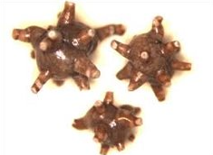
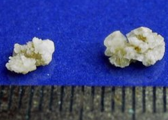
calcium oxalate monohydrate stone
calcium oxalate dihydrate stone
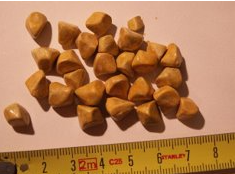
uric acid stone
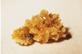
calcium phosphate stone
what are the three types of aminoacidurias
overflow
no threshold
renal aminoacidurias
primary vs secondary causes of aminoacidurias
primary: inherited inborn errors of metabolism or tubular reabsorptive dysfunction
secondary: severe liver disease or generalized renal tubular dysfunction
overflow aminoaciduria
there’s an increase in amino acid excretion so the renal threshold is exceeded so amino acids spill over into the urine
no-threshold aminoaciduria
there’s an increase in amino acid excretion but they aren’t being absrobed at all so everything (meaning AA) ends up in the urine
aminoaciduria
amino acids aren’t breaking down due to missing/defective enzymes
renal aminoaciduria
the levels of amino acids are normal but the tubules are defected causing a lack of absorption so they end up in the urine
cystinuria
an inherited tubular reabsorption problem where cystine (arginine, lysine, and orthotheine) ends up in the urine
cystinosis
an inherited recessive disorder that disrupts the metabolism of cystine so it accumulates in the body/tissues
maple syrup urine disease
an inherited condition where a person lacks enzyme or has defective enzymes so they accumulate leucine, isoleucine and valine
S/S of maple syrup urine disease
severe ketacidosis
seizures
mental development issues
maple syrup urine smell
phenylketonuria
an inherited conditions where a person lacks enzymes or has defective enzymes so phenylalanine accumulates
S/S of phenylketonuria
feeding difficulties
severe mental deficiencies if eating food with phenylalanine
musty/mousy odor
alkaptonuria
an inherited disease due to the lack of an enzyme of defective enzymes so homegenistic acid accumulates in the body
alkaptonuria S/S
darkening of tissues
arthritis due to build up of acid
brown-black color of urine when exposed to air
tyrosinuria
an inherited condition or transient after borth condition due to an immature liver that can’t breakdown tyrosine
S/S of tyrosinuria
spindle/needle like prisms in sheaths, grey-black in color
albinism
a genetic metabolic disorder where a person cant produce melanin or don’t produce normal amounts of melanin
S/S of albanism
no pigmentation in hair, skin, or eyes
sensitive to sunlight
poor eyesight
increased risk of sunbrun or skin cancer
melanuria
a genetic disorder where there’s an overproduction of melanin or melanoma
S/S of melanuria
dark brown/black urine when exposed to air
what hereditary pattern do nearly all aminoaciduria disease have
autosomal recessive