Unit 4 Lab Exam
1/281
There's no tags or description
Looks like no tags are added yet.
Name | Mastery | Learn | Test | Matching | Spaced | Call with Kai |
|---|
No analytics yet
Send a link to your students to track their progress
282 Terms
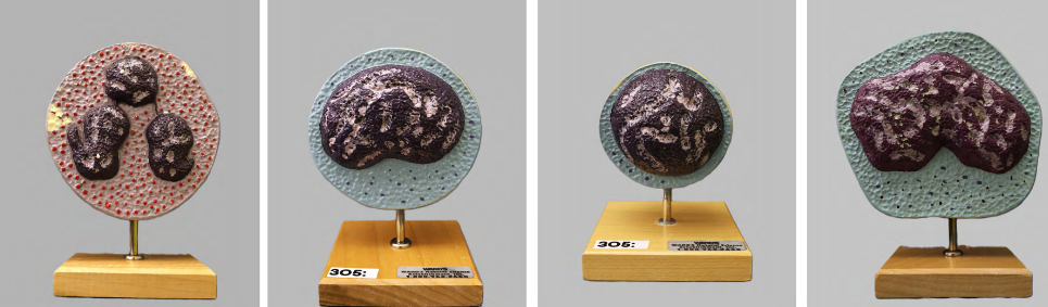
left corner
neutrophil

middle left
‘large’ lymphocyte

middle right
‘small’ lymphocyte

right corner
monocyte

left corner
eosinophil

middle left
basophil

middle right
erythrocytes
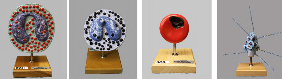
right corner
thrombocytes (platelets)

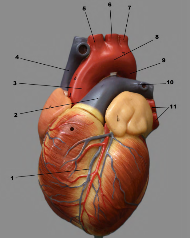
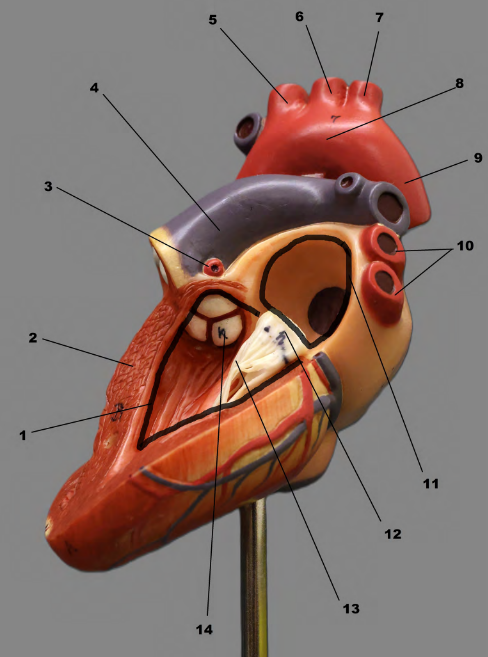
1
base

2
right auricle

3
ligamentum arteriosum

4
left auricle

5
anterior interventricular sulcus (a groove/depression)

6
apex
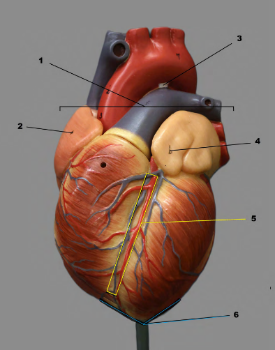
Note The anterior interventricular sulcus is a what? on the anterior surface of the heart between the ventricles.
groove/depression

Note Sulci on the heart's surface have blood vessels that do what?
'sit' 5 in the grooves/depressions.

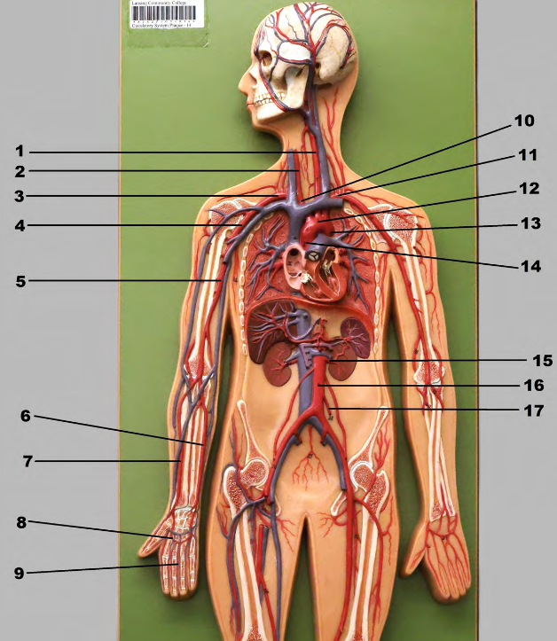
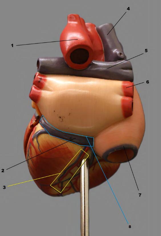
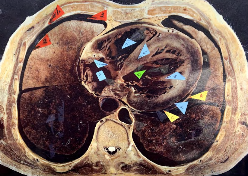
1
cardiac vein

2
pulmonary trunk

3
ascending thoracic aorta

4
superior vena cava

5
brachiocephalic trunk

6
left common carotid artery

7
left subclavian artery

8
aortic arch
9
descending thoracic aorta

10
pulmonary artery

11
pulmonary veins

Note

1.
descending thoracic aorta
2.
coronary sinus

3.
posterior interventricular sulcus (groove/depression)

4.
superior vena cava

5.
pulmonary artery

6.
pulmonary vein

7.
inferior vena cava

8.
coronary sulcus (groove/depression)
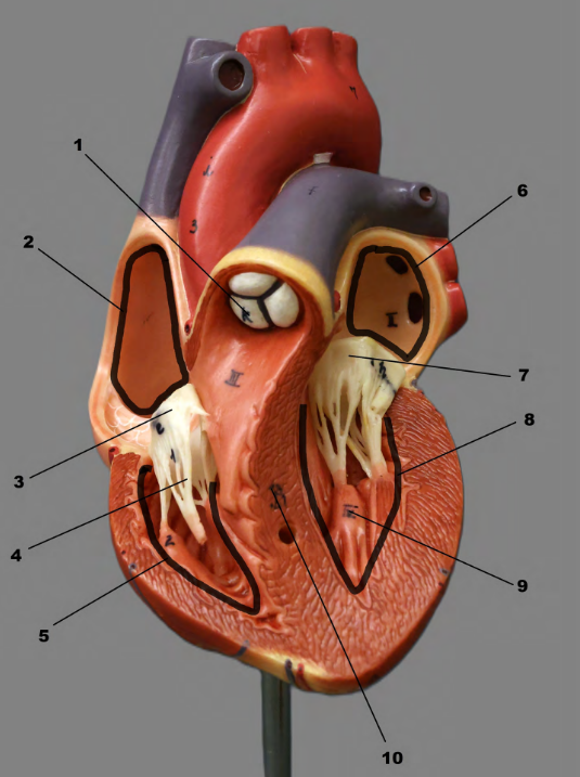


1
left ventricle

2
interventricular septum

3
left coronary artery

4
pulmonary trunk
5
brachiocephalic trunk

6
left common carotid artery

7
left subclavian artery

8
aortic arch

12
bicuspid valve

13
chordae tendineae

14
aortic semilunar valve

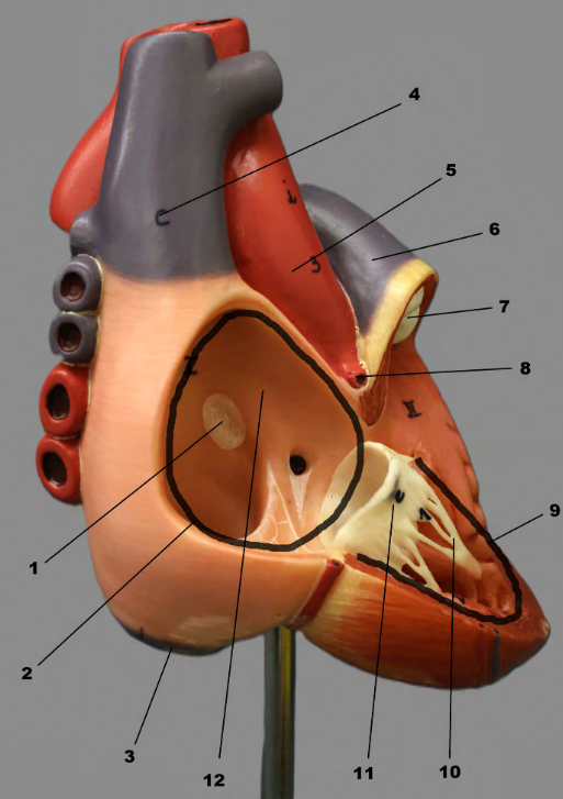
epicardium or visceral pericardium

myocardium

endocardium
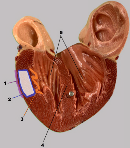
interventricular septum

5
trabeculae carneae

Notes


B


right ventricle
Personal note: One way to memorize this is specifically the right ventricle is that it’s closest the sternum, meaning it will be compressed first when doing CPR.


12

Notes
The fibrous pericardium is the most superficial (outermost) layer of the pericardium; The parietal pericardium touches the inside of the fibrous pericardium
Personal note, not in slideshow: It’s hard to tell which side is left or right, but knowing that the heart sticks out on the left(?) side of the chest, you can know which side it is.