Ultrasound SEM 2 (recent)
 Call Kai
 Learn
Learn Practice Test
 Spaced Repetition
 Match
Match Flashcards
 Knowt Play
1/139

There's no tags or description
Looks like no tags are added yet.
Name | Mastery | Learn | Test | Matching | Spaced | Call with Kai | Chat |
|---|
No analytics yet
Send a link to your students to track their progress
140 Terms
Flow going into the liver
Heptopedial
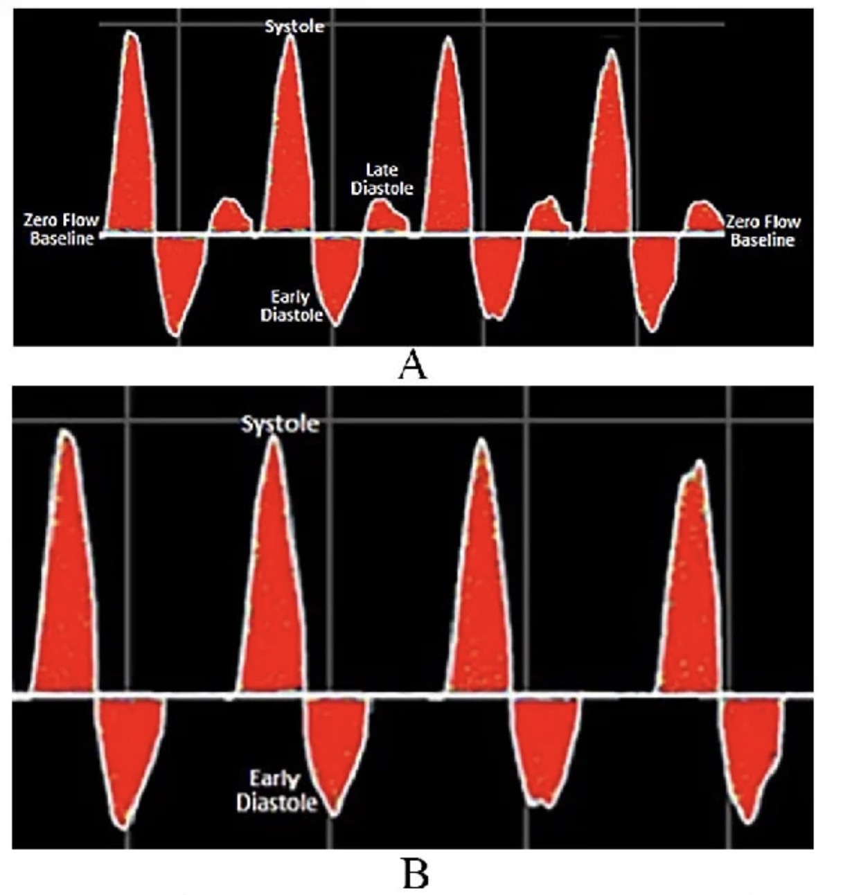
A ) triphasic
B ) biphasic
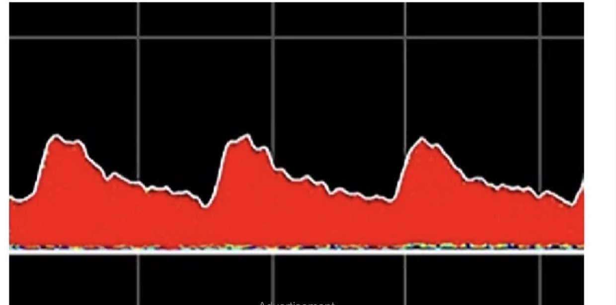
Monophasic
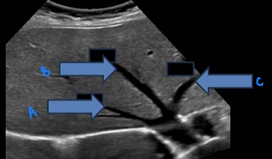
A ) right hepatic artery
B ) middle hepatic artery
C ) left hepatic artery
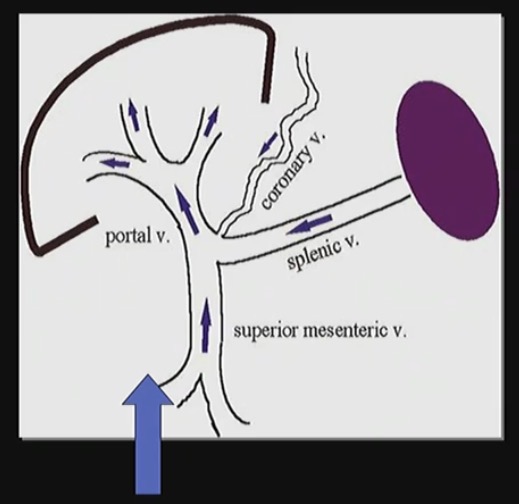
How does the flow of blood come into the liver
The hepatic artery brings ooxygen rich blood into the liver and the hepatic portal vein brings nutrient rich blood from digestive t
Liver’s 5 main functions of the liver
Filtration
Digestion
Metabolism
Protein synthesis
Storage of vitamins and minerals
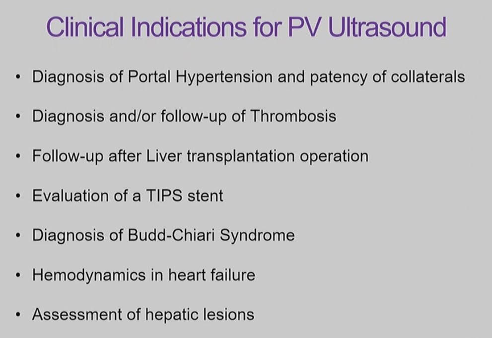
Clinical indications for PV ultrasound

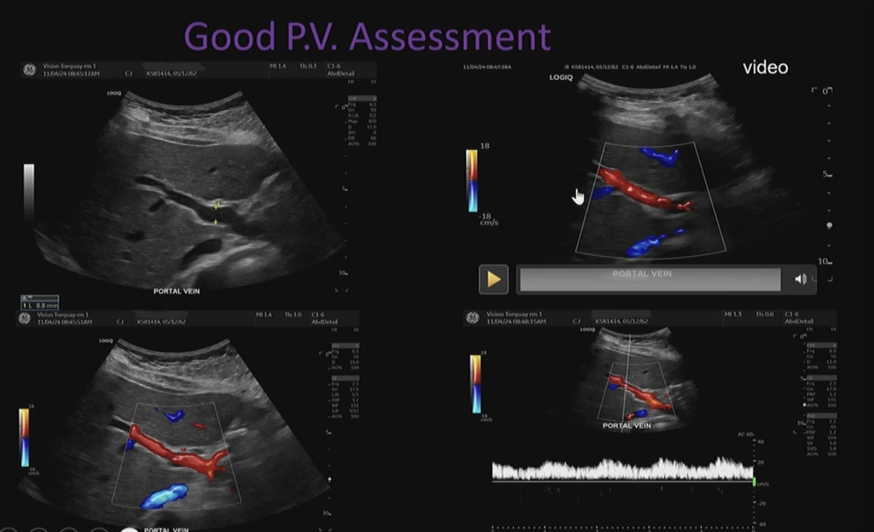
Portal vein ultrasound technique
The patient is in LPO position (or take image intercostally)

The assessment of PV
Checking for thrombosis, or enlargement of vessel paired with Doppler trace.
Uniformity of colour in the vessel.
Angle of insonation
60 degrees or less
if angle isn’t good the vessel has no colour fill
To fix angle can heel toe the transducer or can go intercostally

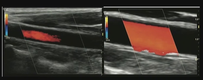
What’s the problem with these images
Colour gain too low
Colour gain too high

Scale set too high (Bring it down to 18)
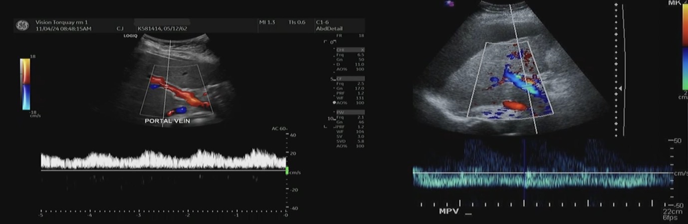
Which is hepatopetal and which is heptofugal
The image on the left is hepatopetal → towards the liver
The image on the right is hepatofugal → away from the liver
Red is towards the transducer
Explain how pulsatility should be shown in portal vein
Should be absent in healthy patient, with flows showing normal gentle undulation
What are some PV pathology
Vascular abnormalities
Portal vein thrombosis
Portal vein hypertension
Portal vein aneurysm
Metabolism disorder
cirrhosis
PV thrombosis
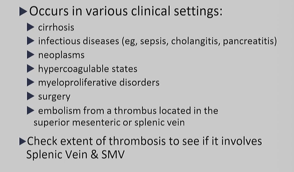
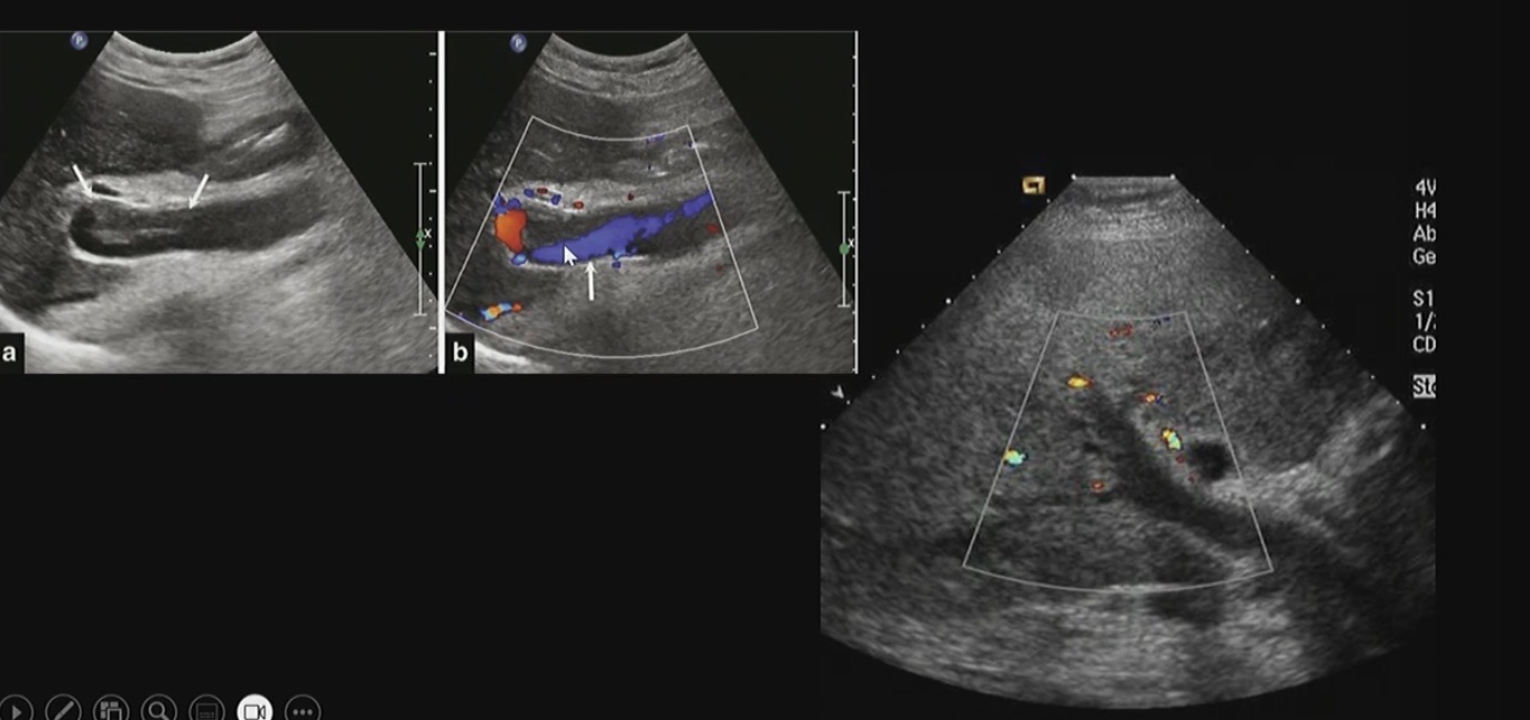
What pathology is this
Thrombosis
Portal hypertension
Elavated pressure in the PV system
Increased resistance to flow (sinusoids)
Flow diverted to system veins through collaterals
Later phase: thrombosed portal vein replaced by collateral or cavernous venous malformation at ports hepatis
Portal hypertension types
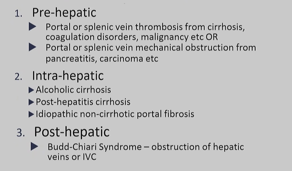
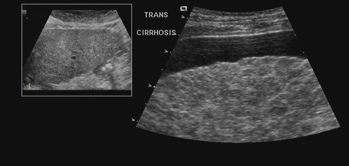
Cirrhosis
enlargement of the liver
Mottled / hetorgonis appears of liver

Portal hypertension

Partial thrombosis
The purpose of the pelvic girdle
Support
Stability
Movement
Attachment
Thyroid anatomy

Typical size of thyroid
Normally length 5 cm, width 3 cm, depth 2cm
Isthmus 2 - 3 mm thick
Thyroid glads weigh about 20 to 30
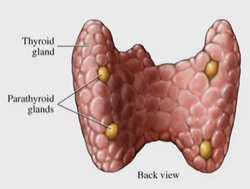
Thyroid physiology
Is an endocrine gland, maintain body metabolism and growth by secreting thyroid hormones.

SCM → sternoclidomastoid
SC → subcutaneous layer
SH → sternohyoid muscle
ST → sternothyoid muscle
LCM → longest Colli muscle

Clinical indications for thyroid US
Palpable mass (thyroid)
? Nodule
? Goitre
Diffuse thyroid enlargement
Pain, voice changes, tightness in the throat
Extra-thyroid mass
As an aid to fine needle aspiration biopsy / core biopsy
What probe should you use for thyroid ultrasound
High frequency transducer 15 hz
Patient Preparation for thyroid US
Explain the procedure
Obtain good patient history
Mindfulness of patient comfort
Pillow under head
Use warm gel
Scanning techniques
Patient is supine, head extended
Palpation of palpable mass / ROI
Both thyroid lobes and the isthmus are scanned in both trans and long planes
Gentle transducer pressure
Imaging documentation of normal thyroid + / - pathology. Use colour Doppler
Cone loops as required
Volume measurements of both thyroid. AP measurement of the isthmus
Diffuse thyroid disease
hyperthyroidism
Hypothyroidism
Thyrotoxicsis
Graves’ disease
Goitre
Thyroiditis
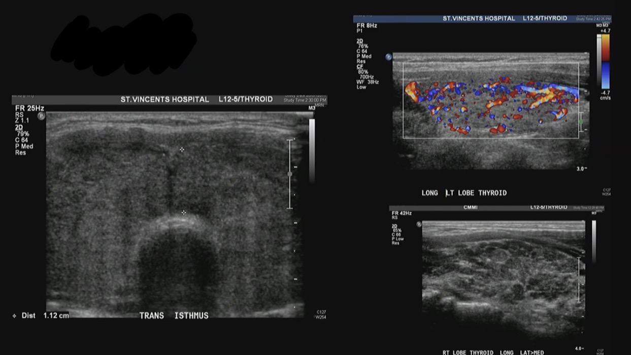
Thyroiditis
increase in thyroid size
Hetrogenous
Increased vascularity
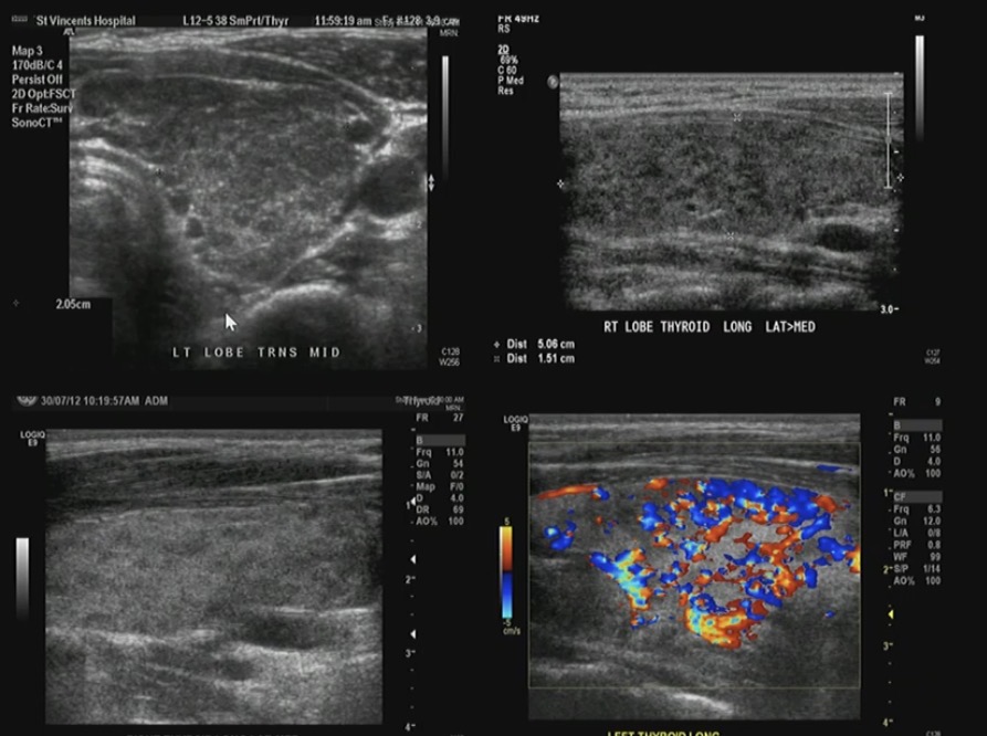
Graves’ disease
enlarged thyroid
Hetrogenous (patchy)
Increased vascularity
(Similar to thyroiditis)

Goitre
Enlarged
Usually multi nodular
How would you describe nodules in US
Echogenicty
Sharpness of border
Calcifications
Taller than wide
Invasion
Chaotic vascularity
Halo
Nodes

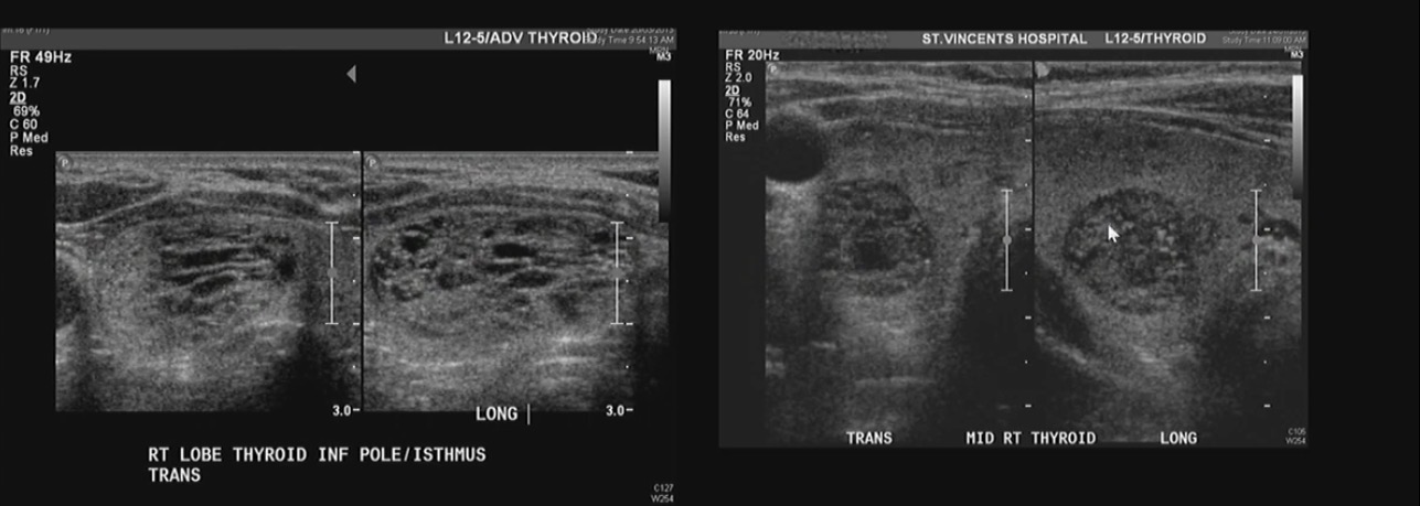
Honeycomb apperence
Typical begin spongiform nodule
Cyst within the nodule
Breast anatomy
Paired superficial structure between skin and superficial muscle
Part of the integumentary system
Accessory reproductive function - breast feeding
Consists of adipose tissue, glandular tissue and fibrous connective tissue.
Breast anatomy
Skin, subcutaneous tissue
Nipple ad aerolar
Fibroglandular tissue
Intramammary fat
Coopers ligaments
Intramammary ducts
Pectoralis muscles costal bones

What are the four quadrants of the breast

What are the three layers of breast that can be distinguished on ultrasound
Pre mammary zone
Mammary zone
Retro mammary zone

Premammary zone
Between the skin and parenchyma
contains fat - hyperechoic compared to parenchyma
Coppers ligaments - echogenic linerar bands
Mammary zone
Parenchyma
fibroglandular tissue - hyperechoic
Ducts - hyperechoic / anechoic tubes running through
Retro mammary zone
Between parenchyma and muscle
contains fat - hyperechoic compared to parenchyma
Coppers ligaments ligments - echogenic linear bands
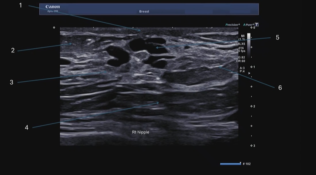
1) skin
2) intermammary fat
3) fibroglandular tissue
4) retro mammary fat
5) ducts
6) copper ligaments
Clinical indications for breast US
Palpable lump
Correlation with mammography
Breast pain
Nipple discharge
PHX breast ca
Fox breast ca
Breast implant
US equipment for breast
Hugh frequency transducer 15mHz
Patient prepration for breast US

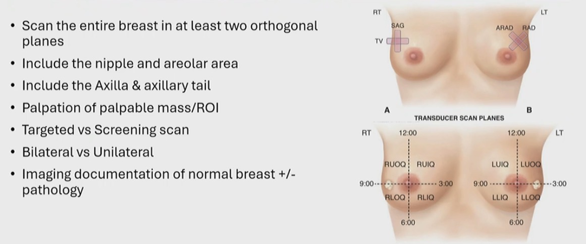
Scanning techniques for US
Breast pathology
Cyst
Fibroadenomas
Malignancy
Mastitis
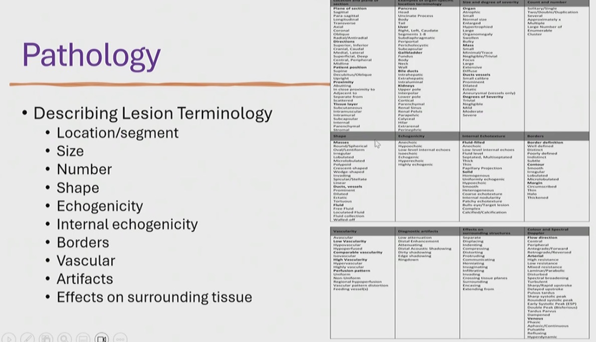
How to describe Breast lesions

Breast cyst
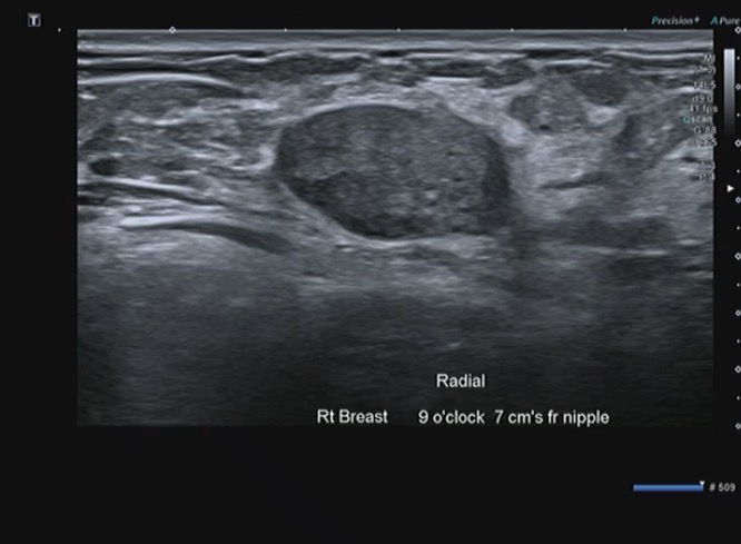
Fibroadenoma
most common soild benign lesion
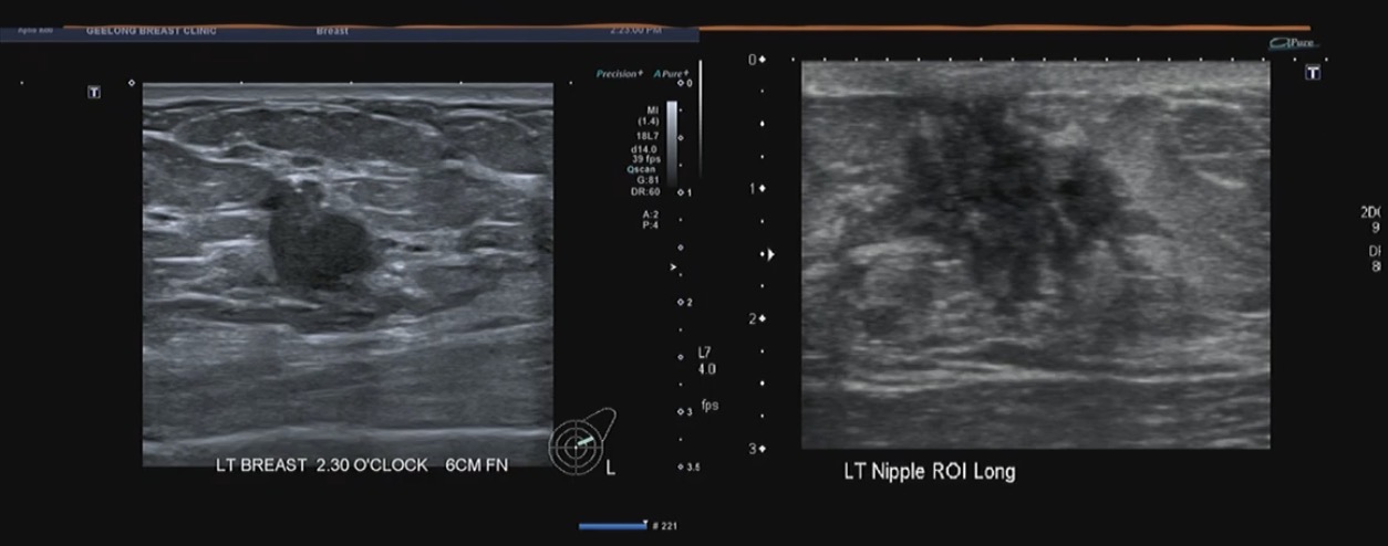
Malignancy
Taller than wide
Non identified lesions
Posterior shadowing
If put colour on you see vascularity
Invading the tissue
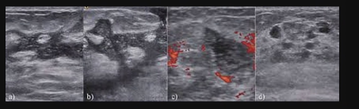
Mastitis
Dilated ducts
hyper vascular → indicates inflammation in breast
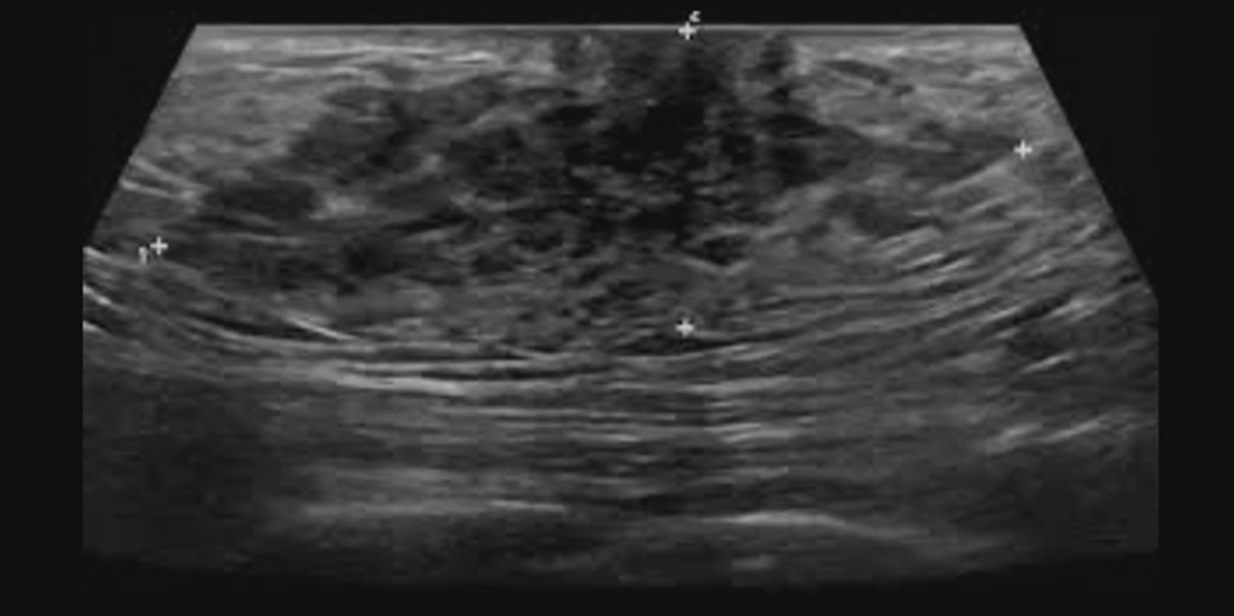
Gynaecamastia
Lump under the nipple area
In men
Ill defined
Hyperechoic

Scrotal anatomy
A - Tunica vaginalis (parietal layer)
B - Tunica vaginalis (visceral layer)
C - Tunica albuginea
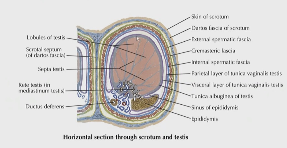

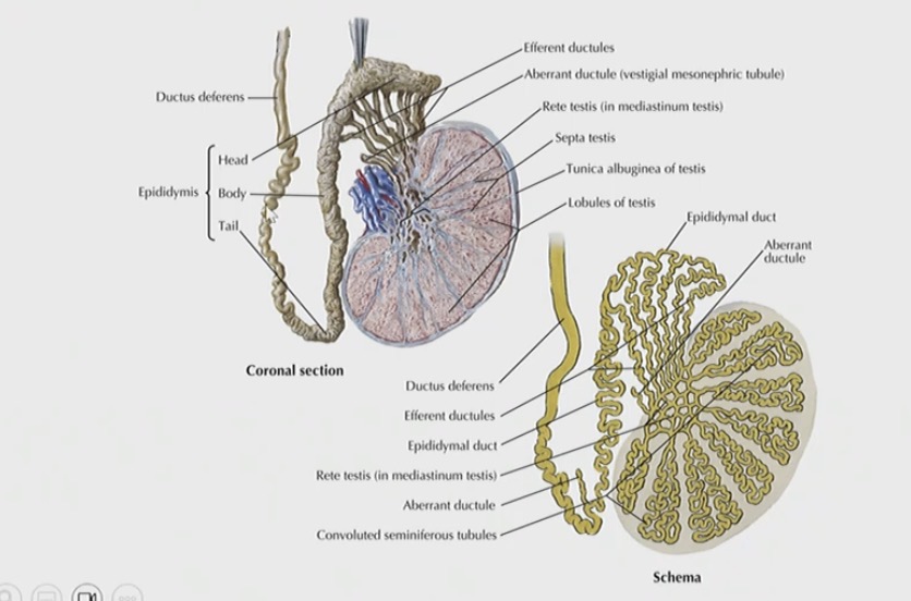
Image 1 (head of the epididymis)
Image 2 (body of the epididymis)
Image 3 (tail of the epididymis)
Clinical Indications for Scrotal US
Testicular pain
Palpable lump
Scrotal swelling
Trauma
Follow up with previous lump
Follow up of microlithiasis
Location of undescended testis
What transducer for scrotal exam
High frequency transducer 15 MHz
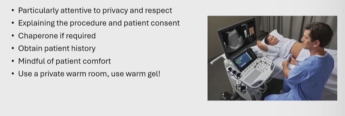
Patinet preparation scrotal US
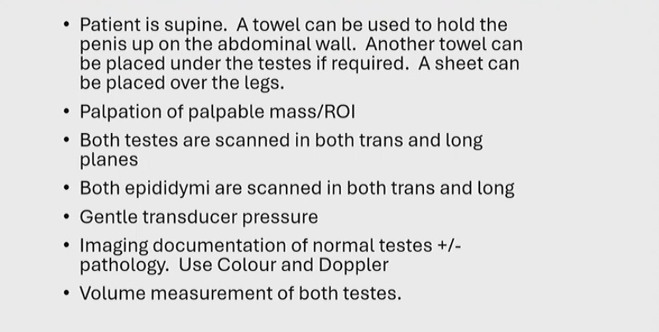
Scanning technique for scrotal US
Common scrotal pathology
Cyst
Calcification
Microlithiasis
Hydroceles
Varicoceles
Epididymitis
Torsion
Trauma
Intra and extra testicular lesions
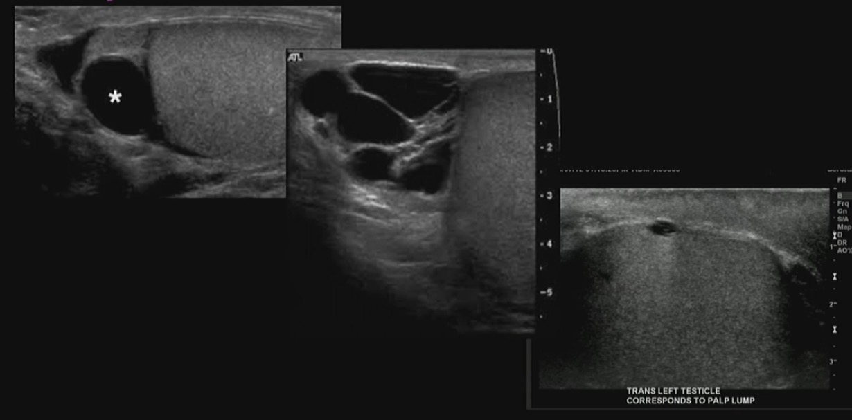
Scrotal cyst
anechoic posterior shadowing

Microlithiasis
intratubular calcifications

Hydrocele
Large collection of fluid in scrotum
Posterior enhancement
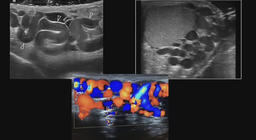
Varicocele
Collection of abnormally dilated and tortious veins in the scrotum
More than 3 cm
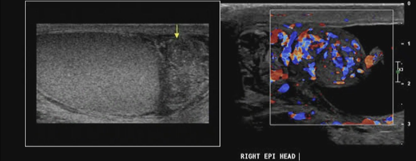
Epidymitis
inflammation of the epididymis
Hetrogenous
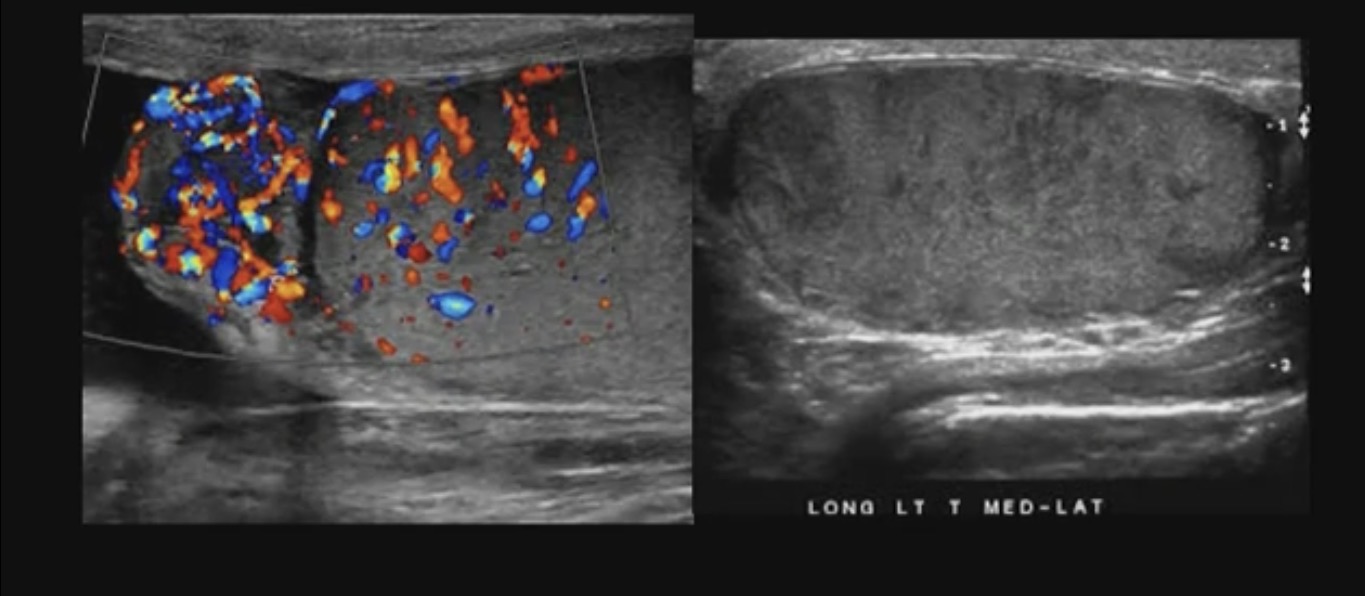
Epididymo-orchitis
Infection / inflammatory process of epeidimus and the testice
Increased vascularity
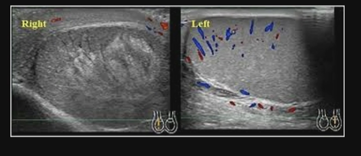
Testicular torsion
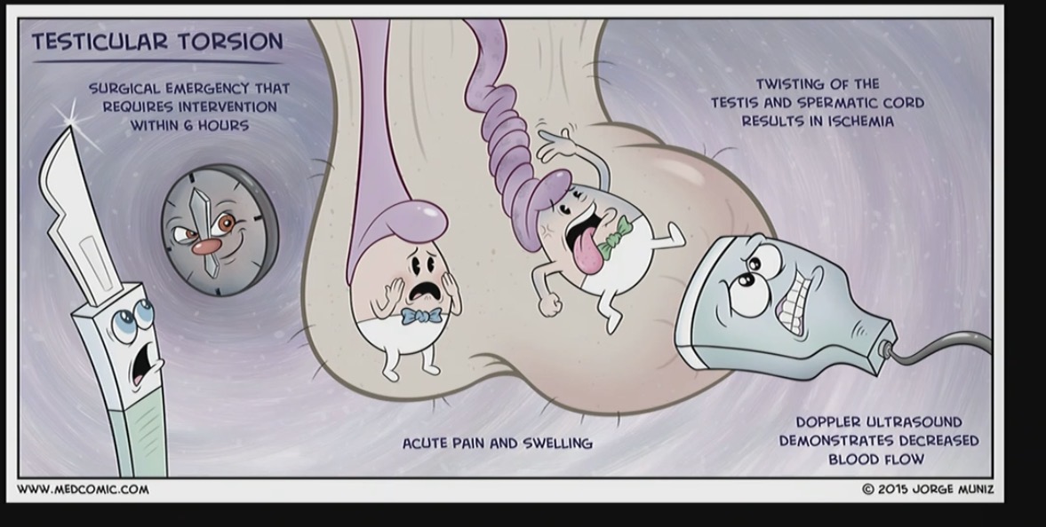
When the cord that provides blood flow to the testice twists and cuts of the blood supply
No blood flow

Trauma
acute swelling / hematoma
Rupture of the testicle
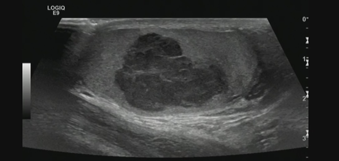
Testicular malignancy
What are the different types of skeletal muscle and there anatomy

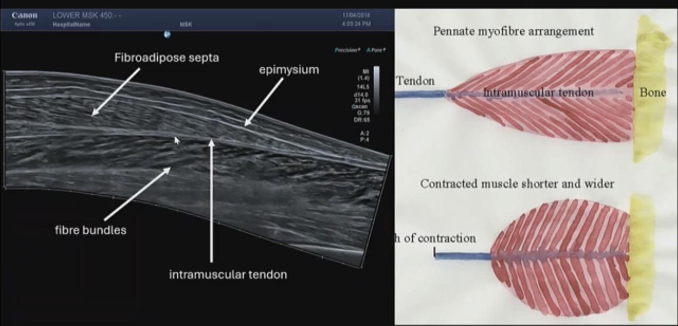
Appearence of muscle on US
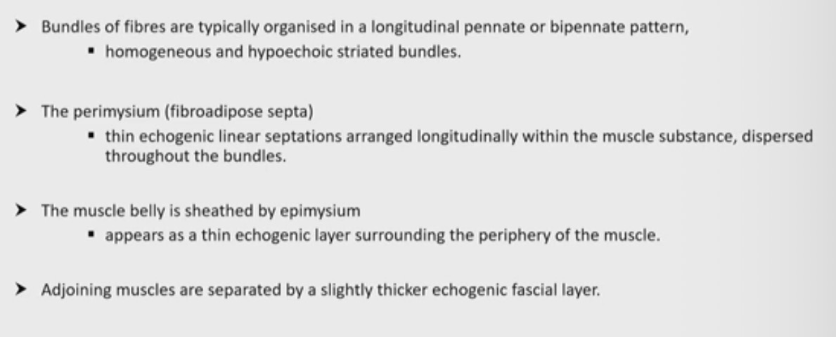

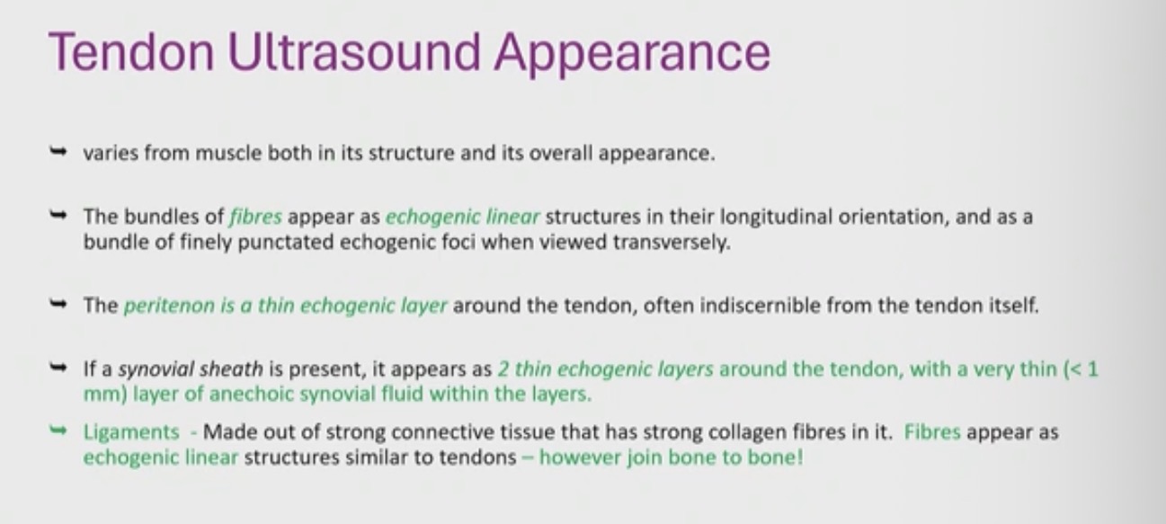
Tendon appearance on US
Ligaments → bone to bone
Tendons → bone to muscle
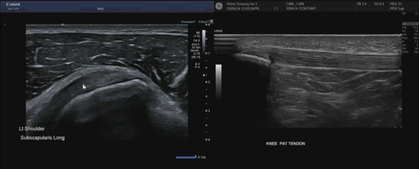
Tendon
Nerve ultrasound appearance
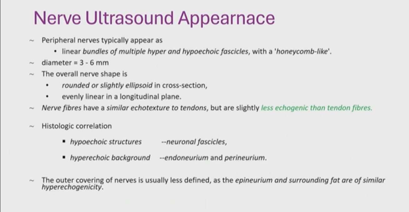

Which one is the nerve

MSK US what transducer would you use
High frequency transducer - 15Mhz
Clinical indications for MSK
Trauma - direct or indirect
Overuse injuries
Lumps and bumps
Numbness and pain
Joint pathology

Tendonopathy
Heterogenous tendon thickened
Hyper echoic bursa above the tendon
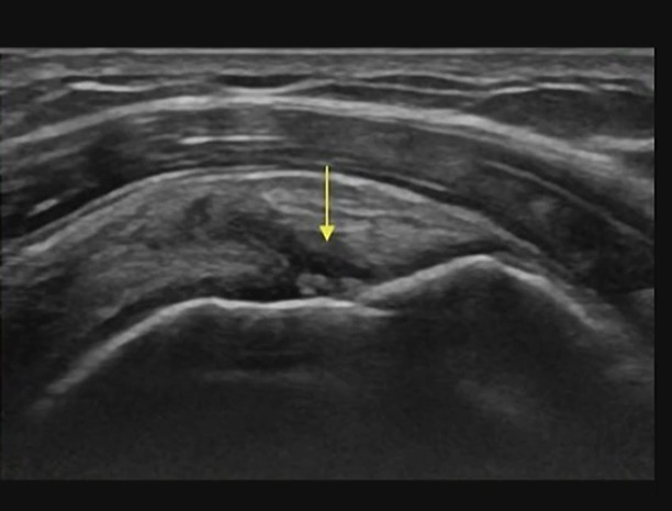
Articulatar surface tendon tear
supraspinatus tendon
Partial thickness tear
Boney irregularity
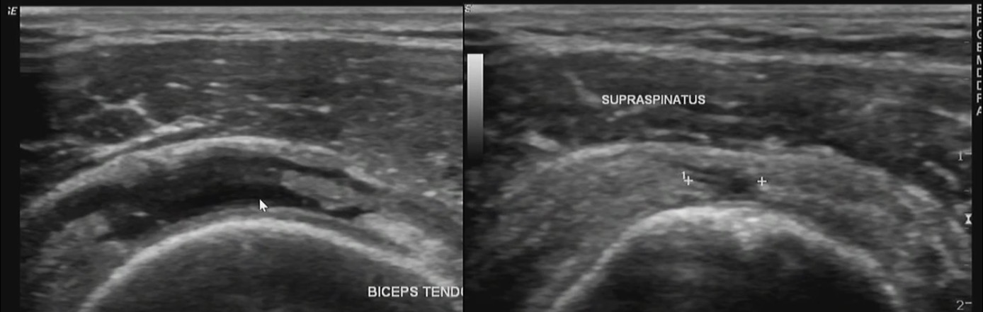
Full thickness tear of the bicep
Partial thickness tear

Completely ruptured long head of bicep tendon
Completely retracted tendon
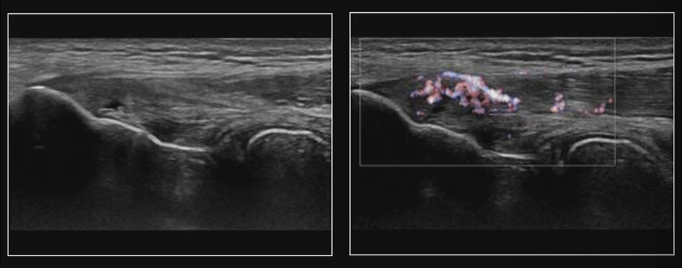
Tennis elbow / common extensor tendonopathy
increased vascularity to the elbow
Patchy heterogenous tendon with hyper echoing areas (should be homogenous)
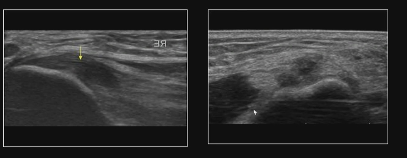
Inflammation of common extensor
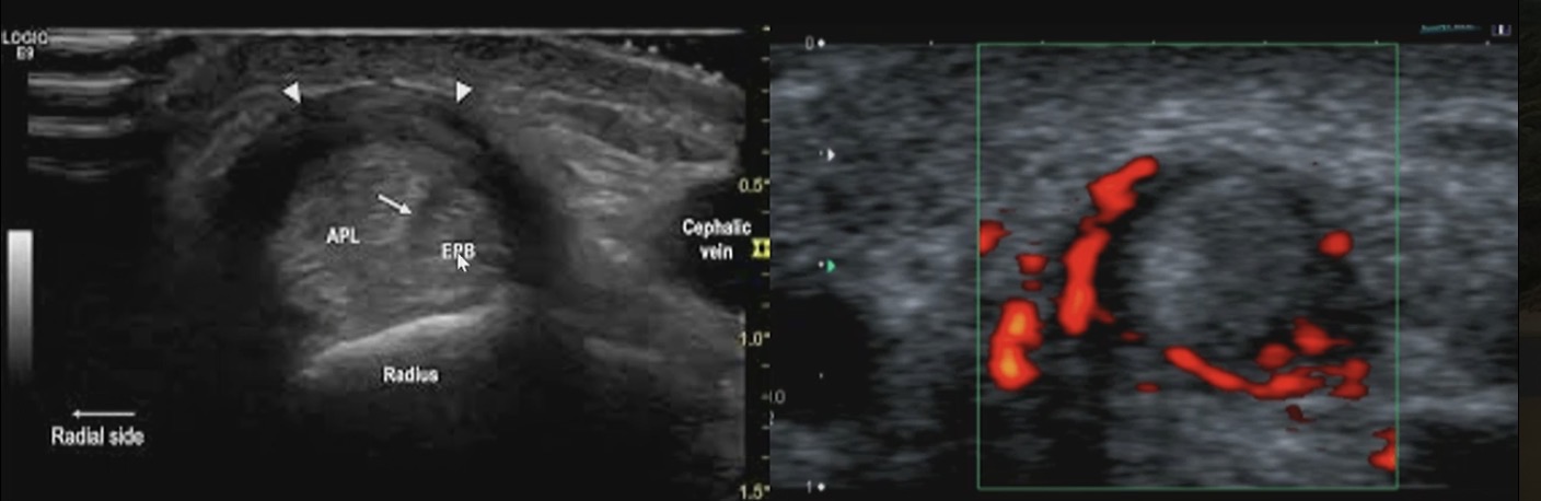
???
thickened sheath (hypoechoic)
Increased vascularity
Chronic patellar tendonosis
Reason for a US of First Trimester
Determine gestational age
Localisation and viability of pregnancy
Fetal number
Fetal anatomy
Nuchal translucency

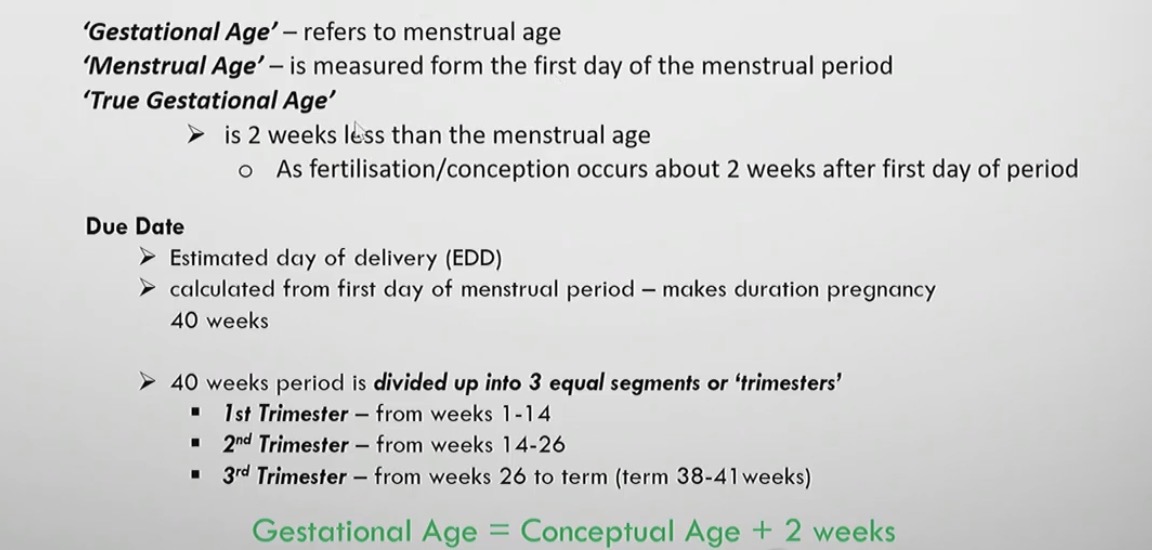
What are these terms
Gestational Age
Menstrual Age
True Gestational Age
Determining gestational age in very early pregnancy
the gestational sac is the first structure visualised 5 weeks (MSD)
The crown-rump length (CRL) is used to predict gestational age
Towards the end of 1st trimester we can use BPD
In the second and third trimester we determine gestational age with:
Head circumference
Biparietal diameter
Abdominal circumference
Femur length
Humeral length
First trimester clinical indications
Unsure of dates - positive BHCG
Pelvic pain
Vaginal bleeding
Suspected Misscarriage
Suspected FDIU
Suspected Ectopic Pregnacy
Suspected Multiple Pregnacy
Abnormal Pregnancy developments
1st trimester scan technique
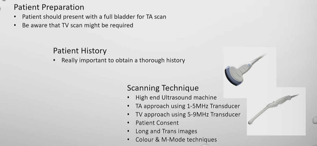
Patient prep
Patient history
Scanning technique
TV → transvaginal scan
TA → trans abdominal scan
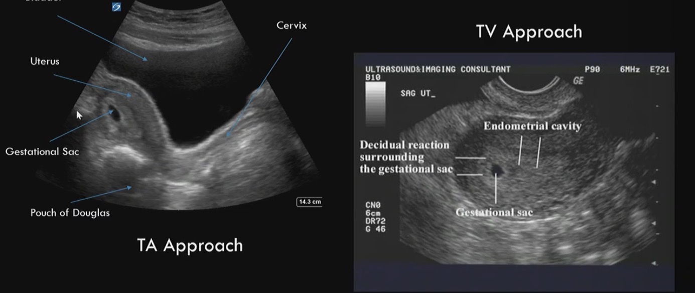
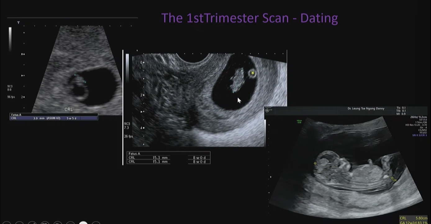
When was this taken
1st trimester scan
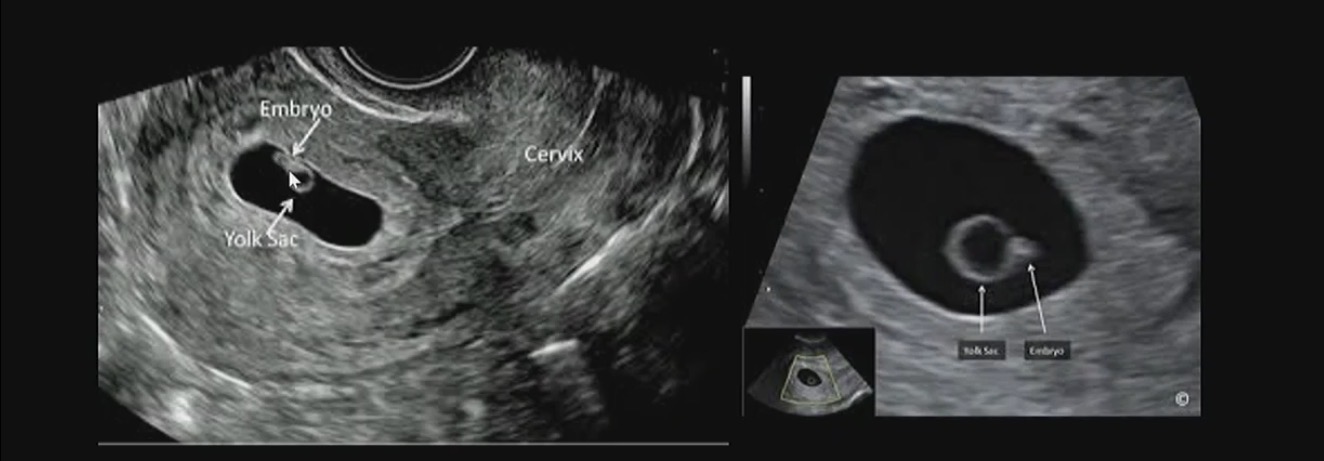
How can we tell if
Embryo with heart beat to tell its a viable
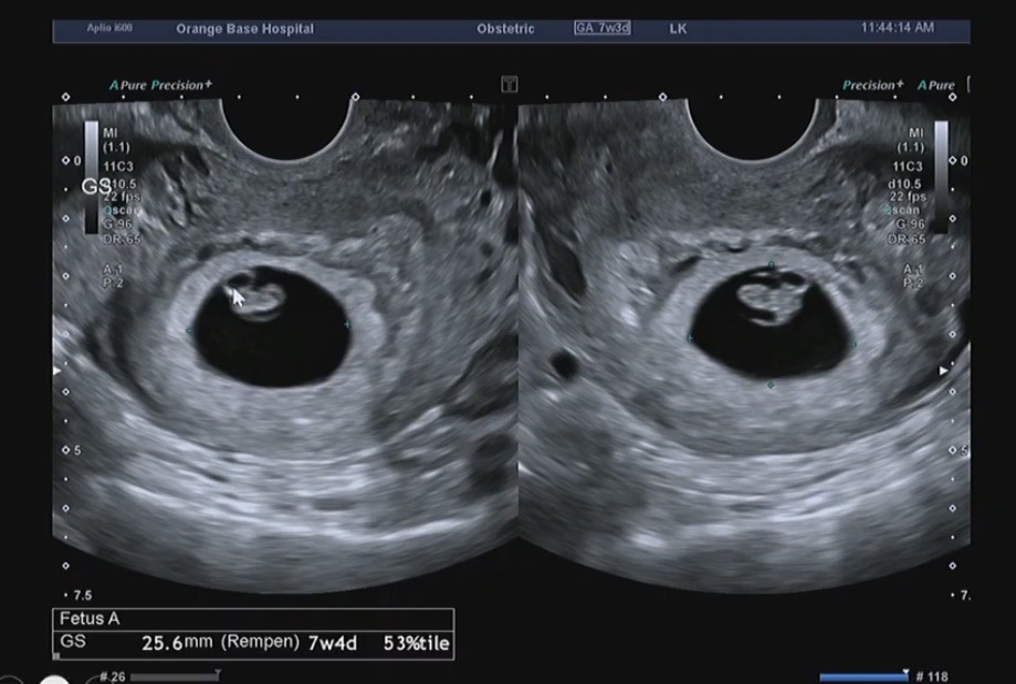

What do we use to measure fetal heart rate
100 beats per min under 6 weeks
Use M-mode (motion mode)
Or you can use colour box to show heart rate

How do you date the fetus
From the crown of the baby head to the rump
CRL (crown rump length)