Chapter 2
1/107
There's no tags or description
Looks like no tags are added yet.
Name | Mastery | Learn | Test | Matching | Spaced | Call with Kai | Chat |
|---|
No analytics yet
Send a link to your students to track their progress
108 Terms
reflex
automatic, stereotyped movement produced as the direct result of a stimulus
reflex pathway example with a kneetap
sensory neuron from muscle → spinal cord (back part) → either to brain, to other leg, or to single synapse in reflex
single synapse in reflex → reflex motor output in front part of spine, causing thigh muscle to contract
the electrical signal is sent across…
the synapse, the gap between 2 neurons (presynaptic & postsynaptic)
nerve fiber
bundle of nerve cells
there are between ____ & ____ neurons in the body with ____ connections each
there are between 10 billion and 1 trillion (estimated 86 billion) with 10,000 connections each
parts of neuron
dendrites, cell body or soma, axon, terminal endings/buttons
dendrites
branches off cell body
axon
inside the myelin sheath
terminal endings/buttons
ending of neuron
in what direction do messages travel in the neuron?
from the dendrites/soma downward toward the terminal endings
do neurons take different shapes in different areas of the body?
yes
axodendritic
axon terminal of one neuron synapses on dendritic spine of another
are there other ways neurons can connect besides axodendritic?
yes
myelin sheath
fatty tissue produced by other cells (glia) in the brain/nervous system that protects the length of the axon
what does the myelin sheath allow to occur?
it allows electricity to travel smoothly down the axon
in the _______ the _______ is not protected by the myelin sheath
in the gaps at the Nodes of Ranvier, the electrical signal is not protected
What happens when the electrical signal is not protected?
ion exchange
How does a neuron fire? What is the nerve impulse?
action potential
How does a firing neuron cause the next neuron to fire? How do they communicate?
neurotransmitters
action potential is…
voltage
action potential steps
Starts with electrical resting potential: inside of the cell is 70 mV more negative than outside due to Cl- ions inside & Na+ ions outside (resting potential = -70 mV)
Stimulation of the neuron causes the opening of ion gates that let in Na+ ions, which makes the inside more positive: -70, -69, -68, -67…
When enough Na+ ions get in for the potential to be reduced to -55 mV, suddenly the ion gates to the cell membrane are flung open, allowing Na+ to rush in.
So much Na+ enters that the potential shoots all the way up to +40 mV, so the inside is now positive relative to the outside (the action potential)
The ion pumps work to reduce the potential back to -70 mV by pushing positive ions out (K+ because Na+ goes out slower) then another pump takes Na+ back out and puts K+ back in
Na+ goes ____ & K+ goes ____, until it switches
Na+ goes in and K+ goes out
-55 mV is a ________
threshold; below that voltage there is no action potential
firing is ____ or ____
all or none, it won’t happen if it doesn’t meet the threshold
does more intense stimulation cause a more intense action potential?
no, it just causes more frequent ones (up to 1000/sec) and in more neurons
action potential travels down _______________ by _____________
action potential travels down the length of the axon from the cell body by depolarizing neighboring areas
how fast does action potential travel?
50 to 100 m/sec
the myelin sheath _______________ the speed of action potential and ion exchange
the myelin sheath speeds these processes up
Herman Van Helmholtz
measured the speed of a nerve
Where does action potential happen?
at the Nodes of Ranvier
How does communication across the synapse happen?
neurotransmitters
Communication across the synapse (neurotransmitters) steps
synapse; terminal endings of presynaptic neuron: may relay impulse to dendrites of postsynaptic neuron
terminal buttons contain little sacs (vesicles_ of chemicals (neurotransmitters) and at action potential, vesicles burst and release neurotransmitters into synapse
receptor molecules on membrane of dendrite are like little locks to be opened: neurotransmitters are the keys, and this is what opens ion gates to allow Na+ inside in the first place
These are based on shape (lock & key model)
neurotransmitter life cycle stages
Synthesis
Storage
Release
Receptor Interaction
Inactivation
Reuptake
Degradation
serotonin
neurotransmitter responsible for long term good feelings
SSRI (selective serotonin reuptake inhibitors) block reuptake of serotonin; anti-depressants
dopamine
neurotransmitter responsible for short term good feelings
Neurotransmitters may open a gate to to let _____ inside. Is this excitatory or inhibitory?
Neurotransmitters may open a gate to let Na+ inside. This is excitatory (more likely to fire) because the potential is getting smaller, toward -55 mV.
Neurotransmitters may open a gate that pushes ________ out. Is this excitatory or inhibitory?
Neurotransmitters may open a gate that pushes positive K+ ions out. This is inhibitory (less likely to fire) because the potential inside the cell is getting larger (-70, -71, -72).
inhibition of a reflex example
swearing ups pain tolerance
spatial summation
simultaneous signals coming from multiple presynaptic neurons being received by a single postsynaptic neuron; several weak signals from different locations → 1 large signal
temporal summation
single presynaptic neuron rapid-firing signals to a post-synaptic neuron; several weak signals at different times from one location → 1 large signal
EPSP
Excitatory Post-Synaptic Potential; increases chance of action potential
IPSP
Inhibitory Post-Synaptic Potential; decreases chance of action potential
reciprocal inhibition example
when extensor muscle is excitated, flexor muscle is inhibited & vice versa
disinhibition examples
alcohol: temporarily damages higher brain areas, resulting in loss of inhibition of impulsive behaviors
disconnecting spine from dog’s brain
withdrawal reflex might be inhibited by the higher-level knowledge that although a heated dish of food hurts, avoid letting go until you reach the table
Nervous System Parts
Central vs. Peripheral
Peripheral: Somatic vs. Autonomic
Autonomic: Sympathetic vs. Parasympathetic
Central NS (CNS)
brain, spinal cord
contains the hindbrain, midbrain, and forebrain
Peripheral NS
away from center
everything besides the brain and spinal cord
includes somatic division and autonomic division
Somatic NS
body
nerve fibers connecting to muscles and senses
Autonomic NS
self rule
vital functions: heart rate, breathing, digestion, reproduction
contains sympathetic and parasympathetic branch
Sympathetic NS
“with feeling”
excited states
energy-consuming
fight or flight
arousal: mobilizes for emergency (speeds heart and lungs, inhibits digestion & sexual function)
Parasympathetic NS
“goes with the sympathetic”
vegetative, calm states
energy conserving or storing
rest & digest or feed & breed
calm: maintains normal functioning (slows heartrate and lungs, etc.)
Sympathetic NS functions
dilates pupil
relaxes bronchi
accelerated heart beat
inhibits activity
contracts vessels
Parasympathetic NS functions
contracts pupil
constricts bronchi
slows heart beat
stimulates activity
dilates vessels
brain parts bottom to top/inside to outside/old to new
hindbrain, midbrain, forebrain
hindbrain parts
medulla oblongata ("oblong marrow”)
pons (“bridge”)
cerebellum (“little brain”)
medulla
part of hindbrain
breathing
heartbeat
blood circulation
pons
part of hindbrain
arousal and attention
facial expressions
cerebellum
part of hindbrain
integration of muscles to perform fine movements
no coordination or direction of these movements
balance
cat transected above the hindbrain
low-decerebrate cat
can move but not act
midbrain
forms movements into acts
controls whole body responses to visual and auditory stimuli
contains the tectum and tegmentum
tectum (“roof”)
superior colliculus (“upper hills”)
inferior colliculus (“lower hills”)
tegmentum (“floor”)
substantia nigra (“black substance”)
red nucleus
responsible for responses to visual stimuli
superior coliculous
responsible for responses to auditory stimuli
inferior coliculous
forebrain parts
thalamus (“inner chamber”)
hypothalamus (“below the inner chamber”)
basal ganglia
limbic (“border”) system
cerebral cortex/neocortex
thalamus
sensory & motor relay center (to various cerebral lobes)
hypothalamus
controls responses to basic needs (food, temperature, sex)
responsible for the 4 F’s
4 F’s of Motivation
fighting & flighting (sympathetic), feeding & mating (parasympathetic)
basal ganglia
regulates muscle contractions for smooth movements
exerts inhibition for movement and relaxes that body part when the signal is sent
translates signal in brain into movement
limbic system
hippocampus (memory)
amygdala (emotion)
The amygdala is responsible for what emotions?
fear, rage, fight-or-flight
cerebral cortex/neocortex & four lobes
four lobes:
frontal: front; decision making, personality
parietal: near top & back of brain; sensory perception, spatial awareness
occipital: back of brain; visual processing
temporal: sides of brain near temples; auditory processing, memory
seat of “higher” intellectual functions
cat transected above the limbic system
acts normal, with purpose, just is clumsy
Dopamine is sent to the forebrain to… & from…
Dopamine is sent to the forebrain to allow basal ganglia to work. It is sent from the substantia nigra.
Parkinson’s Disease attacks…
things that make dopamine, which doesn’t allow basal ganglia to work and prevents smooth movements
CS
conditioned stimulus
CR
conditioned response
US
unconditioned stimulus
UR
unconditioned response
determine the US, UR, CS, & CR for Pavlov’s dogs
unconditioned stimulus: food
unconditioned response: salivate
conditioned stimulus: bell
conditioned response: salivate
cerebral hemispheres (cerebrum)
corpus callosum
cerebral cortex
corpus callosum
large band of neural fibers that connects hemispheres
each hemisphere controls opposite side of body
largest “commissure” or pathway between brain hemispheres, but not the only one
cerebral cortex (= skin or bark)
1 to 3 mm thick, 3ft² if flattened out
higher motor, sensory, and intellectual functions
Phineas Gage
pole through frontal lobe (likely pre-frontal area)
personality change: responsible and gentle → more argumentative
loss of inhibition of impulsive behaviors due to the lack of social constraints normally imposed through the action of his missing pre-frontal brain areas
Paul Broca
Broca’s area: region in brain responsible for speech
Carl Wernicke
Wernicke’s area: region responsible for comprehension
high-decerebrate cat
transected just above the midbrain
can act, but without regard to environment, without purpose
The patient with the damaged amygdala knew the blue slide had been followed by the loud noise, but did not show a fear response (Galvanic skin response) to it. On the other hand, the patient with the damaged hippocampus did show the fear response to the blue slide, but did not remember that the blue slide had been followed by the loud noise during the experiment.
This is because the amygdala is responsible for a fear response and the hippocampus is responsible for memory.
A "double dissociation" is strong evidence that these two brain regions are involved in separate independent functions -- and indeed, that the the two functions ARE independent
US: loud noise, UR: Galvanic skin response (fear)
CS: blue slide, CR: fear
US: unconditioned stimulus
food in mouth
input to a reflex
UR: unconditioned response
salivation to food
output of reflex
CS: conditioned stimulus
bell
initially results in investigatory response, then habituation
after conditioning, results in CR
CR: conditioned response
salivation to bell
response to CS
measure amplitude, probability, latency
Franz Joseph Gall (1758-1828) 4 discoveries
Cortex is a functioning tissue, not just protective covering (bark/skin)
Commissures (connecting pathways) exist between brain hemispheres, other than the corpus callosum
There is a crossing of ascending nerve pathways from the spinal cord to contralateral hemispheres of the brain
Distribution of and distinction between grey matter and white matter tracts
a. grey matter: neuron cell bodies doing information processing
b. white matter: mostly myelinated axons sending signals over longer distances
Franz Joseph Gall Phrenology - 4 Items
The brain is the material instrument (organ) through which the mind holds intercourse with the outer world
The mind has a mix of separate mental abilities, all in its own specific place
The size of each center/organ corresponds with the functional efficiency of each faculty
The development of the organ is reflected in the shape, size, and irregularities of the cranium
Believed could measure bumps on the skull to predict mental traits
Why wasn’t Franz Joseph Gall honored in textbooks?
He believed the degree of development of a mental organ could be measured by the relative size of the corresponding brain area
He believed the skull fits the brain as a glove fits a hand
blindsight
brain damage to the visual cortex that leads to blindness
patients could not hold a card so that its orientation matched a slot, but they could put the card perfectly into a slot
they can in a sense, see, but they are not aware of seeing
dual-center theory: hypothalamic control centers
one part of the hypothalamus (lateral region) served as the “go” center for eating, while the ventromedial region served as the “stop” center.
rat with ventromedial hypothalamus lesion will…
be a fat rat because it can’t stop eating 💔 #vomit
motor cortex
region in brain that controls voluntary muscle movements, coordinating and executing motor functions throughout the body
cerebral cortex in the frontal lobe

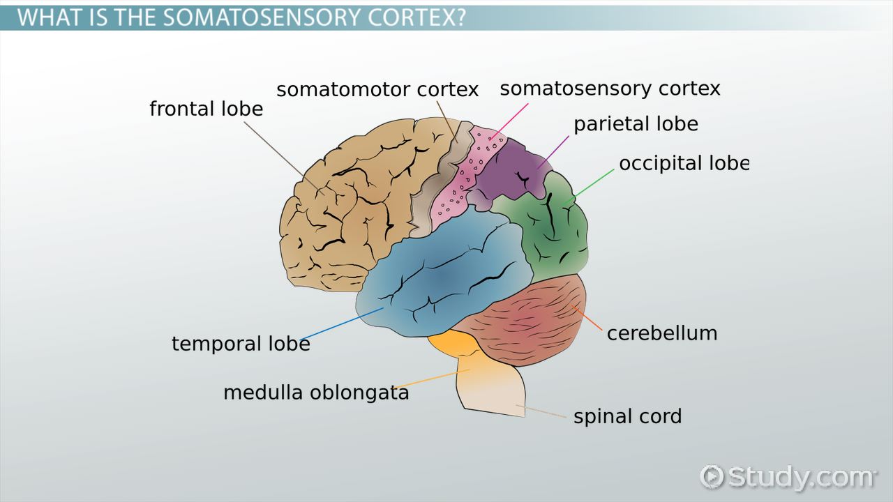
somatosensory cortex
part of the brain that processes sensory information from the body, such as touch, temperature, and pain, providing humans with a sense of physical self
located in the parietal lobe
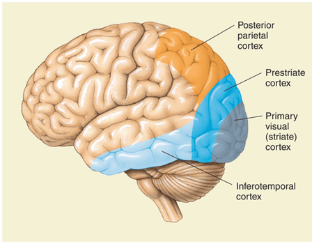
visual cortex
region in brain responsible for processing visual information received from the eyes, allowing humans to perceive and interpret visual stimuli
located in occipital lobe
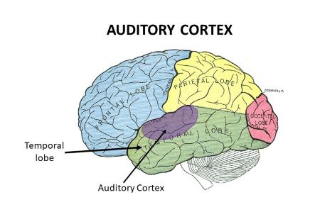
auditory cortex
part of brain that processes auditory information, allows perceiving and interpreting of sounds, including speech and music
located in the temporal lobe
prefrontal cortex
part of brain involved in higher cognitive functions, decision making, personality expression, and social behavior regulation
pre-frontal lesion
damage to prefrontal cortex
deficits in social behavior regulation, loss of planning, moral reasoning, unable to take action
apraxia
failure in sequencing components of actions, inability to organize movements
caused by frontal lesions just forward of the motor cortex
not paralysis, caused by motor cortex lesion
can’t tie shoes, brush teeth
“no doing”