Week 7 - Tools for Mechanotransduction + Lab on a Chip
1/29
There's no tags or description
Looks like no tags are added yet.
Name | Mastery | Learn | Test | Matching | Spaced | Call with Kai |
|---|
No analytics yet
Send a link to your students to track their progress
30 Terms
Tools for testing ECM
1. Hydrophobicity = Wettability
2. Topography = SEM
3. Stiffness = micro-indentation, AFM
Tools for testing Integrin
adhesion rupture force (AFM)
Tools for testing FAC
Fluorescence Resonance Energy Transfer
Tools for testing Actin-myosin traction force
Micropillar/microneedle
Traction Force Microscopy
Tools for testing Nuclear lamina
protein expression = immunostaining
Tools for testing DNA
trancription into mRNA (Polymerase Chain Reaction)
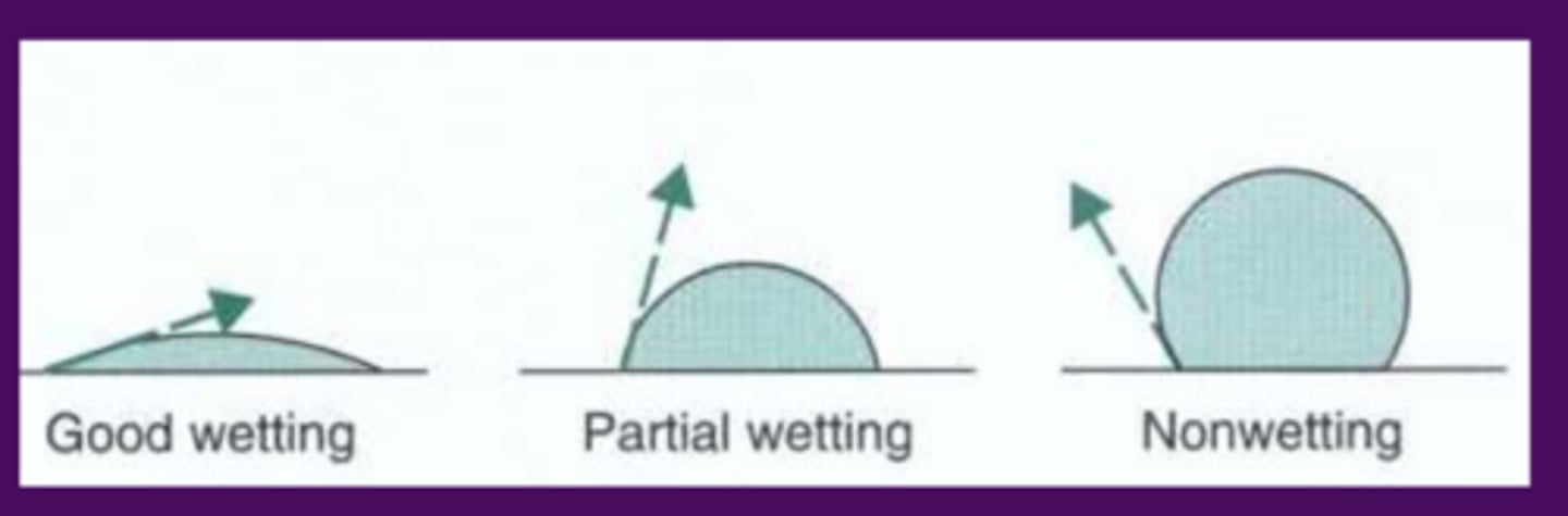
Wettability
measures hydrophobicity
put water drop on surface and measure angle
larger angle = more hydrophobic
Scaning electron microscopy (SEM) for ECM
measures topography
can view surface to see structure of ECM
Testing stiffness of ECM (3 types of tests)
Macro scale tests = compression, tensile, rheology tests
Micro scale tests = microindentation
Micro/nano scale tests = atomic force microscop (AFM)
Microindentation (ECM testing)
measures stiffness of ECM
compresses surface to test stiffness
micro scale
Atomic force microscope (ECM testing)
measure stiffness of ECM
compresses surface and measures how much surface bent with laser (to test stiffness)
nano (atomic) scale
5 things an Atomic force microscope measure?
force
stiffness
height of contact (at each point = roughness)
adhesion force
rupture force
[and more]
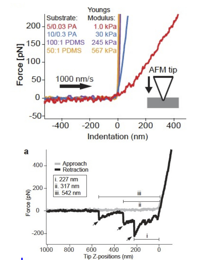
AFM graph interpretation
slope of curve (indentation vs force → top graph) shows stiffness of material
steeper = stiffer
Area between line for Approach and Retraction represents the adhesion force
the closer the lines are the less adhesions (bottom graph)
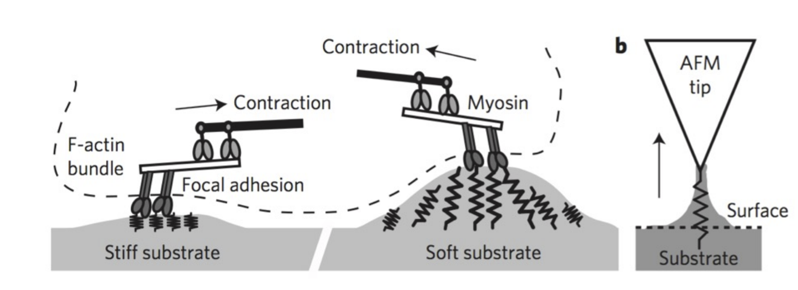
Atomic Force microscope (integrin measurments)
Rupture of integrin-mediated focal adhesion can be measured (rupture length + force)
Measured by detecting degree of conformational change (change in stiffness)
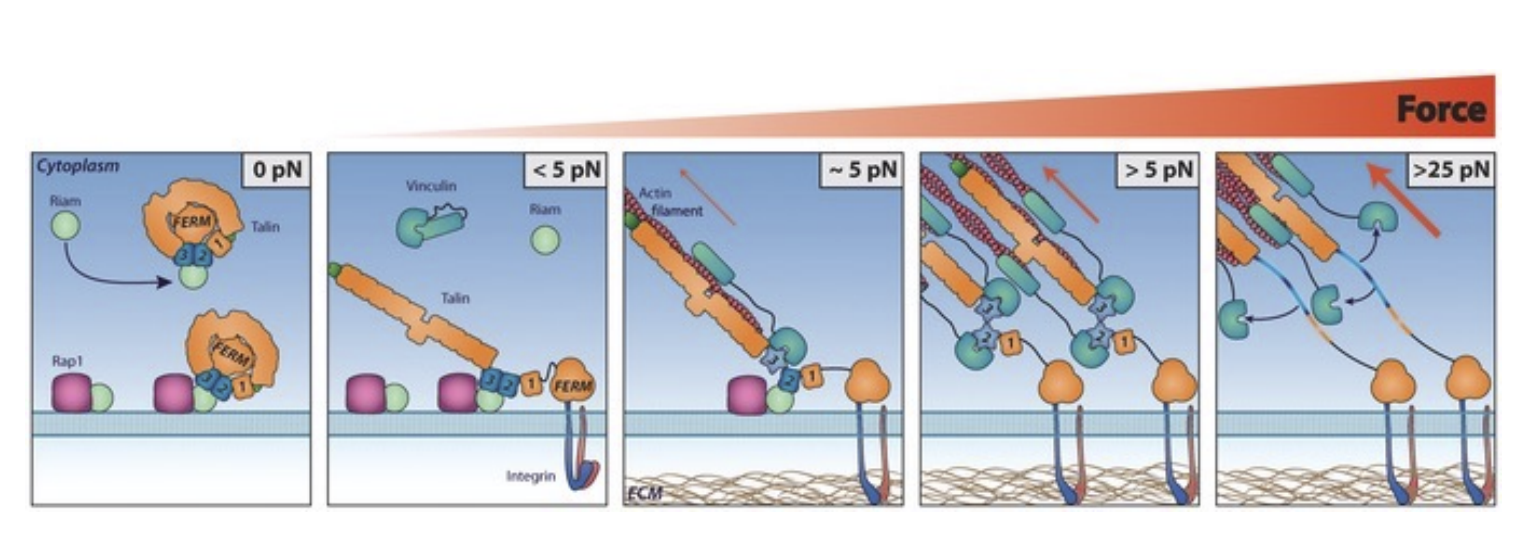
Fluorescence Resonance Engergy transfer (FRET) for testing FAC
Conformational changes in Vinculin and Talin
they stretch based activation from ECM forces
Fluorophores attached to Focal Adhesion Complex emit light when activated
when protein is unfolded
emitted light can cause nearby fluorophores to emit light
= determines how close fluorophores are (if no other fluorophores are emitted = FAC is spread out)
Measuring traction force (micropillar/microneedle)
Put cells on array of pillars
Force = k(spring constant) * displacement
large displacement of cell = large traction force
Variable (in spring constant) = stiffness, diameter, length
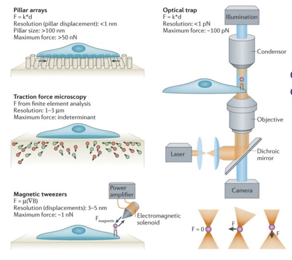
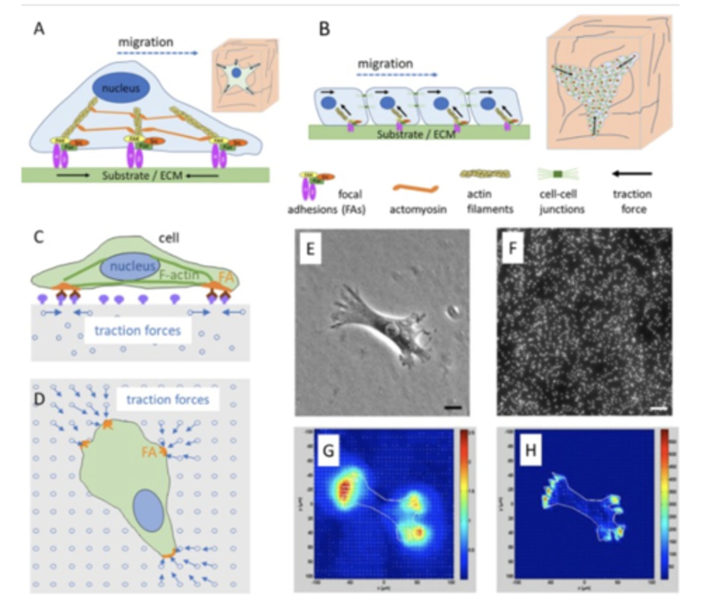
Traction force microscopy
Measure forces applied by cells (on hydroge) based on displacement of fluorescent beads
Micropillar pros and cons (vs TFM)
Pros:
- no need for FEM
- no need for beads
- high resolution
Cons:
- no z axis movement
- discontinued focal adhesion
Traction Force Microscopy pros and cons (vs micropillars)
Pros:
- x/y/z axis data
- continuous focal adhesion
Cons:
- required equipment
- FEM
FEM
finite element method (analysis)
simulating the effects of mechanical stimuli on cells
uses computer
Lab on a Chip
devices that integrate laboratory functions on a single chip
capable of handlign extremely small fluid volumes
Organ on a chip
3d multichannel and microfluidic cell structure device on microchip
mimics human organ (liver/lungs)
Organ on a chip (in relation to animal study)
usually used before animal/human study
see if drug is efficacious, before testing on animal/humans to see how it interacts with the system
Human on a chip
system of several organ on a chips
better mimics human system
Pros of Lab on a chip (7)
low fluid volume = low cost
more specific/defined
faster analysis = more analysis per vol
better control over process
easy to modify conditions
inter-connectivity with chips (more tissue types)
data supports human clinical trial
Cons of Lab on a chip (4)
not perfect as animal study for whole system
not fully developed yet
never reaching complexity of real tissue
detection principles may not always scale down positively (signal:noise ratio)
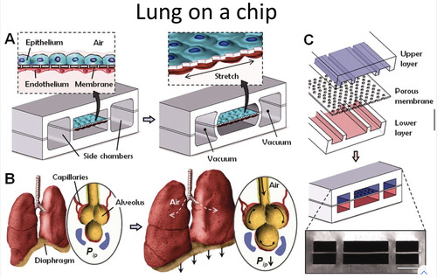
Lung on a chip (how does it mimic human lungs)
Attach side chambers to vacuum = stretch tissue
(mimics alveoli stretching)
can measure stretch by viewing movement of pores on the membrane (under microscope)
Mimicing cancer growth
put cancer cell in hydrogel (mimic stiffness of ECM)
let cancer grow = view tumour cell composition
view which cells are present in tumour (can be influenced by stiffness of hydrogel)
What can we determine/measure when Mimicing cancer growth?
proportion and cell types in tumour
tumour composition (cells in center vs periphery)
epithelial to mesenchymal transition
Cancer tissue mechanics (metastasis)
Local invasion:
- cells migrate through collagen fibres
- requires aggressive phenotype
Intravasation (and extravasation):
- cells squeeze through cell-cell adhesions
- ruptures nuclear envelope
Circulating tumour cells