Respiratory System
 Call Kai
 Learn
Learn Practice Test
 Spaced Repetition
 Match
Match Flashcards
 Knowt Play
1/87

Earn XP
Description and Tags
A&P Exam 3
Name | Mastery | Learn | Test | Matching | Spaced | Call with Kai | Chat |
|---|
No analytics yet
Send a link to your students to track their progress
88 Terms
Functions of the respiratory system
Provides O2 to blood, gets rid of CO2 in blood, speech, defends against inhaled microbes, traps and dissolves blood clods, converts angiotensin to influence arterial concentrations
Describe conversion of angiotensin
Endothelial cells of pulmonary capillaries contain ACE → converts angiotensin I into angiotensin II → vasoconstricts arterioles
ACE must be in the lungs because 100% of blood goes through lungs
Anatomy and function of respiratory system
Nasal cavity - warms, filters, and moistens air
Pharynx - pathway for air, liquid, food
Esophagus - travels to stomach to deliver food and liquid
Larynx - part of airways and has vocal cords
Epiglottis is a flap that covers the glottis, which goes to the lungs, prevents food from going into lungs
Cartilaginous C rings that hold airway open
Explain ciliary mucus
Epithelial surfaces at end of bronchioles have cilia that flow up towards pharynx, secretes watery mucus/goblet cells
Particulates matter and dust that’s moved by cilia to pharynx where it’s swallowed, keeps lungs clear
Order branching from top to bottom
Conducting zone (anatomical dead space, no gas exchange): Trachea → bronchi → bronchioles → terminal bronchioles →
Respiratory zone (gas exchange): respiratory bronchioles → alveolar ducts → alveolar sacs
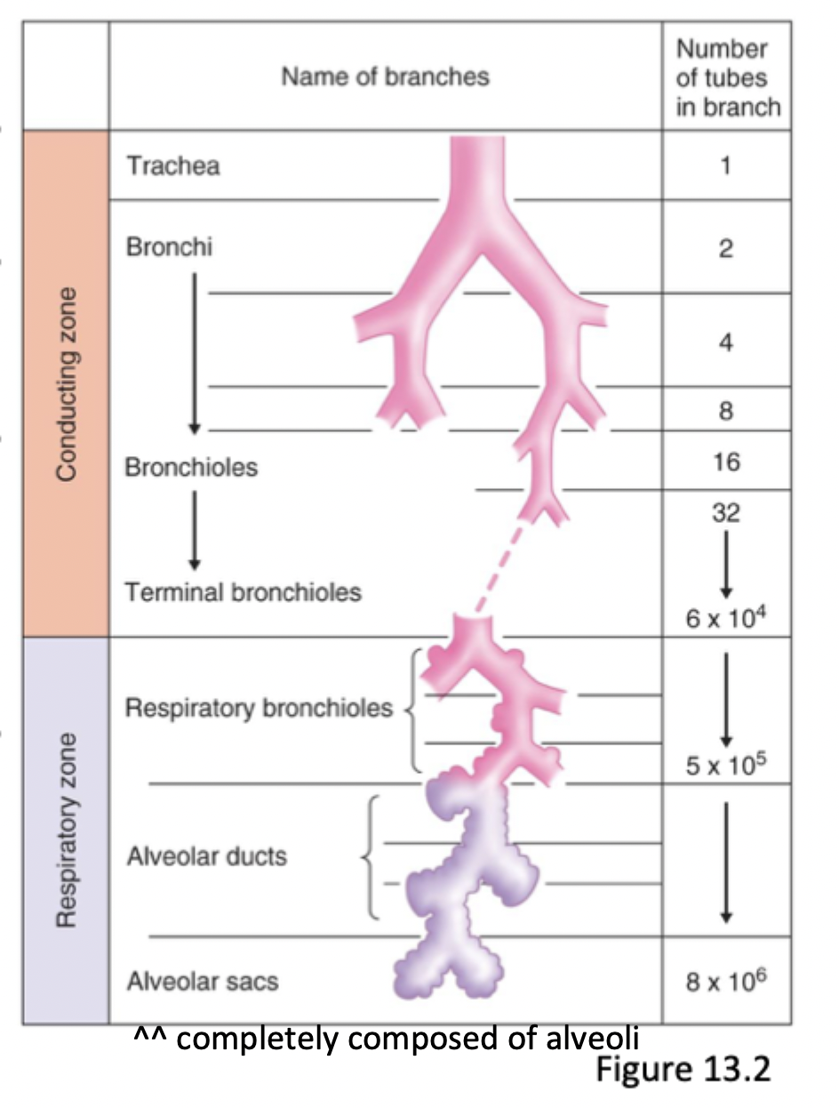
Describe alveoli
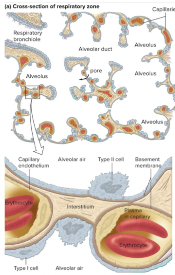
Tiny hollow sacs that are continuous at the ends with a large surface area, main site of gas exchange
Describe the types of alveoli
Type I - flat epithelial cells, single layer thick, make up alveolar walls
Type II - secrete surfactant that prevents collapse of alveoli
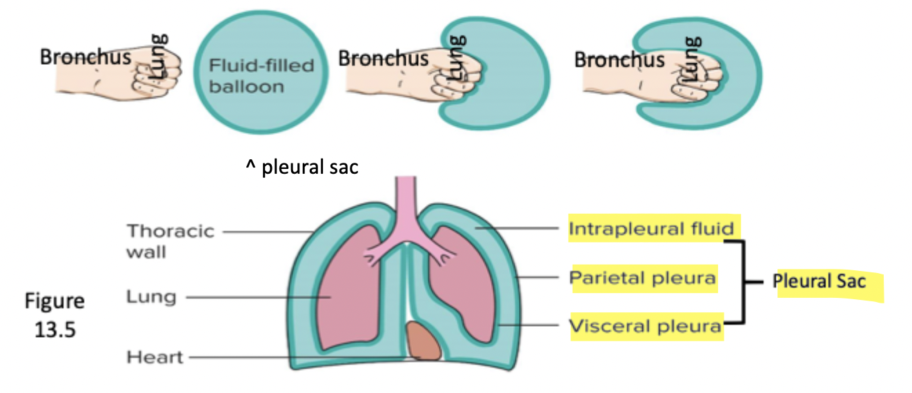
Describe the pleural sac and its parts
Intrapleural sac, parietal pleaura, visceral pleura
Intrapleural sac is very thin, has few mL of fluid that reduces friction
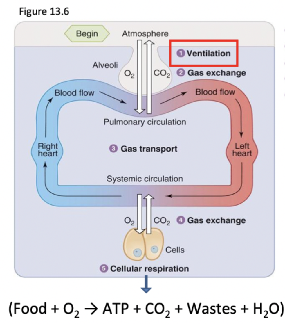
Explain the steps of respiration
Bulk flow between air in atmosphere in alveoli
Exchange of O2 and CO2 in alveolar air and blood in lung capillaries via diffusion
Transport of blood through circulation with bulk flow
Exchange of gas between tissues and blood
Cellular utilization of O2 and production of CO2
What must match in the steps of respiration
The amount of ventilation needed matches the amount of cellular respiration occurring.
Equation for transpulmonary pressure
= P alveolar - P interpleural
Equation for chest wall pressure
= P interpleural - P atmospheric
How does alveolar P change with the lungs
When air comes into the alveoli, the lungs expand, P alv < P atm.
When air leaves the alveoli, the lungs become smaller, Palv > P atm
What is Boyle’s Law
States that at constant temperature and at a fixed number of gas molecules, pressure is inversely proportional to the volume of the container
Name and explain skeletal muscles used in respiration
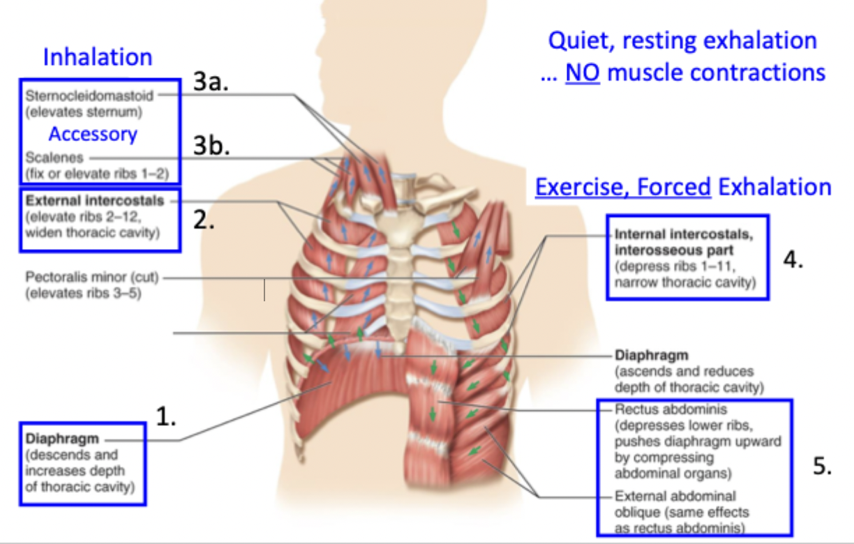
Diaphragm - contacts to move abdomen down and enlarge the thorax
External intercostals - contracts to pull ribs up and out to increase thoracic cavity
Accessory muscles - for deeper and forceful inhalations
Includes sternocleidomastoids and scalenes, pulls ribs up
Internal intercostals - forceful, exercised exhalation and depresses ribs to narrow thoracic cavity
Abdominal muscles - moves diaphragm up and compresses abdominal organs
How is the chest wall aided by elastic recoil
Elastic recoil holds the chest wall inward and downward by attachment to parietal wall of pleural sac, transmural pressure gradients, and surface tension
How are the lungs aided by elastic recoil
Elastic recoil open the lungs to a larger volume by attachment to visceral wall of pleural sac, transmural pressure gradients, and surface tension
What does it mean when the transpulmonary pressure is pos or neg?
Pos - P is greater in alveoli → lungs are bigger and held open
Neg - P is greater in IPS → lung collapses (fatal)
What does it mean when the chest wall pressure is pos or neg?
Pos - P is greater in the IPS → chest wall is stretched and recoils inward
Neg - P is greater in the atm → chest wall caves inward and recoils outward
Define, point out, and equation of functional residual capacity
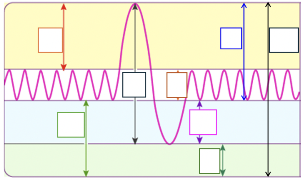
FRC is the volume of air remaining in lungs at the end of a quiet exhale when muscles are relaxed
RV + ERV
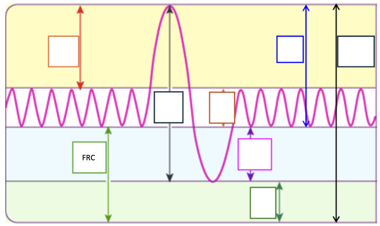
At FRC, _____ _____ of chest wall = _____ ____ of the lung
Outward recoil ; inward recoil
At FRC, _______ pressure = _________ pressure
Alveolar ; atmospheric → no movement of air
Describe pneumothorax
Puncture of chest wall → air from atm goes into the IPS → Pip becomes 0 and loses pressure gradient → Ptrans becomes 0 and lungs are free to collapse
Describe atelectasis
Collapse or blockage of lungs by disease, genetics, infections, ventilation malfunction
Air leaks from lungs → IPS → lungs lose air and collapse
What is meant by pneumothorax and atelectasis being unilateral
Usually affects just one lung at a time because the pleural sacs are different from right and left
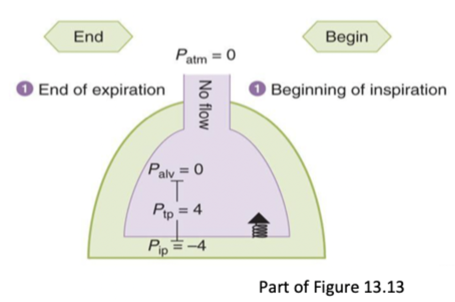
Explain the phase of the cycle at the end of exhalation and beginning of inhalation
At FRC
No air flow
Palv = Patm
Pip = 4
Muscles are relaxed
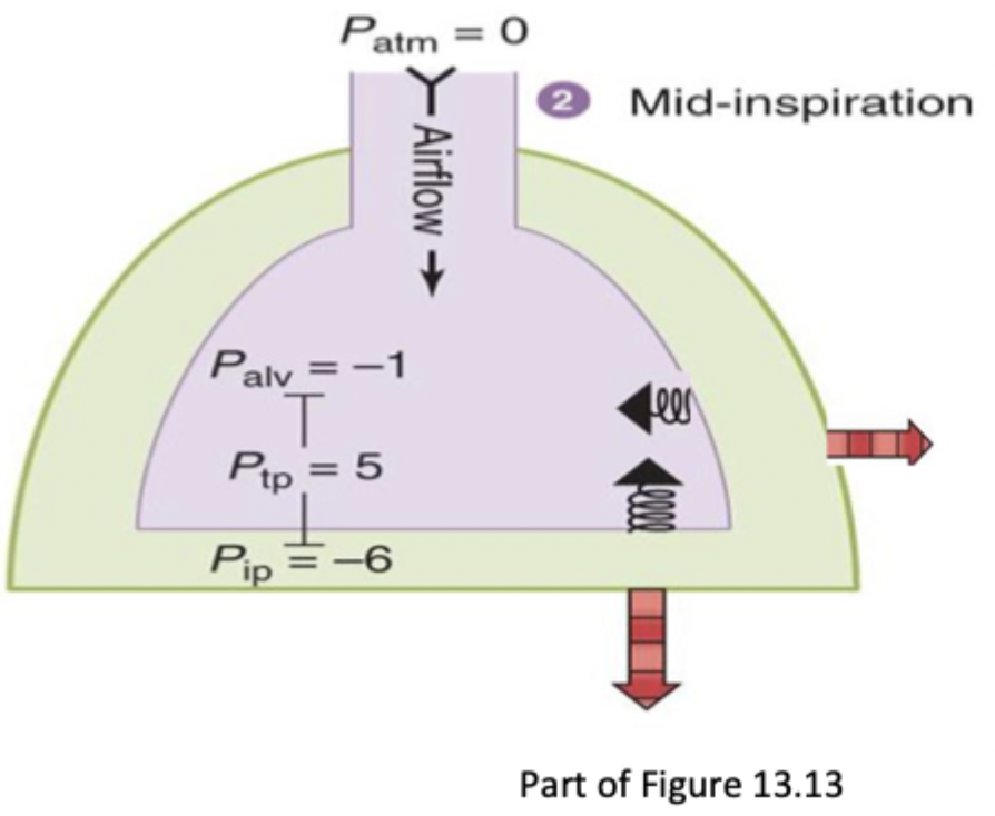
Explain the phase of the cycle at mid-inhalation
Diaphragm and external intercostals are pulling chest wall out to increase volume of IPS and lungs
Air flows from atm → lungs
Pip decreases to -6
Palv decreases to -1
P trans increases (is held more open)
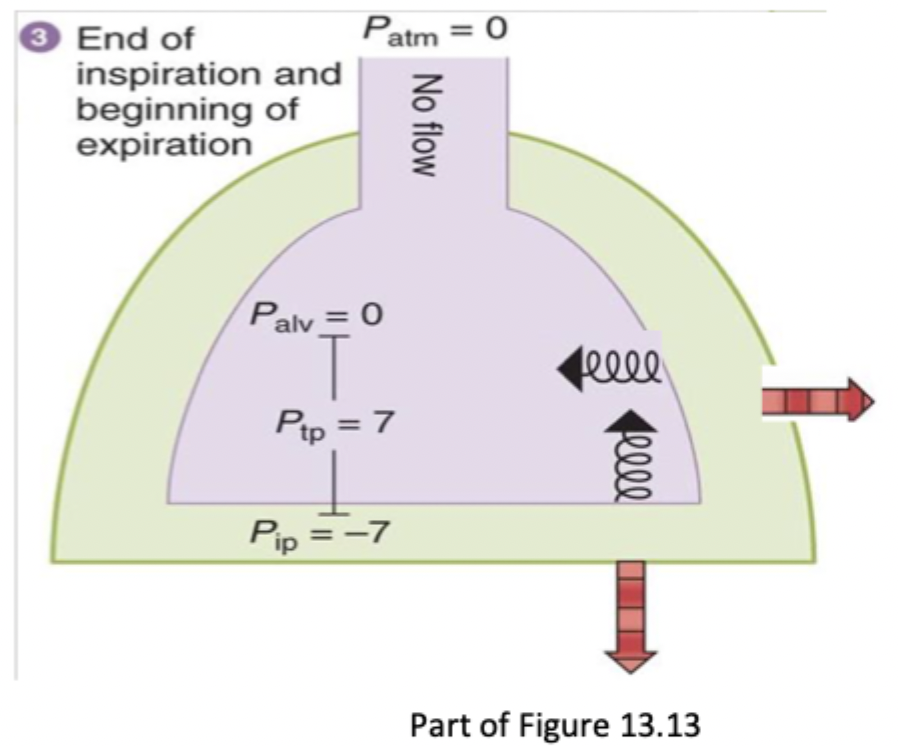
Explain the phase of the cycle at end of inhalation and beginning of exhalation
Diaphragm and external intercostals are no longer contracting, lungs are at largest volume
No air flow
P atm = P alv
Pip = -7
Ptrans = 7
Reached tidal volume
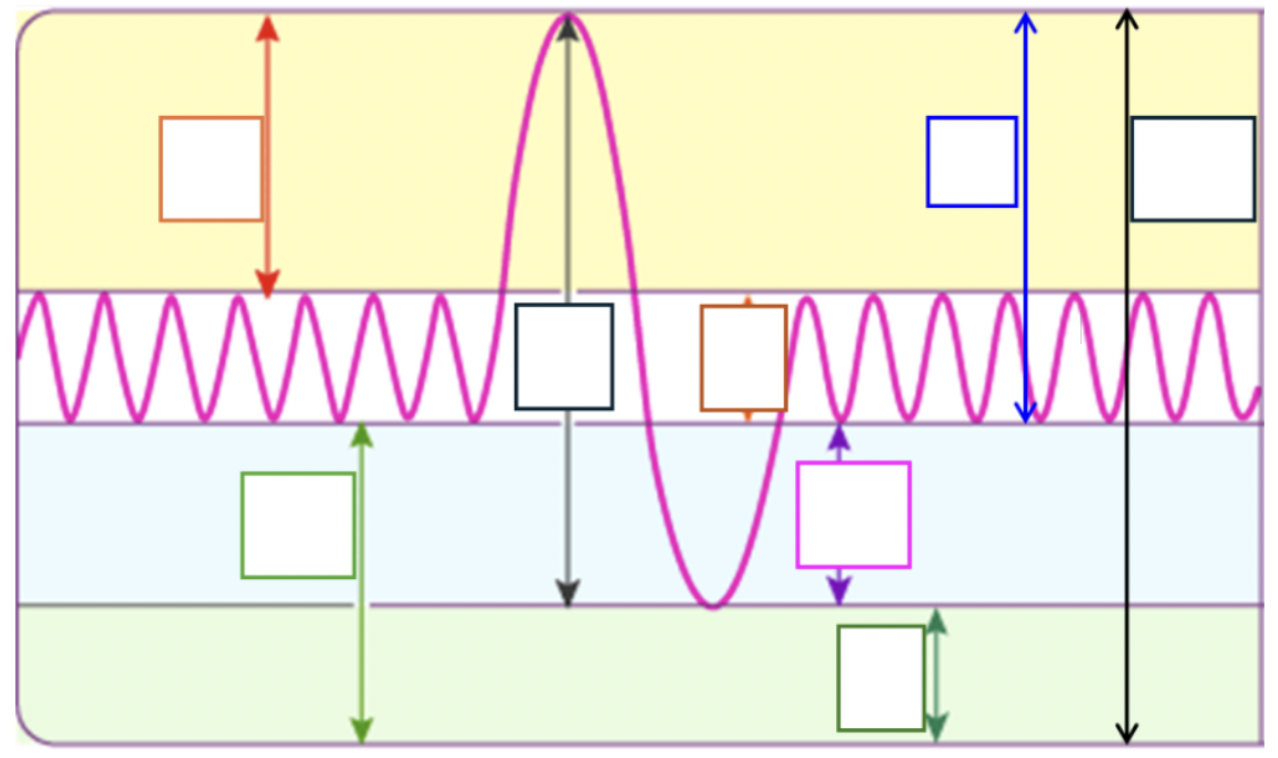
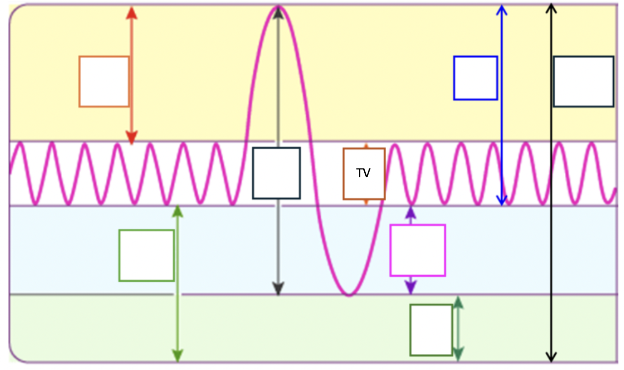
Define, exact #, and point out tidal volume
The amount of air inhaled or exhaled during a single breath, 500 mL
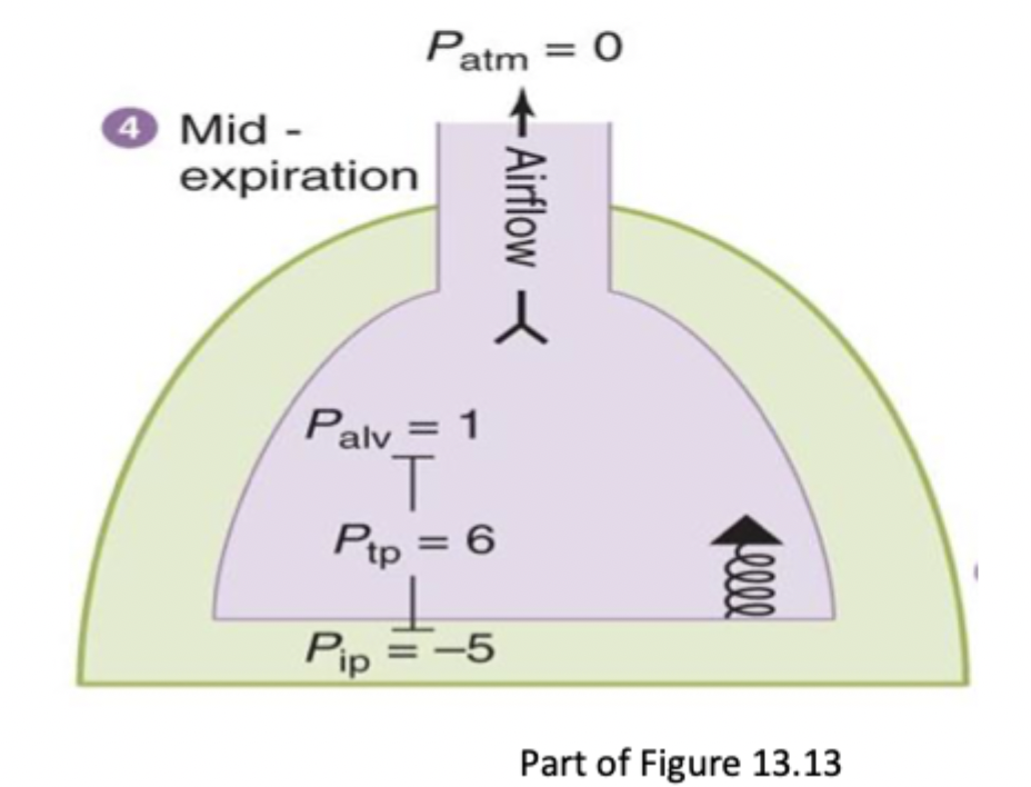
Explain the phase of the cycle at mid-exhalation
Diaphragm and external intercostals are relaxing and thoracic cavity is decreasing
Pip increases to -5
Palv = 1
Ptrans decreased → lungs recoil inward
Air flows out of lungs to atm
Define work of breathing
Effort, metabolic cost to expand lung volume, move air, and quietly exhaled
What two factors affect work of breathing
Lung compliance and airway resistance
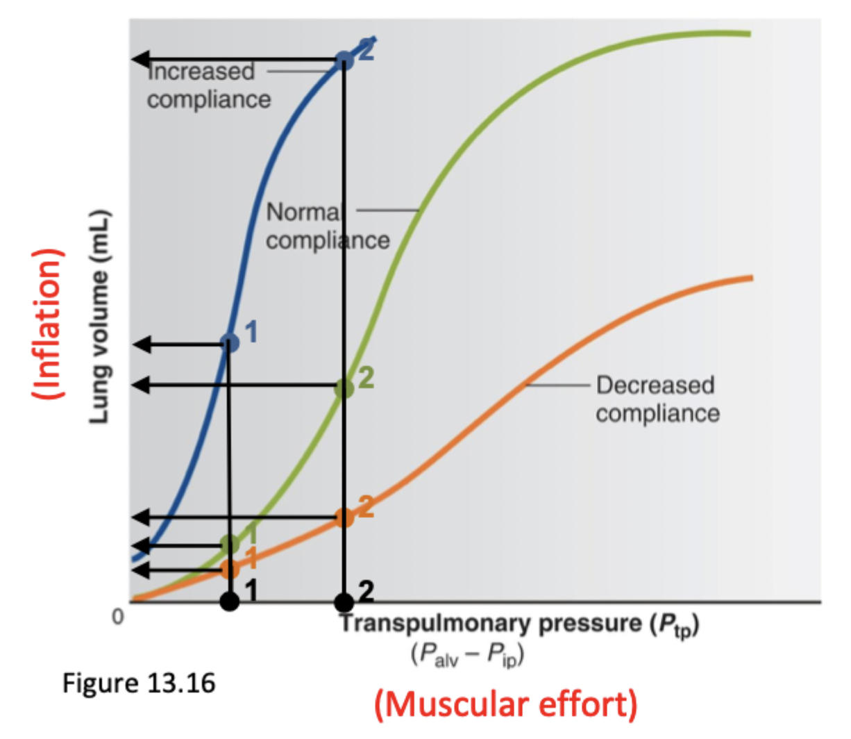
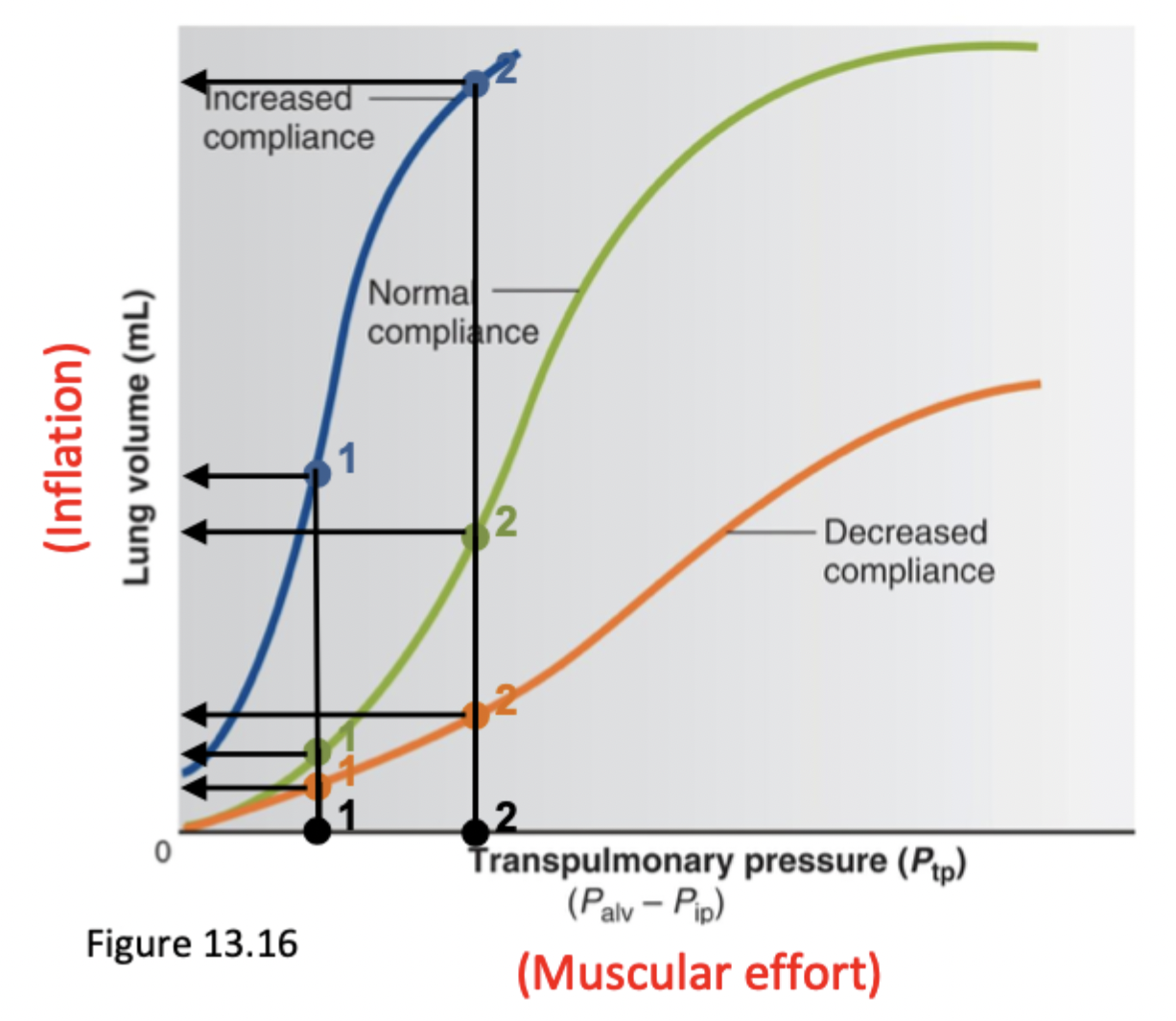
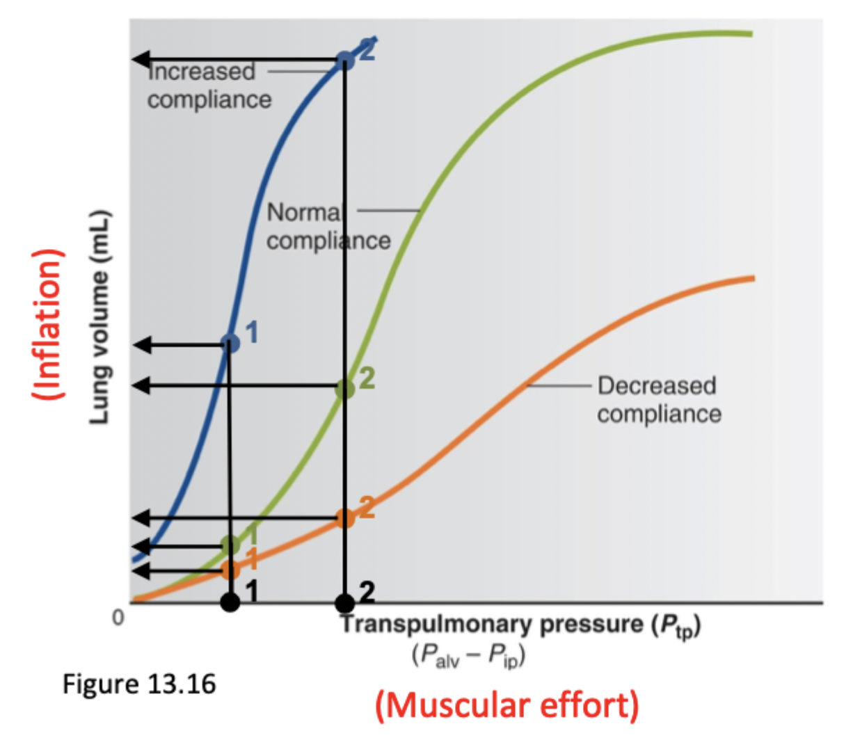
Define and explain lung compliance
The stretchability of lungs
Increasing compliance → easier to inflate. You get a substantial amount of increase of lung inflation when increasing muscular effort
Explain emphysema in lung compliance
Emphysema is floppy lung tissue where elastic recoil is reduced. No elastic recoil = no elastic fibers to pull inward → more difficult to exhale and deflate lungs
Right shift
Explain pulmonary fibrosis in lung compliance
Lung tissue is scarred and stiffer → less compliant → more effort to get air into lungs to inflate them
Explain surfactant in lung compliance
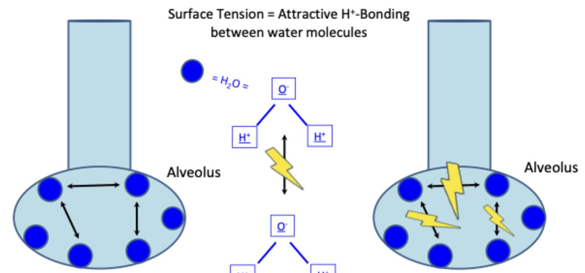
Increases lung compliance by decreasing hydrogen bonding between water molecules and decreases surface tension → easier for lungs to expand
Prevents collapse of alveoli by releasing more surfactant in smaller alveoli to decrease its P that will make it collapse
Released by type II alveolar cells, increased by deep breaths
Explain airway resistance in work of breathing
The larger radius the airways, the less resistance
Explain asthma in airway resistance
Bronchioles constrict → inflammation → lower radius → increased resistance
Explain emphysema in airway resistance
Elastic fibers that hold open airways are lost → bronchial radius narrows → increased resistance
Explain bronchitis in airway resistance
Inflammation and swelling of bronchioles → excessive production of mucus → airways narrow
How much metabolism does it take to breath? With COPD?
3% of total metabolism
With chronic obstructive pulmonary disease → 30% of total metabolism
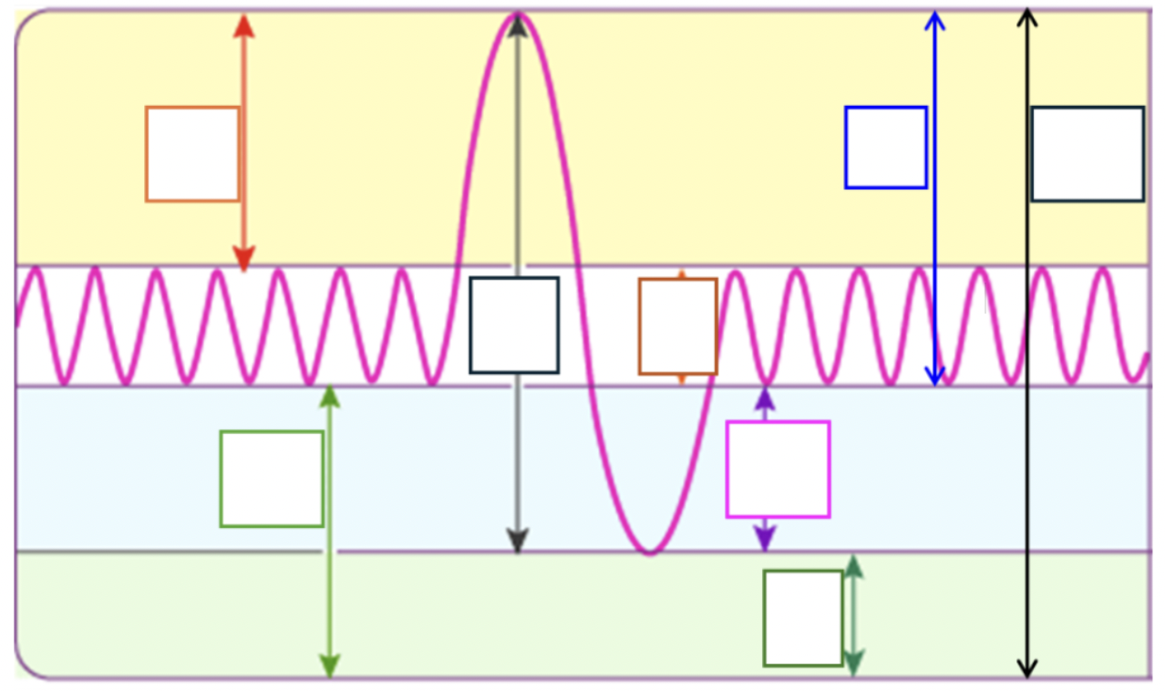
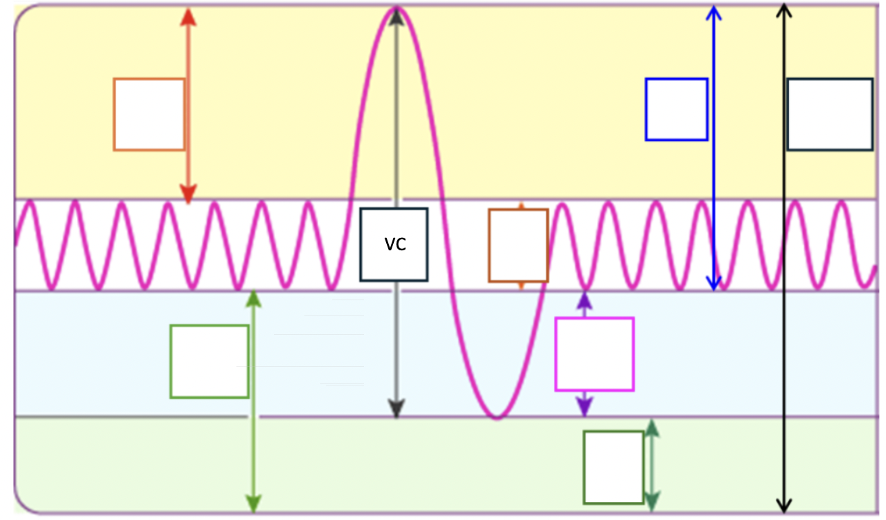
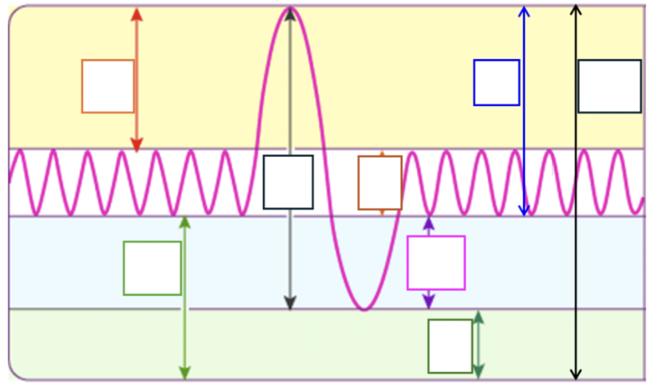
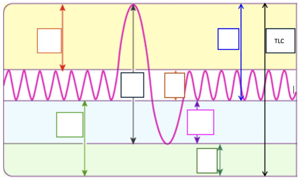
Define and point out inspiratory reserve volume (IRV)
Amount of air inhaled with max effort in excess of tidal volume
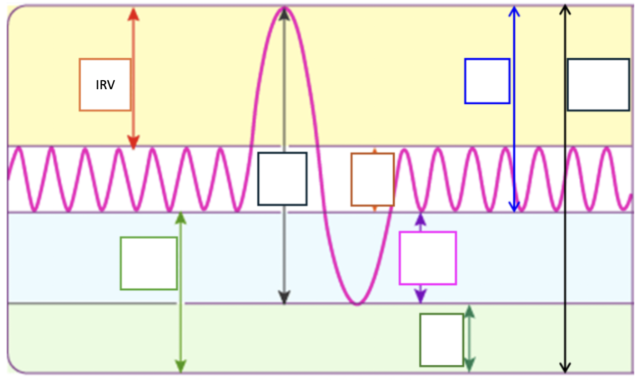
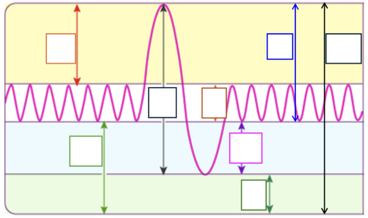
Define and point out expiratory reserve volume (ERV)
Amount of air exhaled with max effort in excess of tidal volume
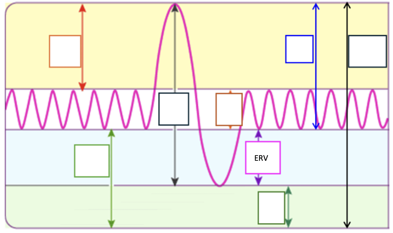
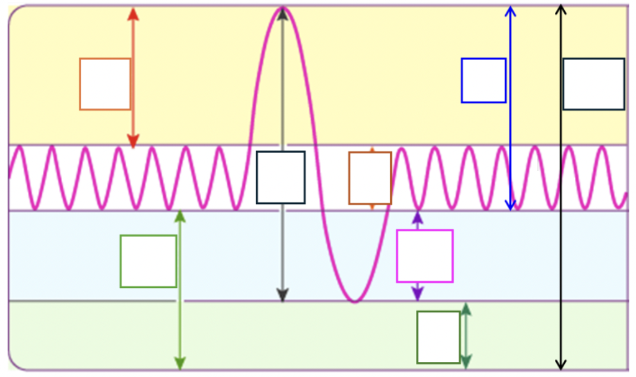
Define and point out residual volume (RV)
Amount of air remaining in lungs after max exhalation
Keeps alveoli inflated between breaths, mixes with fresh air
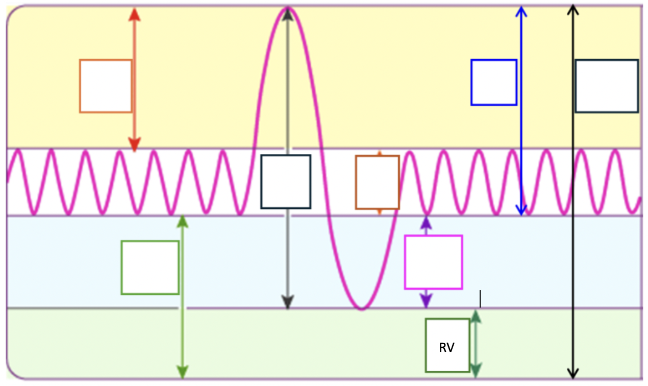
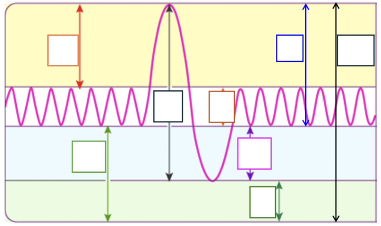
Define, point out, and equation of vital capacity
Amount of air exhaled with max effort after max inspiration
Used to assess thoracic muscle strength and pulmonary function
ERV + TV + IRV
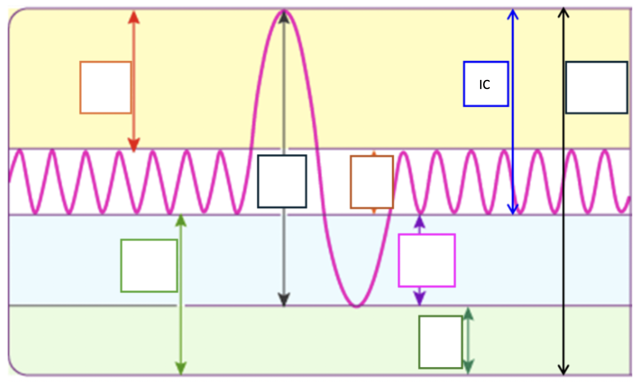
Define, point out, and equation of inspiratory capacity (IC)
Max amount of air inhaled after a normal tidal expiration
TV + IRV
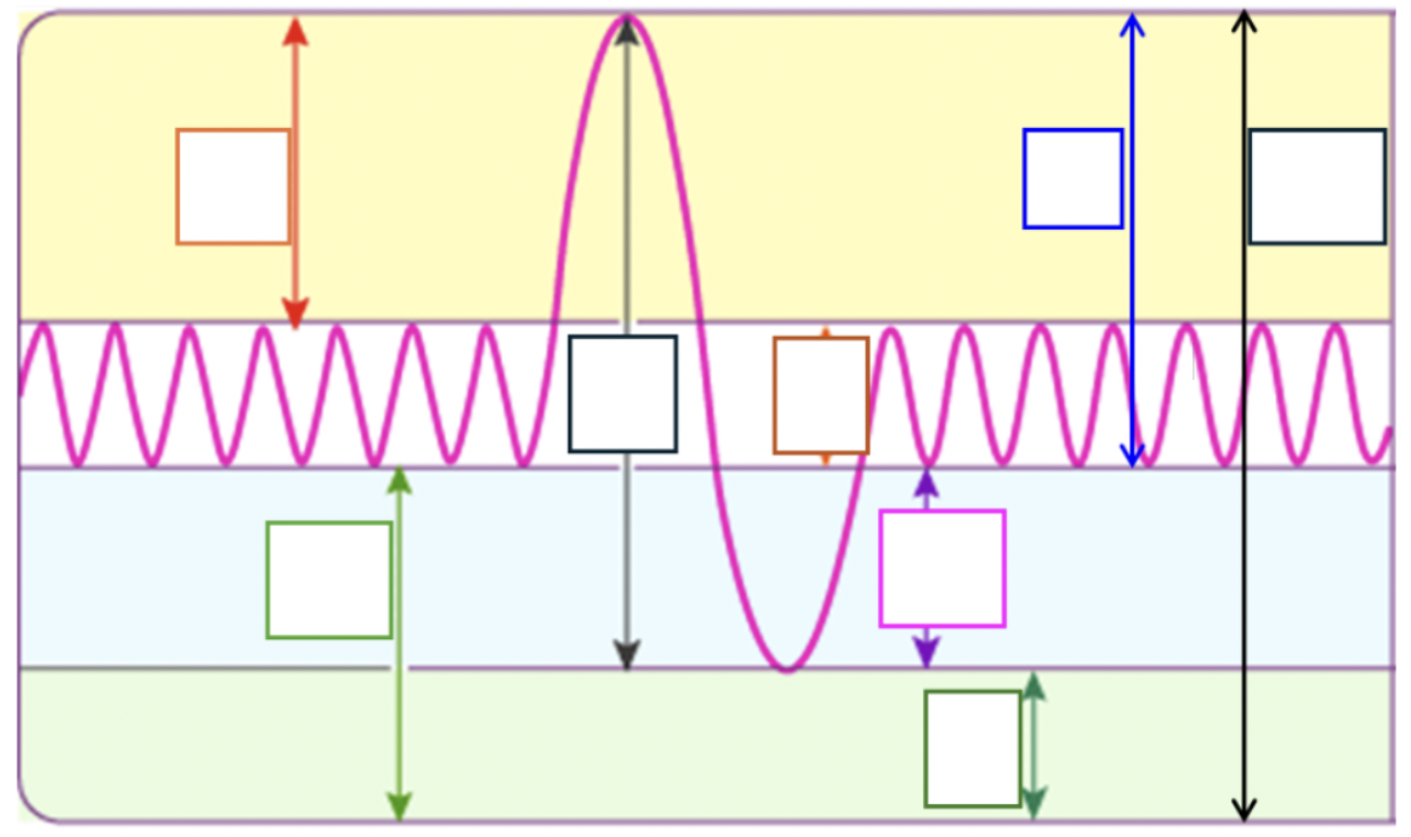
Define, point out, and equation of total lung capacity (TLC)
Max amount of air that lungs can contain
RV + VC
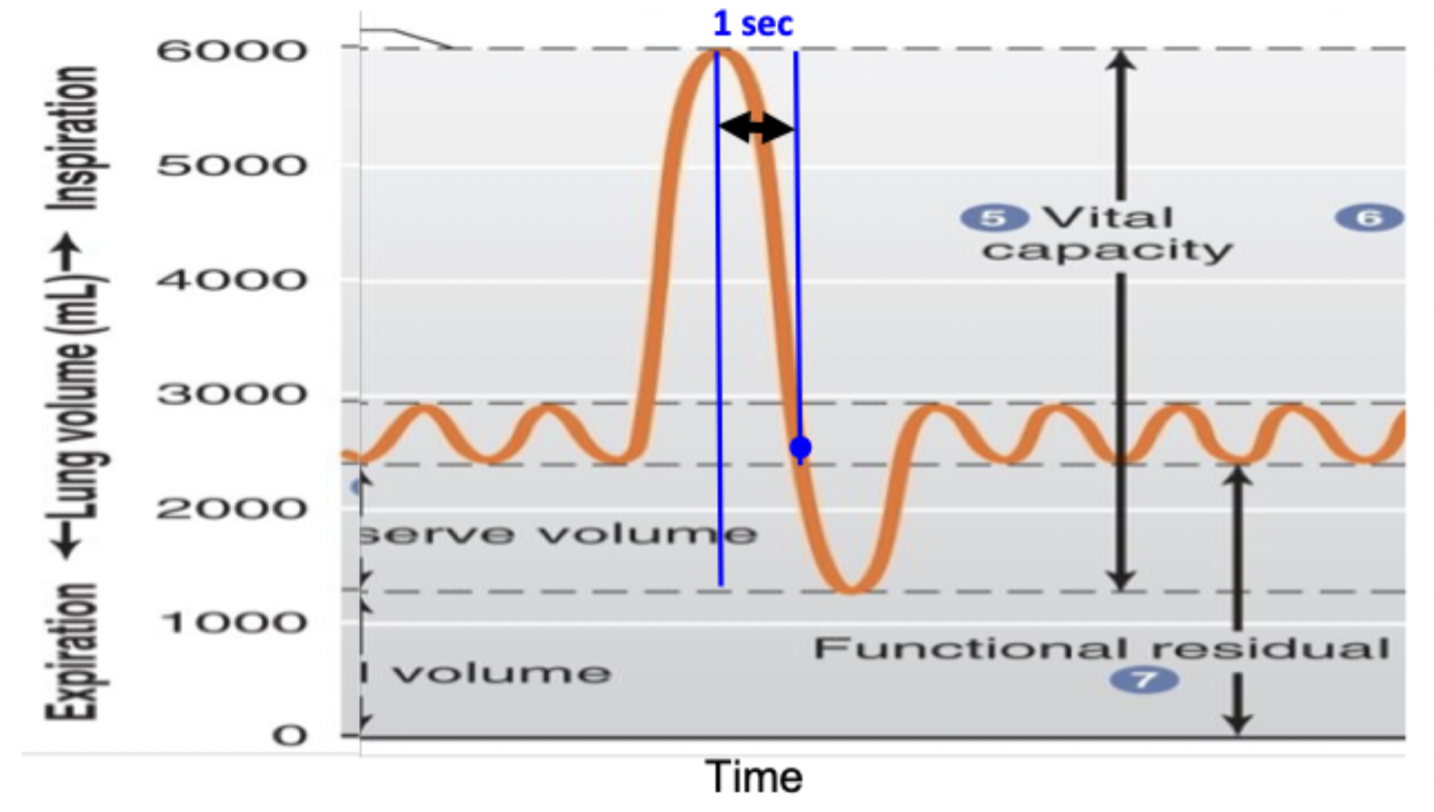
Define forced expiratory volume in one second (FEV1) and its fraction
Person takes max inhalation then exhales maximally as fast as possible
The fraction is the FEV1/VC x 100%
Define alveolar dead space
Volume of air that reaches alveoli but fails to participate in gas exchange due to lack of adequate blood flow → in diseased states
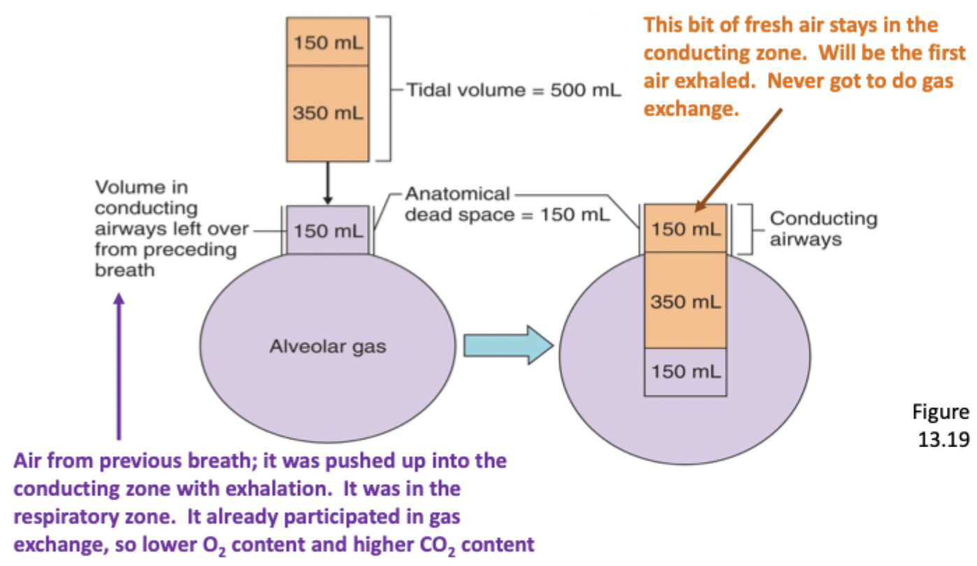
Explain anatomical dead space
A conducting zone volume, naturally doesn’t participate in gas exchange, usually 150 mL
When the tidal volume comes into the conducting airways, the fresh air will push the dead space air into the alveoli, so 150mL of air will be dead space while the rest is fresh air
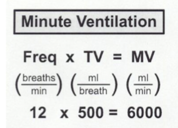
Define + equation of minute ventilation
The total amount of air moved into and out of the lungs per minute
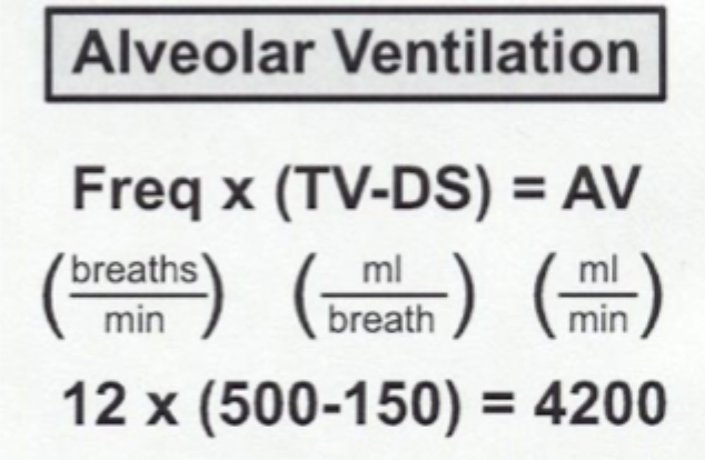
Define + equation of alveolar ventilation
The total amount of FRESH air moving into and out of the lungs per minute that’s available for gas exchange
Will always be less than the minute ventilation
Must match metabolism for homeostasis to be maintained
When person is maintaining homeostasis, what values of O2 are all equal
Net O2 entering lungs
O2 entering blood
O2 entering tissues
O2 burned by cells
Explain pathway of O2 movement (starting with inhalation)
O2 enters the alveoli → leaves lung capillaries at greater vol → left heart → arrives at capillaries where vol lowers → right heart → lung capillaries → leaves alveoli
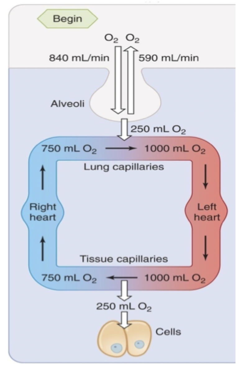
Explain details of net O2 movement
Lots of O2 is breathed out
More O2 is needed to be carried in systemic arterial blood
When person is maintaining homeostasis, what values of CO2 are all equal
CO2 produced by tissues
CO2 diffused into blood
CO2 diffusing into lungs
CO2 breathed into air
Explain the pathway of CO2 movement (starting with production)
CO2 is made in the cells and diffused into the blood → travels to right heart at greater vol → goes to lung capillaries where CO2 is exhaled → travels to the left heart at lower vol → arrives at tissue capillaries
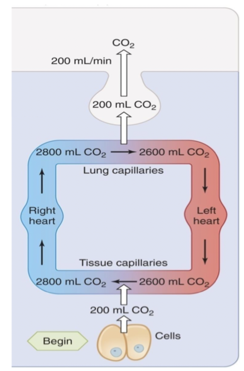
Explain details of net CO2 movement
Not enough CO2 in atmosphere to inhale
Lots of CO2 is needed in both arterial and venous blood for pH balance (buffer system with bicarbonate to maintain pH)
Equation of respiratory quotient (RQ)
= CO2 produced/O2 consumed
Doesn’t 100% match since it depends on which nutrients are being used for energy
List the 4 physical properties of gases
Collisions with walls determines pressure
Dalton’s Law
Only unbound molecules exert pressure
Henry’s Law
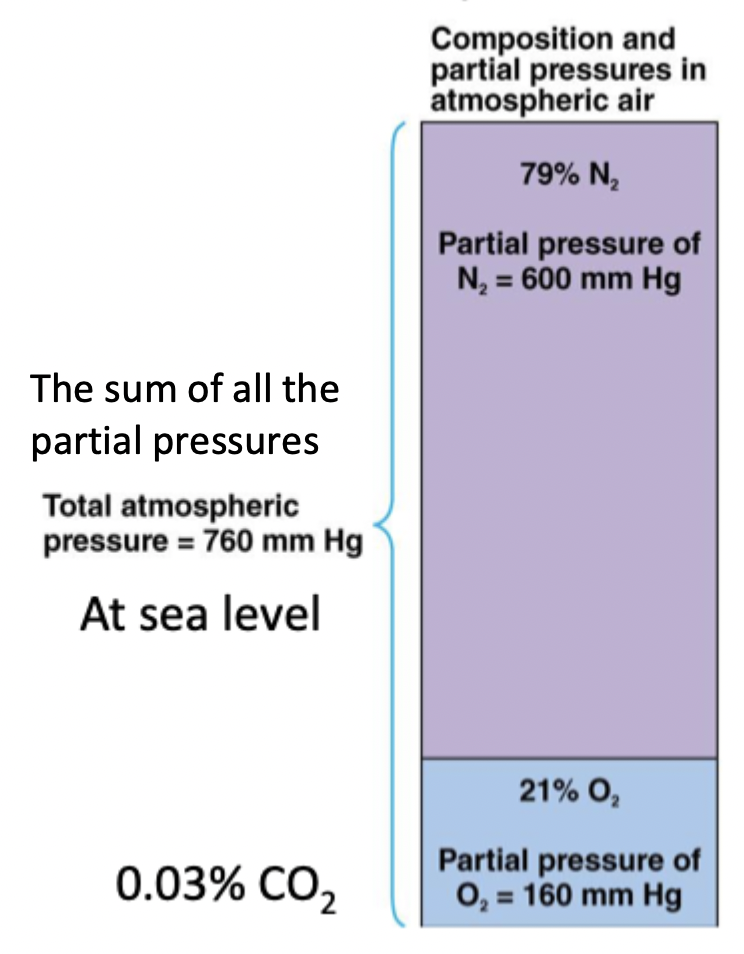
Explain Dalton’s Law with gases
In a mixture of gases, the pressure each gas exerts is independent of each other. The partial pressures can be added up
What percentages of gas makes up the atmosphere at sea level?
79% N2, 21% O2, 0.03% CO2
Makes up 760 mmHg atm
Explain Henry’s Law + equation
The amount of gas dissolved in liquid is proportional to the partial pressure and solubility of the gas where the liquid is in equilibrium.
Higher pressure → dissolves more
Amount of gas = partial pressure * solubility coefficient
List the main typical gas pressures of O2 and CO2 in respiration
O2 → atm = 160 mmHg, alveoli = 105 mmHg, lungs to tissue = 100 mmHg, tissue to lungs = 40 mmHg
CO2 —> tissue to lungs → 46 mmHg, alveoli = 40 mmHg, atm = 0.3 mmHg, alveoli to tissue = 40 mmHg
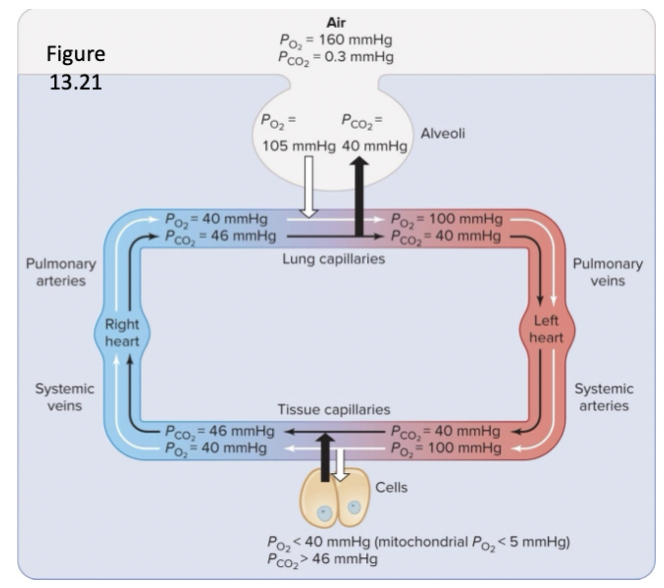
List and describe factors that vary alveolar gas pressures
PO2 of inspired air - breathing air with low PO2 decreases alveolar PO2
Rate of ventricular ventilation - hypoventilation and hyperventilation
Rate of metabolism - increased or decreased metabolism
Explain how rate of ventricular ventilation affects alveolar gas pressures
Hypoventilation - producing CO2 faster than exhaling it → decreases PO2, increases PCO2
Hyperventilation - blowing off CO2 faster than producing it → increases PO2 → decreases PCO2
Explain how rate of metabolism affects alveolar gas pressures
Decreased metabolism → not enough production of CO2 and use of O2 → increased PO2, decreased PCO2
Increased metabolism → too much production of CO2 and use of O2 → decreased PO2, increased PCO2
Explain equilibrium in gas exchange + disease states
Normally - equilibrium occurs 1/3 length of capillary
Diseased - never reaches equilibrium due to decreased rate of diffusion
Pulmonary fibrosis, pneumonia, congestive heart failure
Define matching of ventilation (V) and blood flow (Q) in alveoli
Making sure that each alveolus gets an appropriate amount of air and blood flow to make gas exchange as efficient as possible
Explain matching of ventilation (V) in alveoli
In poor ventilation, the alveoli receives little air but still receives blood → makes that blood O2 poor → low alveolar PO2
Pulmonary arteries will constrict so that the blood can be redirected to other alveoli with more oxygen
Explain matching of blood flow (Q) in alveoli
In low blood flow → alveolus receives lots of air but little blood → wasted ventilation → low alveolar CO2
Bronchioles will constrict to reduce airflow to that alveolus and be redirect to alveoli with better blood flow
How is ventilation-perfusion inequality responsible for drop of PO2 from alveoli to pulmonary venous blood?
Due to the effects of gravity. The lower chest will have increased perfusion but the upper chest will have decreased perfusion. The blood from the upper and lower will mix into the pulmonary vein to make it = 100 mmHg
What are the two methods of O2 transport in blood
Dissolved O2 due to partial pressure gradient → only 1.5% because O2 is poorly soluble
Bound to hemoglobin (98.5%)
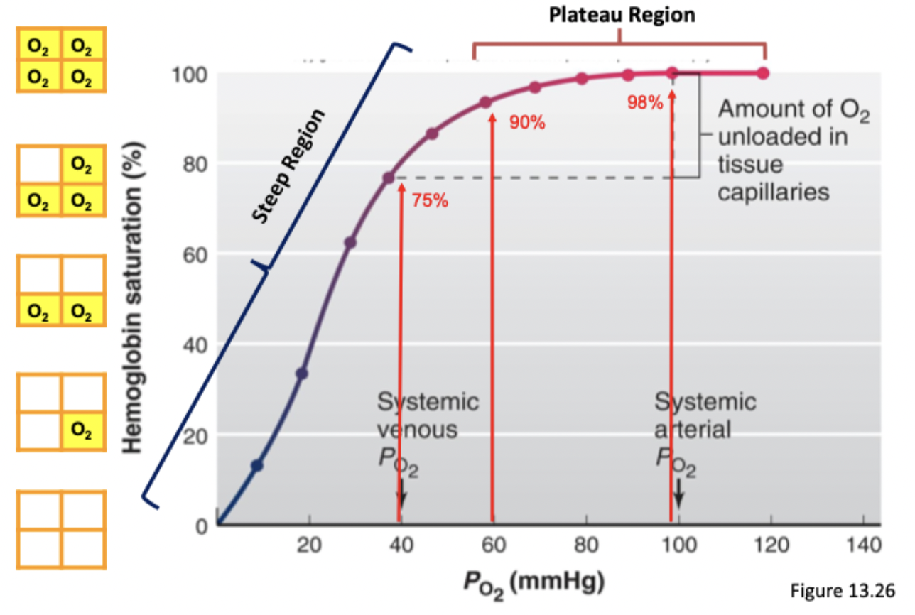
Explain the hemoglobin dissociation curve
Shows how a higher PO2 contributes to a higher hemoglobin saturation
Plateau region (60-100 mmHg) → still has high hemoglobin saturation
Steep region (0-40mmHg) → even a small change in PO2 changes the hemoglobin saturation
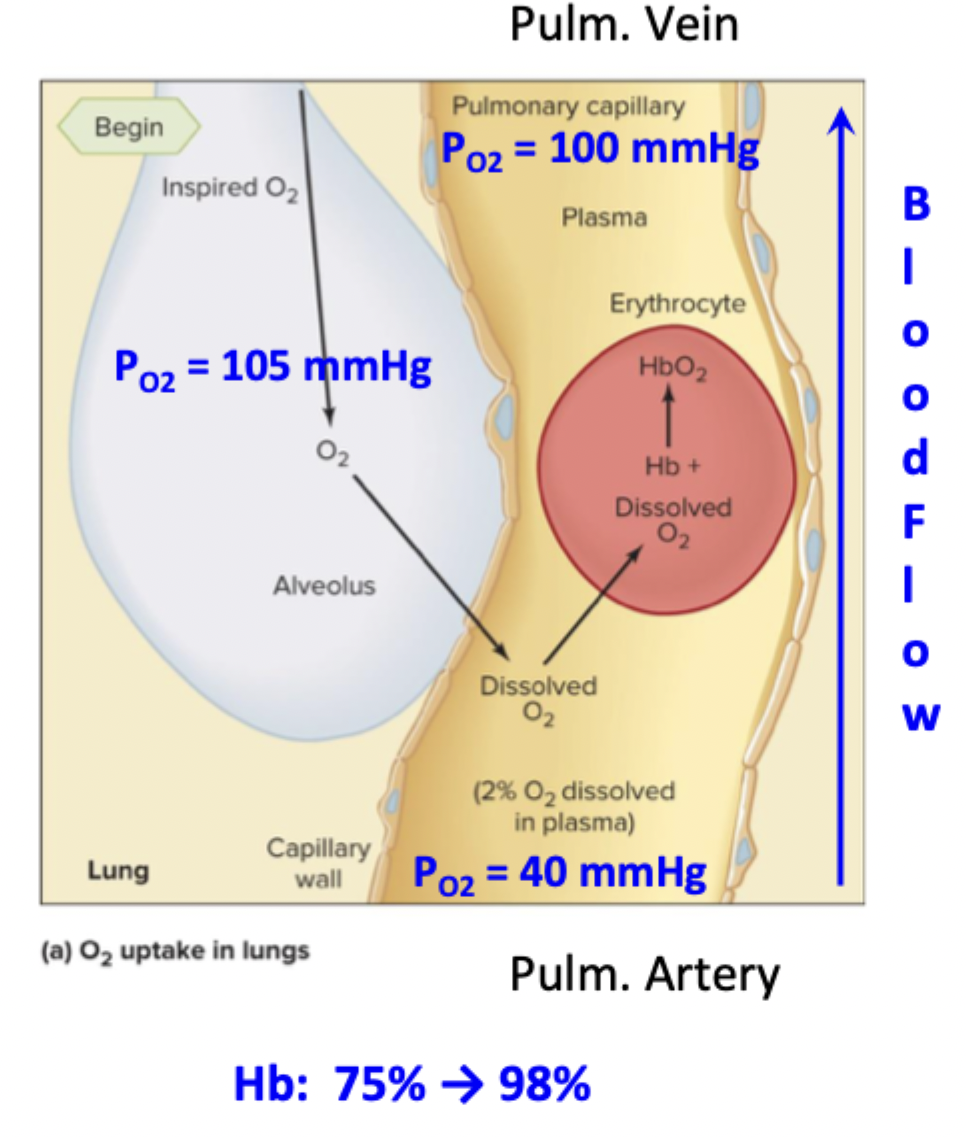
Explain the features of O2 transport from alveolus to blood
O2 in alveolus → dissolved/diffused into plasma → increases PO2 → dissolved O2 binds to Fe in Hb → down pulmonary vein
PO2 in alveolus = 105 mmHg
PO2 at pulm artery = 40 mmHg
PO2 at pulm vein = 100 mmHg
Order of movement in O2 and CO2 transport
Dissolved gases always moves first, then hemoglobin
Then CO2 from bicarbonate
Explain the features of O2 transport from blood to tissues
O2 unbinds from Hb → dissolved into plasma → dissolved into ISF → dissolved into cell cytosol (<40 mmHg) → dissolved into mitochondria (< 5mmHg)
PO2 in syst arteriole = 100 mmhg
PO2 in syst venule = 40 mmHg
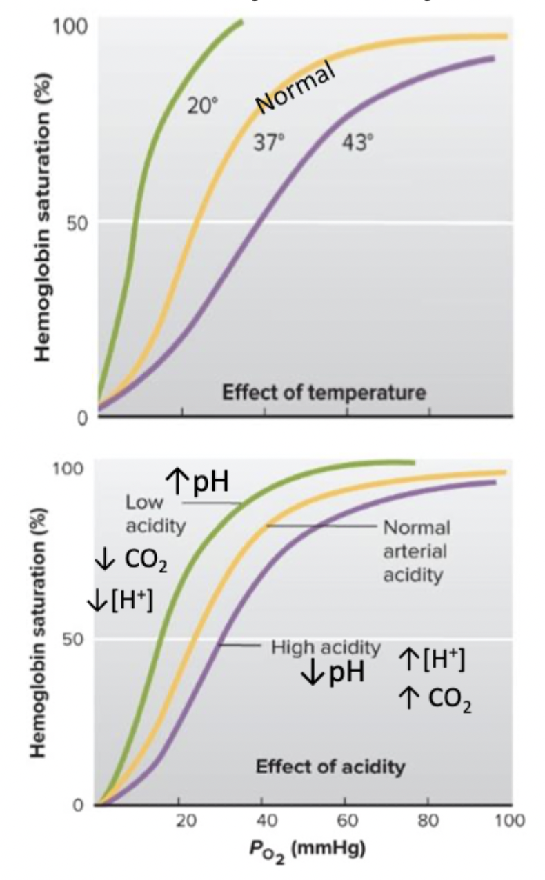
Explain effects of temp and pH on O2 transport
↑ temp → ↑ CO2 = ↑ H+ (acid)
Has a right shift → less affinity and O2 release is promoted
↓ temp → ↓ CO2 = ↓ H+ (acid)
Has a left shift → higher affinity and O2 binding is promoted
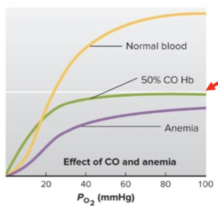
Explain effects of carbon monoxide (CO) on O2 transport
CO binds to the same site on Hb as O2 at a much higher affinity
Reduces content of O2 in blood
Shifts O2-Hb curve to the left
Can suffocate without knowing because PO2 looks normal to the chemoreceptors so you don’t gasp
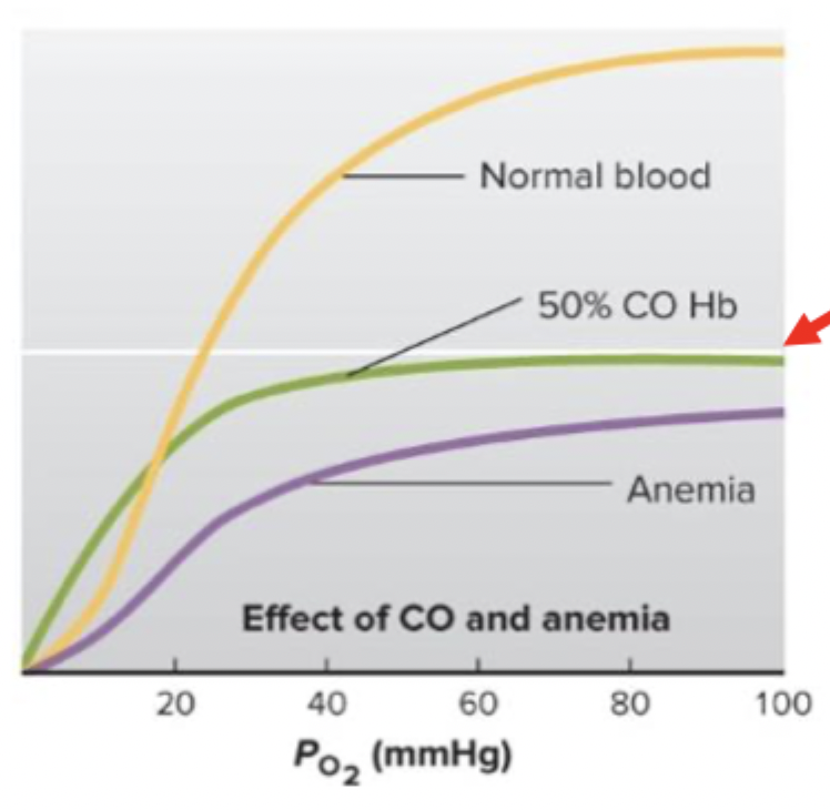
Explain effects of anemia on O2 transport
Significant decreases Hb concentration of the blood → reduces O2 carrying capacity of blood
List and describe 3 methods of transport of CO2 in blood
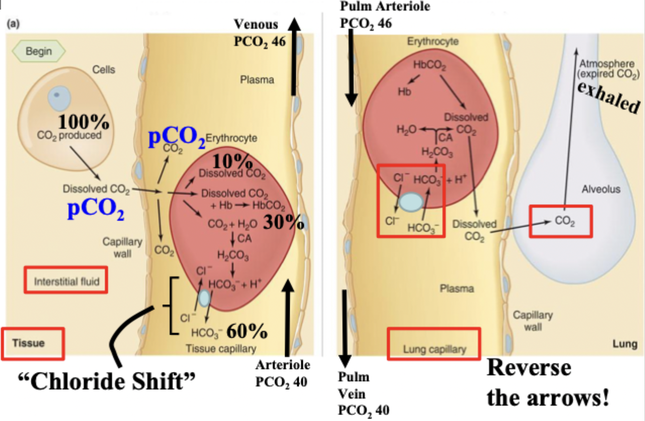
Dissolved - 10% since CO2 is more soluble in plasma than O2
Bound to Hb - 30%, on a different site than O2
As bicarbonate - 30%
Explain CO2 binding to hemoglobin and blood transport
CO2 binds to the “globin” part of hemoglobin
Promoted in venous blood where unbinding of O2 makes Hb have a higher affinity for CO2
Explain transport of CO2 in blood
CO2 produced in cells → dissolved into plasma → binds to Hb/made into bicarbonate → plasma → alveoli
Explain chloride shift
Occurs in Hb with CO2. Cl- will move in while bicarbonate moves out of RBC so that the membrane potential is maintained
Explain role of carbonic anhydrase
Ensures that making CO2 into bicarbonate is fast, makes a lot, and reversible
How is Hb a H+ buffer
It can reversibly bind to H+ to keep the pH at an appropriate range
Describe hypoventilation with CO2
Hypoventilation → ↑ arterial PCO2 → ↑ [H+] → respiratory acidosis
Describe hyperventilation with CO2
Hyperventilation → ↓ arterial PCO2 → ↓ [H+] → respiratory alkalosis