Zoo 250 Exam 3 - SELU John O'Reilly
1/226
There's no tags or description
Looks like no tags are added yet.
Name | Mastery | Learn | Test | Matching | Spaced | Call with Kai |
|---|
No analytics yet
Send a link to your students to track their progress
227 Terms
Myology
Study of skeletal muscles
How many muscles are in the human muscular system?
600
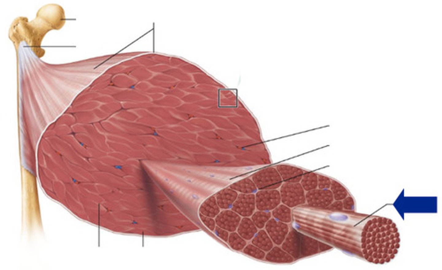
A skeletal muscle consists of more than muscular tissue. What are the connective tissue components? It also has blood vessels.
Muscle (Epimysium), fascicle (Perimysium), fiber (Endomysium), myoblasts
Epimysium + Perimysium + Endomysium = __________
Tendon, which attaches muscle to bone
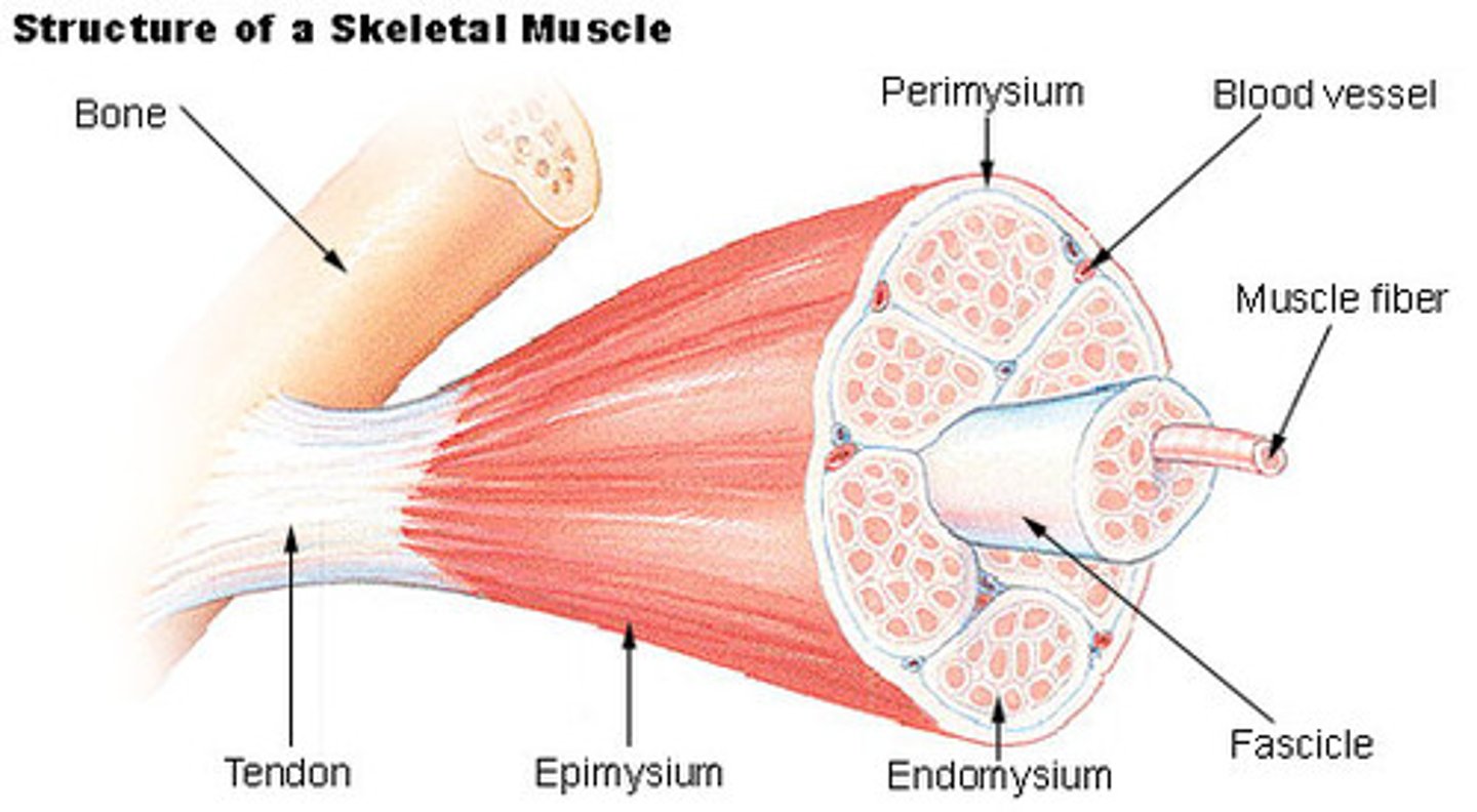
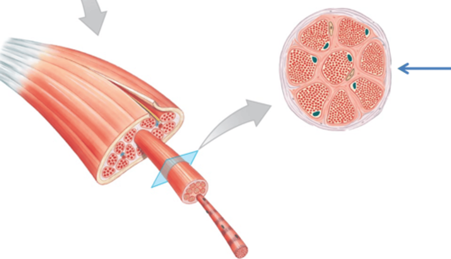
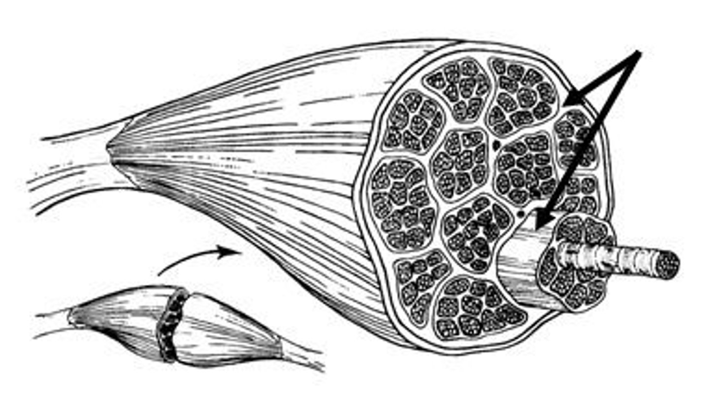
Fascicle
Bundle of fibers
(Skeletal) Muscle
Bundle of fascicles
What is a muscle fascicle made up of?
Bundles of muscle fibers
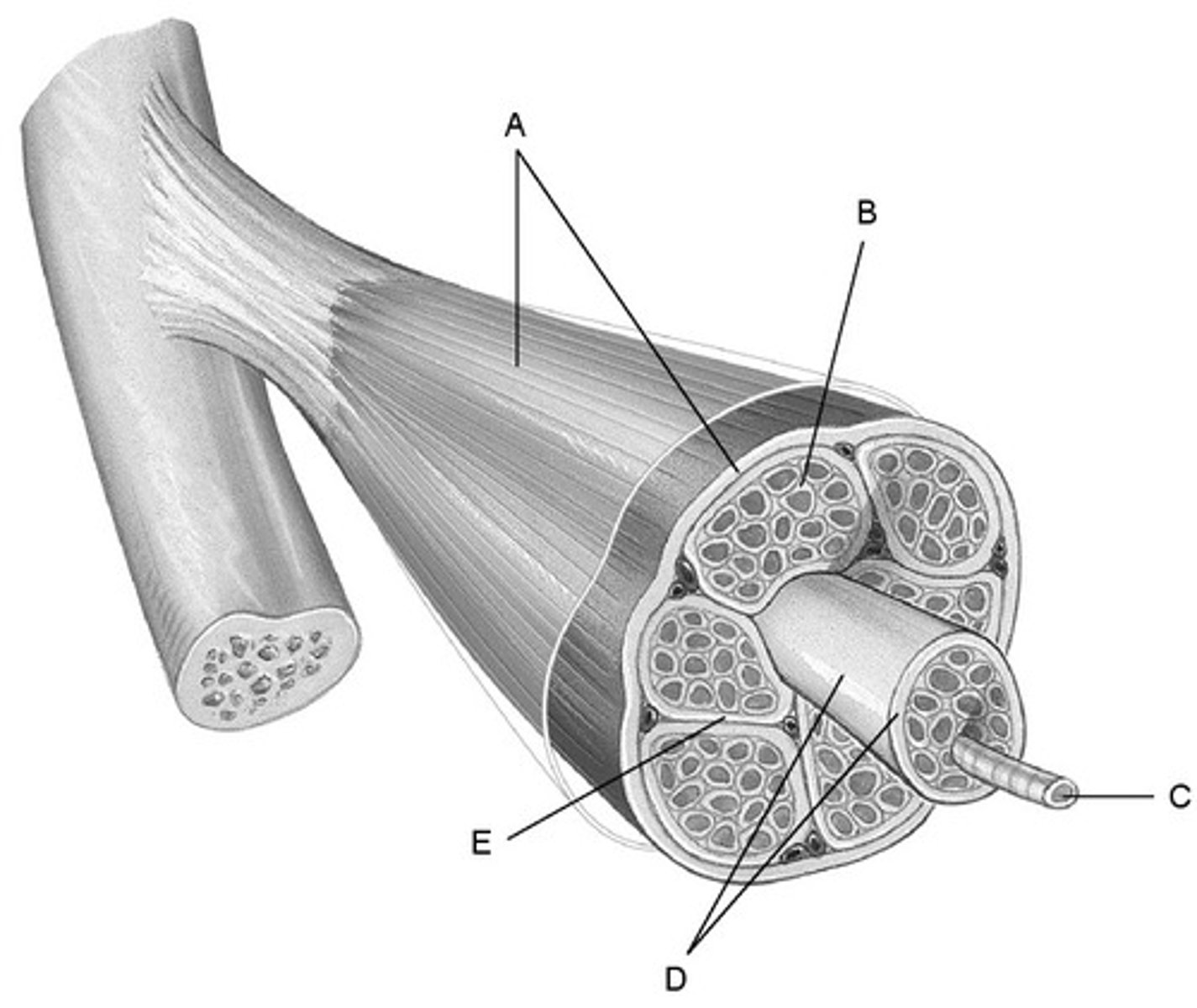
Muscle (Epimysium)
-Fibrous sheath that surrounds the entire muscle
-A

Fascicle (Perimysium)
Thicker connective tissue sheath that wraps muscle fibers together in bundles called fascicles
Fiber (Endomysium)
-Thin sleeve of loose connective tissue that surrounds each muscle fiber
-B
Muscle Fiber
What fuses to produce mature muscle fiber?
Myoblasts
Muscle fibers develop through the fusion of mesodermal cells called __________.
myoblasts

What is the difference between the sarcolemma and the sarcoplasm?
Sarcolemma is the muscle cell membrane. Sarcoplasm is the muscle cytoplasm.
What makes up the sarcolemma and the sarcoplasm?
-T tubules, sarcoplasmic reticulum, terminal cisternae, and triad
-Also myofibrils that contain myofilaments
T Tubules
Continuous with inside and outside of sarcolemma (Muscle cell membrane)
Sarcoplasmic Reticulum (SR)
-Intracellular (Within sarcoplasm- Muscle cytoplasm)
-Has intimate relationship with the T tubules
Terminal Cisternae
Parts of SR associated with T tubules
Triad
Two terminal cisternae + one T tubule
Myofibrils
Bundles of protein
Myofilaments
The protein in myofibrils
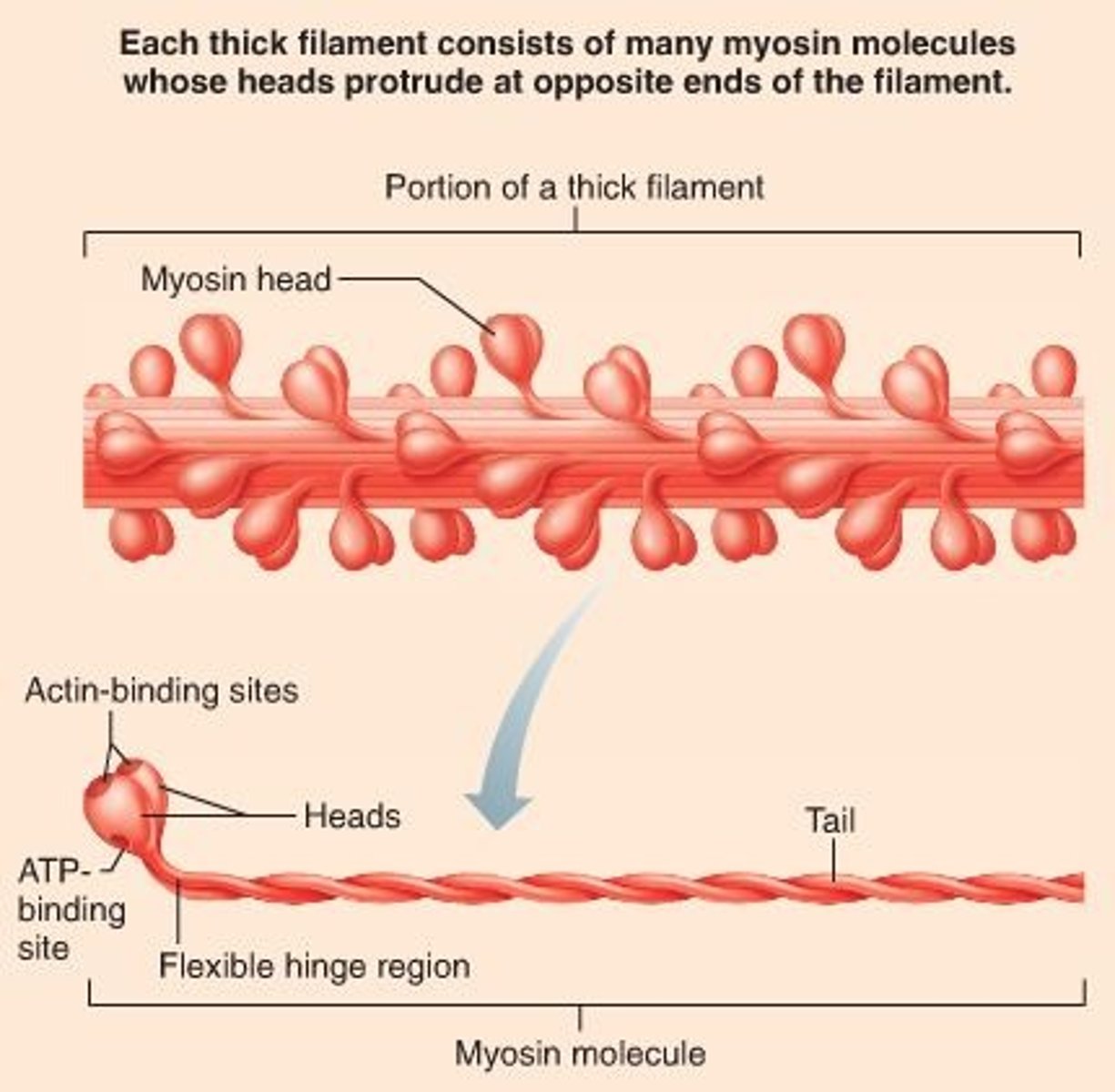
What are thick filaments formed by?
Myosin (Protein), thus generating contraction (Force and movement)
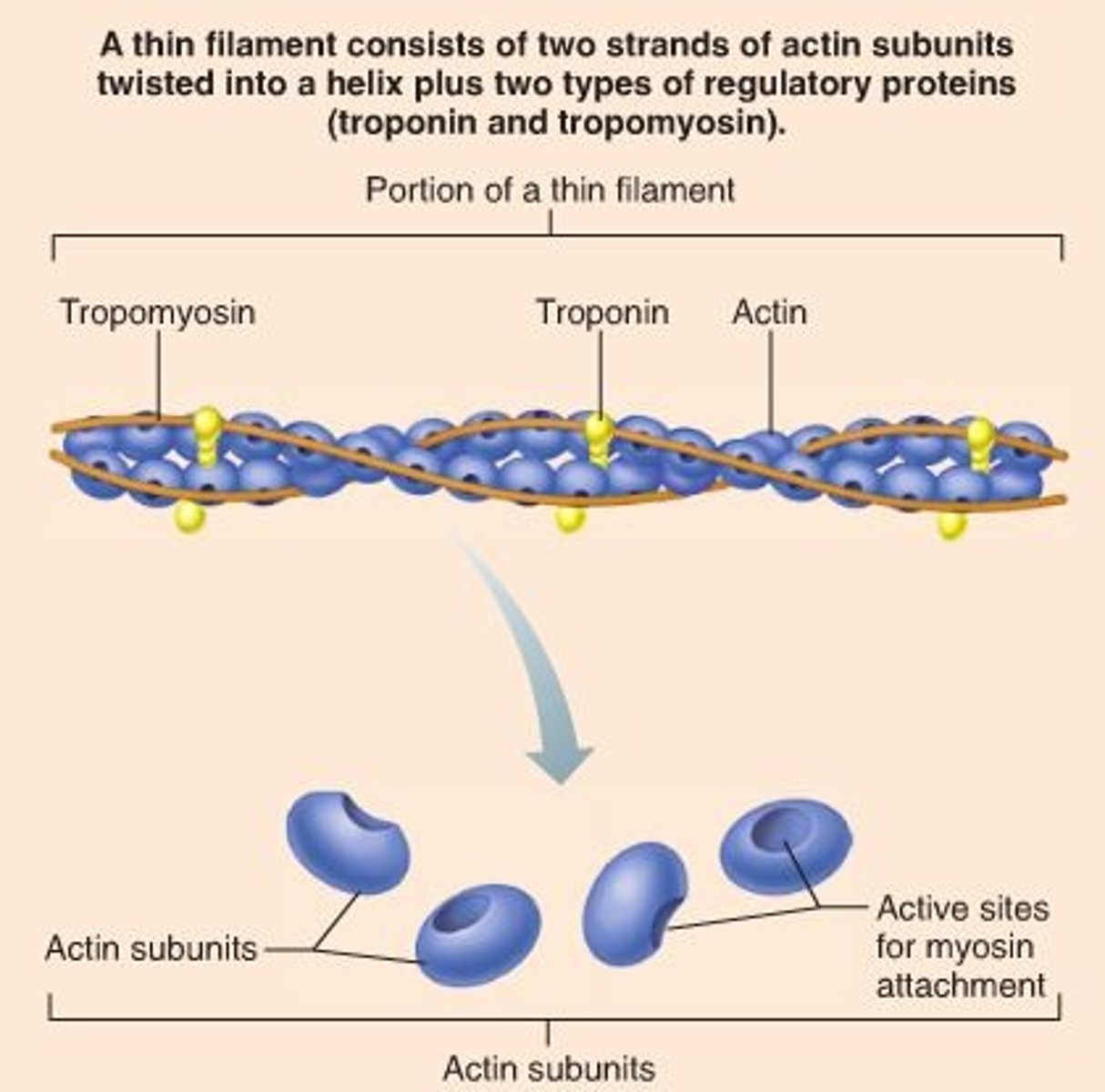
What are thin filaments formed by?
Actin, tropomyosin, troponin
Zone of Overlap
Both thick and thin filaments
What are sarcomeres apart of?
The myofibril
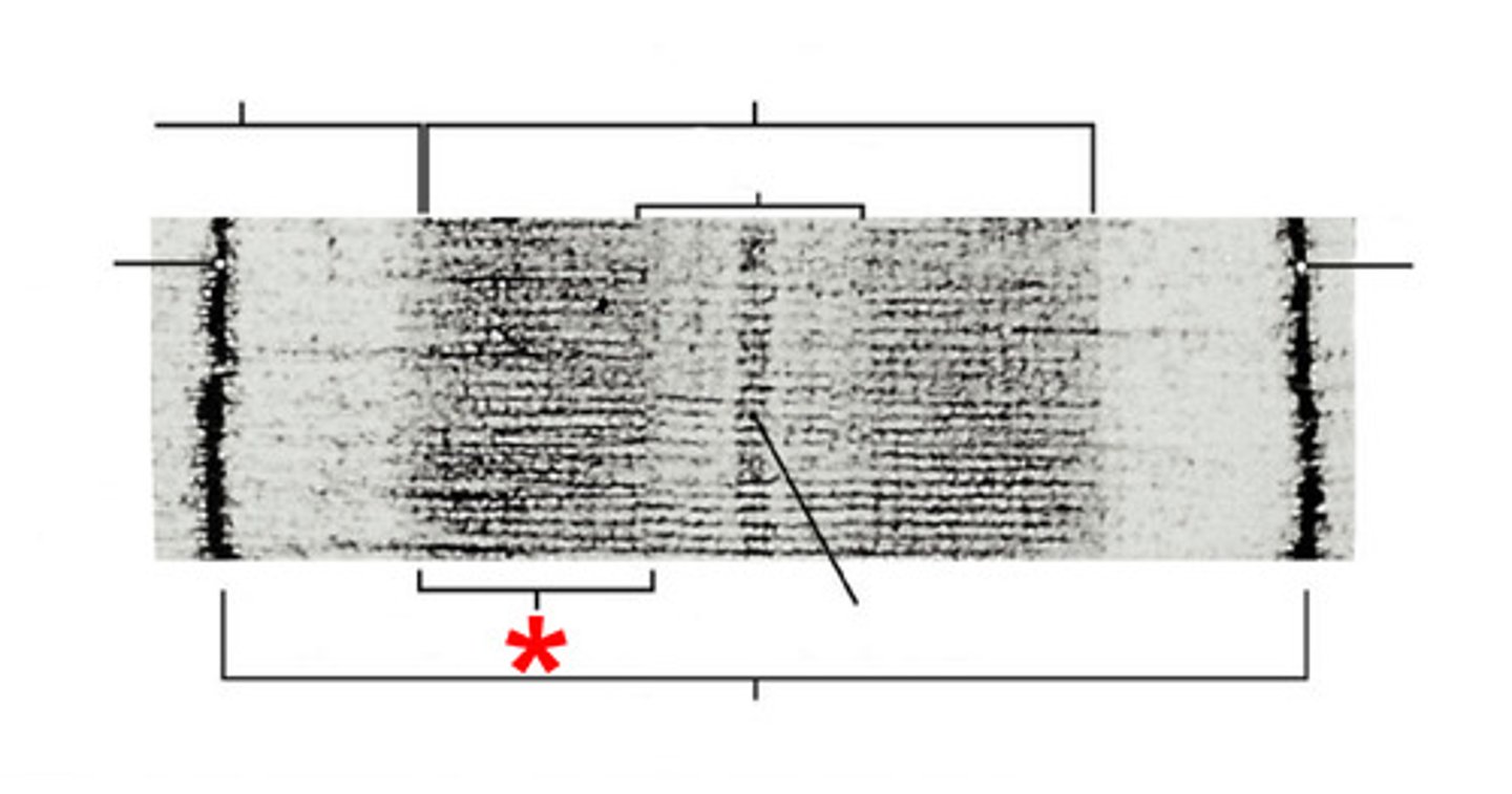
Sarcomere
A structural unit of a myofibril
A (Dark) Band
-Both thick and thin filaments
-Contains M line, H band, and zone of overlap

M Line
Middle of sarcomere (Attachment of thick filaments)

H Band
Only thick filaments
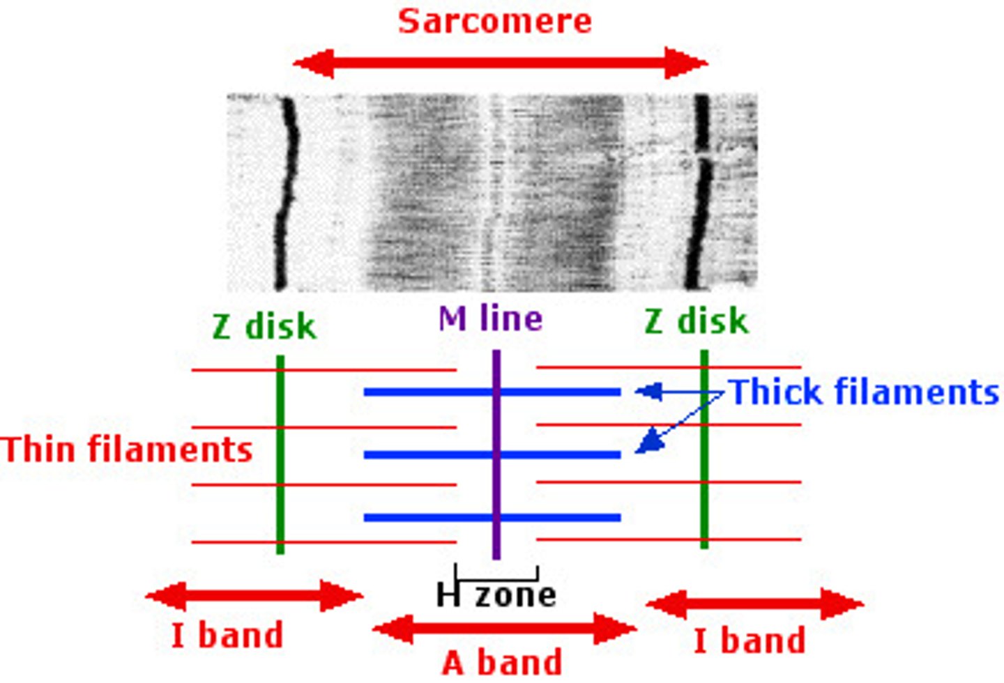
Zone of Overlap
Thick and thin filaments (Both sides of H band)

I (Light) Band
-Only thin filaments
-Contains Z disc

Z Disc
End of sarcomere (Attachment of thin filaments)

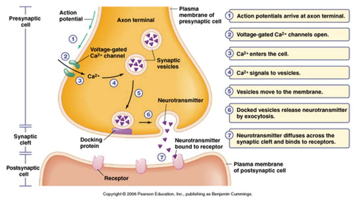
Steps in Neuromuscular Synaptic Transmission - Contraction of the Skeletal Muscle
1. Action potential (AP) reaches nerves in the muscles - Nerve action potential (AP)
1. Ca++ influx (Arrives and goes) into nerve terminal - Makes cells respond in a way they should
2. ACh release from nerve terminal
3. ACh crosses synaptic cleft and binds to ACh receptor on muscle
4/5. Muscle depolarization and AP
6. Muscle AP propagation (Travels) through T tubules
15. Acetylcholinesterase (AChe) breaks down ACh (Acetylcholine) - Stops/terminates information flow and neurotransmitters (Synapse)
ACh
-A chemical released by nerve cells to send signals to other cells
-Charges in your muscle membranes that are from the ions
-Ex. ACh-2
Depolarization
-Loss of polarization
-Any such case in which the voltage (Charge) shifts to a less negative value (More positive)
-Loss of the difference in charge between the inside and outside of the plasma membrane of a muscle or nerve cell
-Activates AP
What creates excitation?
Action potential
Excitation-Contraction Coupling
The physiological process of converting an electrical stimulus to a mechanical response and the start of muscle contraction
Steps in Excitation-Contraction Coupling
1. Muscle AP propagation (Travels) through T tubules (Step 6)
2. Then, activation of DHP receptor (Dihydropyridine receptor) on T tubule
3. Then, activation of RYR (Ryanodine receptor) on sarcoplasmic reticulum (SR)
4. Intracellular Ca++ release from terminal cisternae of SR - Inside cell (Step 7)
5. Then, muscle contraction cycle
-Ca++ is pumped back into SR
What happens when Ca++ is pumped back into SR?
Calcium levels around the actin and myosin filaments are reduced and muscle relaxes
Muscle Contraction Cycle
-The energy released during ATP hydrolysis changes the angle of the myosin head into a "cocked" myosin head position, ready to bind to actin if the sites are available
-Intracellular Ca++ release from terminal cisternae of SR (Step 7)
1. Ca++ binds to troponin of the thin filaments (Step 8)
-(Ca++ allows troponin-tropomyosin to move to reveal active site)
2. Troponin-tropomyosin moves > Exposure of active site on actin (Step 9)
3. "Cocked" myosin head binds active site on thin filament actin, forming a cross-bridge formation between the myosin and actin (Step 11)
4. Myosin head pivots towards M line. This is called the power stroke. (Step 12) - Muscle contraction
5. Myosin head binds to new ATP, which breaks the cross-bridge (Step 13) - Muscle relaxation (Needs ATP) and troponin-tropomyosin covers active site on actin. Myosin can no longer bind to actin, and the muscle fiber ceases to produce or maintain tension. (Step 18)
What is needed for muscle relaxation?
ATP
Muscle Contraction Cycle
-After ATP is needed for muscle relaxation, troponin-tropomyosin covers active site on actin (Step 18) - End of contraction
6. Myosin head hydrolyzes ATP to ADP + Pi (Step 10) - Contraction begins again
-Energy (ATP) needed for muscle contraction
-"Cocked" myosin head (Step 10)
-Then, the process starts again
What is needed for muscle contraction?
ATP (Energy)
Motor Units
Motor nerve (motorneuron) and all muscle fibers that it innervates
A motorneuron may innervate more than one __________ __________, but each muscle fiber is only intervated by one __________.
muscle fiber, motorneuron
Motor Unit Recruitment
-Additional motor units added to accomplish an increase in contractile strength in a muscle
-Recruits neurons (Motorneurons)
Example of Motor Unit Recruitment
When muscles receive information from internal or external stimuli, muscle fibers are stimulated by their respective motor neuron. Impulses are passed from the central nervous system down neurons until they reach the motor neuron, which releases the neurotransmitter acetylcholine (ACh) into the neuromuscular junction. Then, the excitation-contraction coupling, and muscle contraction cycle. Contractions begin. Some examples include twitch, treppe, incomplete tetanus, and complete tetanus.

Isometric Contraction
Muscle develops tension, muscle does not shorten nor lengthen
Isotonic Concentric Contraction
Muscle tension remains constant, muscle shortens
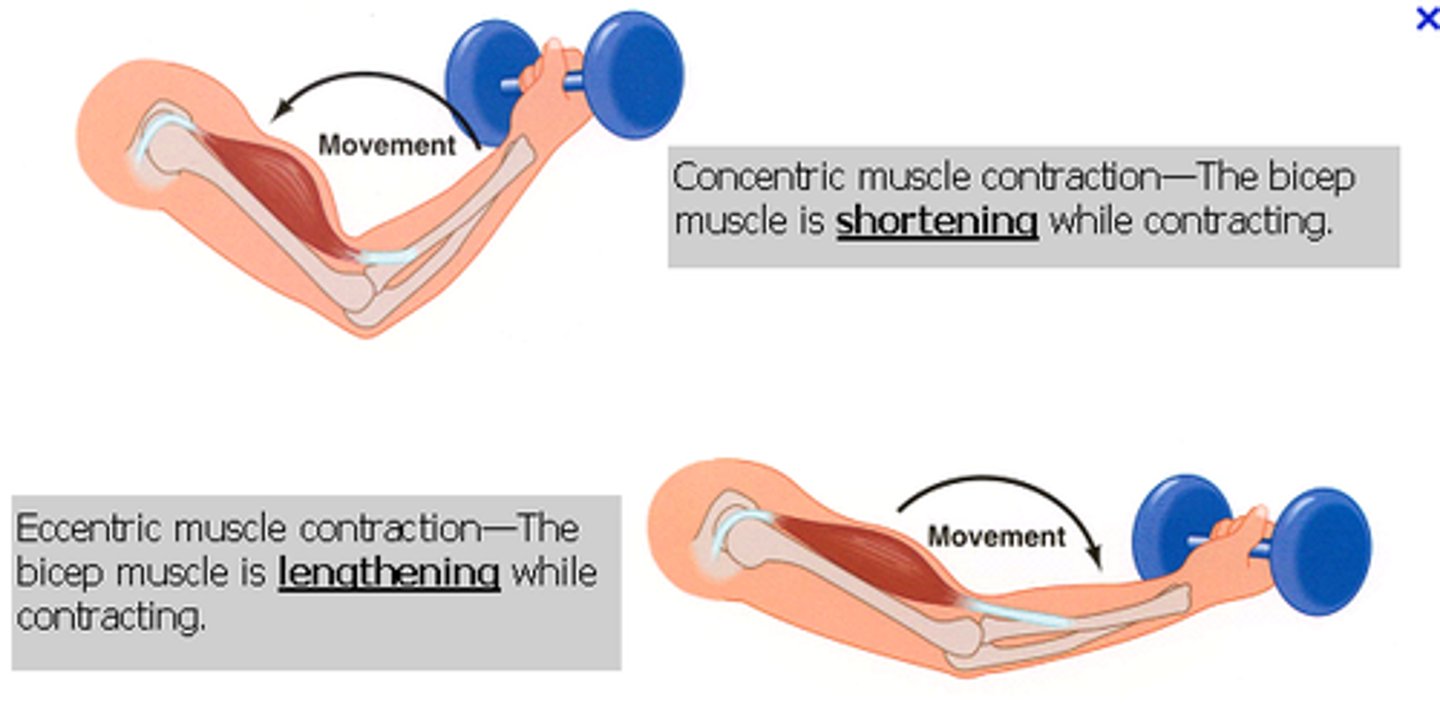
Isotonic Eccentric Contraction
Muscle tension remains constant, muscle lengthens

Myokinase
Transfers Pi from one ADP to another, converting the latter of ATP that myosin can use
Creatine Kinase
Obtains Pi from a phosphate storage molecule, creatine phosphate, and donates it to ADP to make ATP. This is a fast acting system that helps to maintain the ATP level while other ATP-generating mechanisms are being activated.
_____ and _____, collectively called the phosphagen system, provide nearly all the energy used for short bursts of intense activity.
ATP, CP
Phosphagen System
Two enzymes, myokinase and creatine kinase, generate ATP in the absence of oxygen. Myokinase borrows phosphate groups from ADP, and creatine kinase borrows them from creatine phosphate, to convert ADP to ATP.
As the phosphagen system is exhausted, the muscles transition to __________ fermentation to generate ATP by glycolysis.
anaerobic
After 40 seconds or so, the respiratory and cardiovascular systems "catch up" and deliver oxygen to the muscles fast enough for __________ respiration to once again meet most of the ATP demand. Aerobic respiration produces much more ATP than glycolysis does.
aerobic
Muscle Metabolism
-Adenosine Triphosphate (ATP) and creatine phosphate (CP)
-Aerobic (Oxygen) and anaerobic (No oxygen) metabolism
-Muscle fatigue
As glucose and glycogen are depleted, what becomes the more significant fuel for ATP?
Fatty Acids and Oxygen
Low Activity
-Fatty Acid + O2 > ATP
-Glucose > Glycogen
-Creatine > Creatine Phosphate
If fatty acids and oxygen are already obtained by the muscle, __________ will not be needed, so it can be stored as glycogen.
glucose
Moderate Acticity
-Fatty acids + O2 > ATP
-Glycogen > Glucose > ATP
If fatty acids and oxygen are not obtained by the muscle, glucose will be needed, so it can be taken from storage as ___________ and used as glucose > ATP.
glycogen
High Activity
-Glycogen > Glucose > ATP
-Creatine Phosphate > Creatine + ATP
Both __________ and __________ produce ATP, but glycolysis cannot generate ATP as quickly as __________ __________.
glycogen, CP, creatine phosphate
Muscle Fatigue
-The progressive weakness and loss of contractility that results from prolonged use of the muscles
-Anything can cause it
-Ex. If you hold a heavy book at arms length for a minute, you will feel your muscles growing weaker and soon you'll be unable to hold it up
-Ex. Trying to take lecture notes from a fast-talking professor produces fatigue in hand muscles
What are the 2 muscle fiber types?
Slow (I) and Fast (IIB)
Cross-Section Diameter
-Slow- Small diameter
-Fast- Large diameter
Tension (Force)
-Slow- Low tension (Force)
-Fast- High tension (Force)
Contraction Speed
-Slow- Slow speed
-Fast- Fast speed
Resistance to Fatigue
-Slow- High resistance = Slow to fatigue
-Fast- Low resistance = Fast to fatigue
Color
-Slow- Red
-Fast- White
Myoglobin Content
-Slow- High
-Fast- Low
Glycogen Content
-Slow- Low
-Fast- High
Metabolism
-Slow- Oxidative (Aerobic- Oxygen)
-Fast- Glycolytic (Anaerobic- No oxygen)
What are the muscle tissue types?
Skeletal, cardiac , and smooth
Skeletal Muscle Tissue
-Striated
-Linear myofibrils
-Multiple nuclei (Multinucleated)
-No gap junctions
-Activation = Motor nerves (Motorneurons)
Cardiac Muscle Tissue
-Striated
-Linear myofibrils
-Single nucleus
-Gap junctions
-Activation = Pacemaker cells, autonomic nervous system (ANS) > Effect on heart rate
Smooth Muscle Tissue
-Non-striated
-Non-linear myofibrils
-Single nucleus
-Gap junctions
-Activation = Local stimuli, autonomic nervous system (ANS) > Pressure, food coming down your esophagus
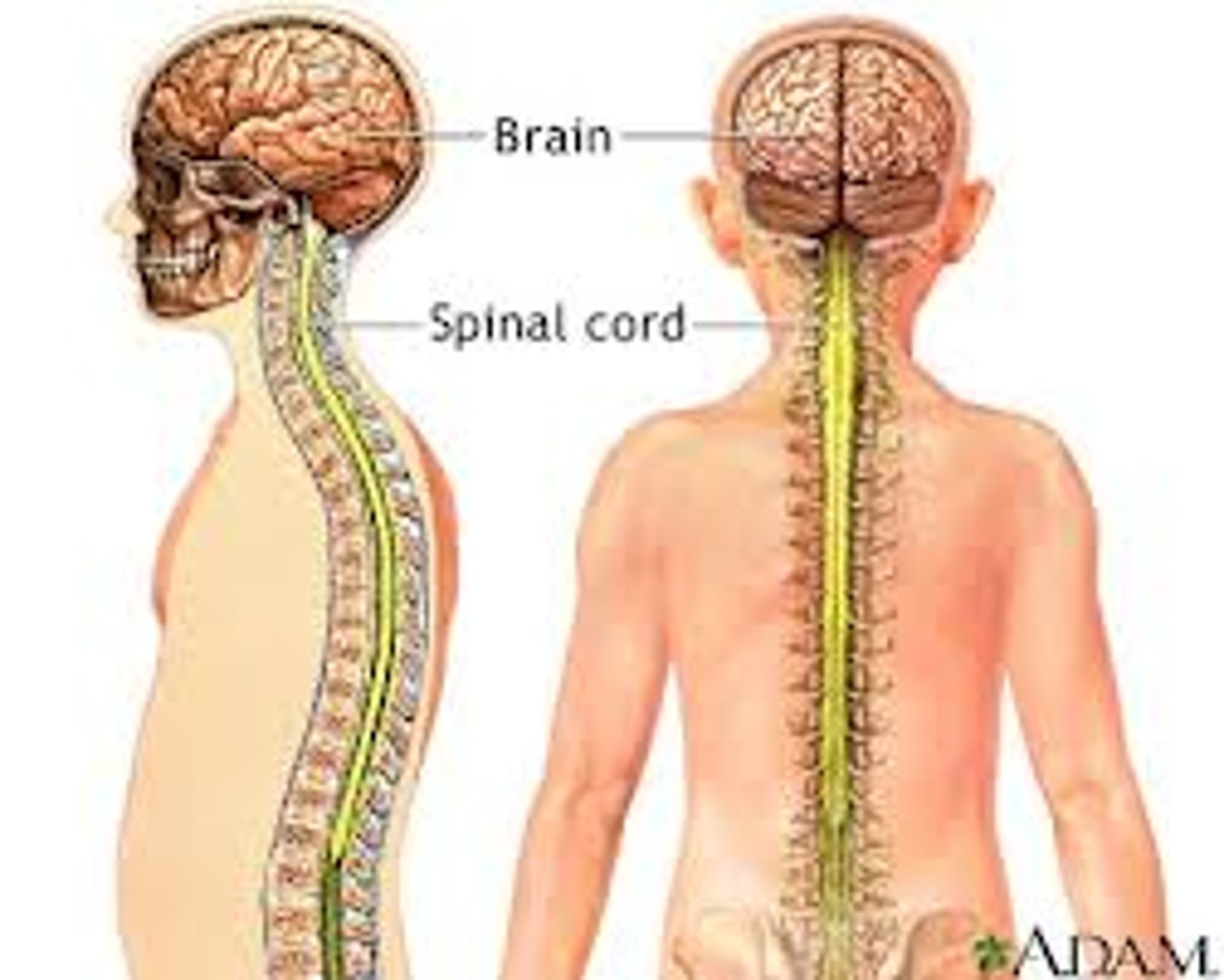
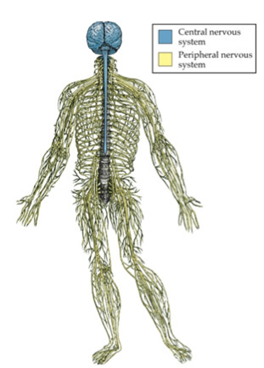
What does the central nervous system (CNS) consist of?
Brain and spinal cord within the dorsal body cavity, which are enclosed and protected by the cranium and vertebral column
What does the peripheral nervous system (PNS) consist of?
Consists of all the rest; it is composed of nerves outside of the dorsal body cavity
What 2 divisons is the PNS functionally divided into?
Sensory and motor divisions
Sensory (Aka afferent) Neurons
Information carried to the CNS (Brain)
Motor (Aka efferent) Neurons
Information carried from the CNS (Brain)
Also, within the CNS:
-Afferent information to the brain
-Efferent information from the brain
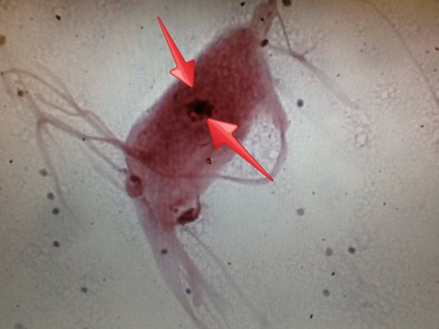
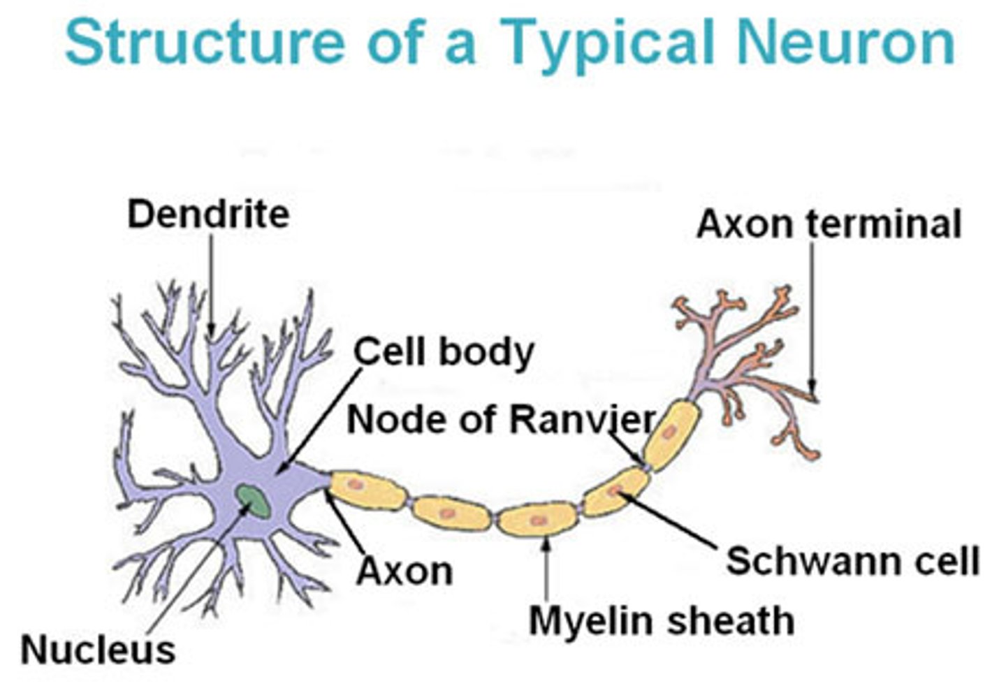
What is the control center of the neuron?
Neurosoma, also called the soma or cell body
The cell body (soma) has a centrally located nucleus. __________ __________ occurs within the cell body (soma).
Cell maintenance
Dendrites
Information input from other neurons are received through dendrites on the cell body (soma)
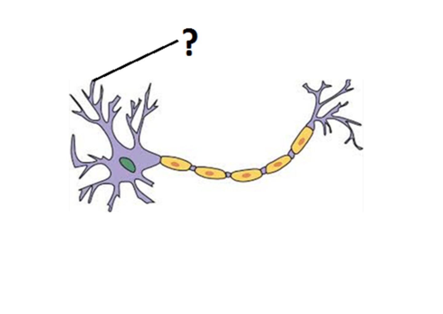
The more __________ a neuron has, the more information it can recieve and incorporate into its decision making.
dendrites
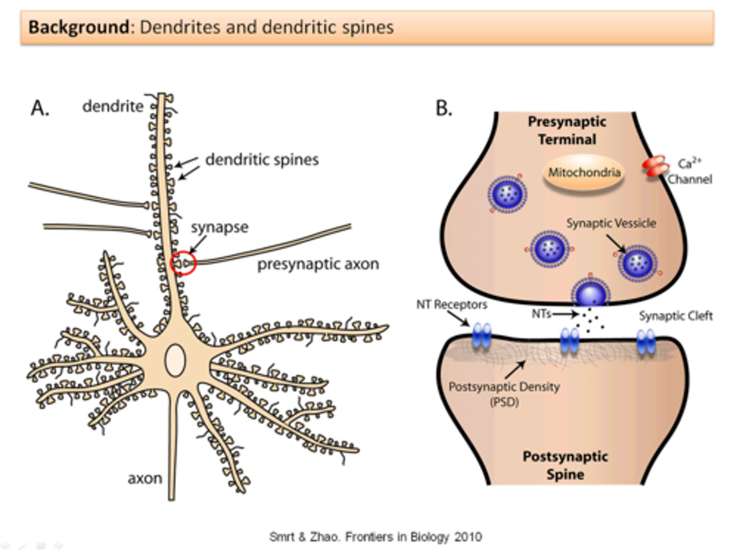
Dendritic Spines
-Small membranous bump/lump/knob from a neuron's dendrite that typically receives synaptic input from other cells
-Dendritic spines serve as a storage site because they can hold alot more
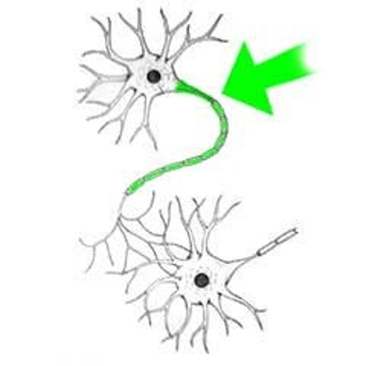
Axon
-Information output to other neurons flow along the cell body's (soma's) axon
-Located in middle of neuron, where the signal from AP is trasferred from the dendrite to the axon terminal
-AP going down
What does the trigger zone consist of on an axon?
-Axon Hillock- Part of the cell body (Soma) of a neuron that connects to the axon. Where the axon (nerve fiber) originates.
-Initial Segment- The short section of nerve fiber between axon hillock and the first glial cell
-They both play an important role in initiating a nerve signal
Action Potential
-Neurons way of transporting an electrical signal from one neuron to the next
-Creates excitation
Where does action potential generation (initiation) occur?
Trigger zone (Axon hillock and initial segment)
Myelin Sheath on Axon
A spiral layer of insulation around a nerve fiber formed by the oligodendrocytes in the CNS and Schwann cells in PNS

The myelin sheath is segmented. Each gap between segments is called a _____ _____ __________ or __________ __________ _____.
Node of Ranvier or myelin sheath gap
Do nodes of Ranvier have myelin?
No
Presynaptic Terminal (Knob) on Axon
Specialized area within the axon of the presynaptic cell that contains neurotransmitters enclosed in synaptic vesicles
At the distal end, an _____ usually has a terminal arborizaton-an extensive complex of fine branches.
axon
Each branch ends in a bulbous _____ __________, which forms a junction (__________) with the next cell.
axon terminal, synapse
Synapse
Intercellular communication