VT111 Lec. 7 Appendicular Skeleton
1/35
There's no tags or description
Looks like no tags are added yet.
Name | Mastery | Learn | Test | Matching | Spaced | Call with Kai |
|---|
No analytics yet
Send a link to your students to track their progress
36 Terms
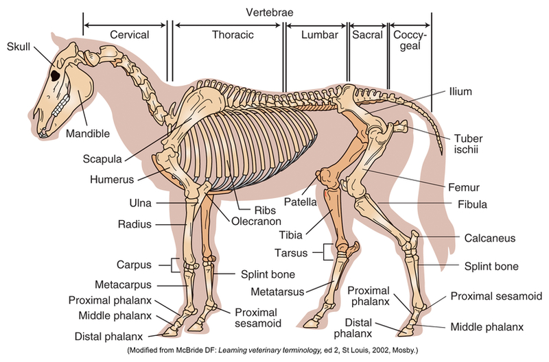
Appendicular Skeleton
Thoracic limb:
Scapula
Humerus
Ulna
Radius
Carpal bones
Metacarpal bones
Phalanges
Pelvic limb:
Pelvis
Femur
Patella
Fabellae
Tibia
Fibula
Tarsal bones
Metatarsal bonesPhalanges
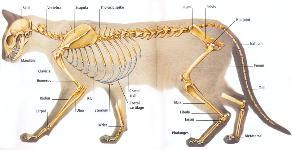
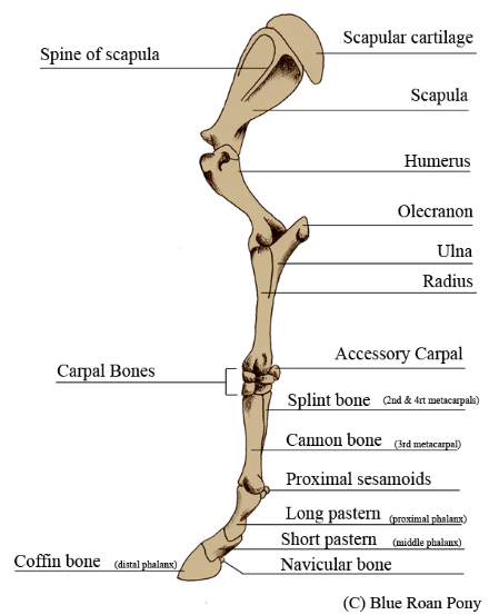
Thoracic Limb
No direct bony connection with axial skeleton
No clavicle to join the scapula with sternum
(Incomplete) Pectoral girdle
Complete is common only in primates also rabbits; not in dogs or cats
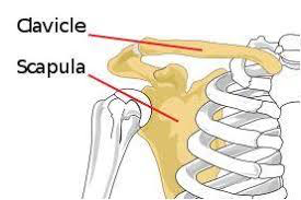
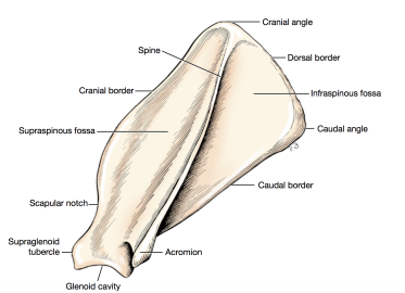
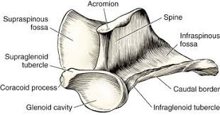
Scapula
Flat and triangular
Lateral surface
Spine
Distal end: acromion
Distal end
Glenoid cavity
Coracoid process
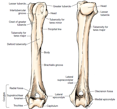
Humerus
Proximal:
Head → scapula
Tubercles: greater and lesser
Middle:
Shaft (diaphysis)
Distal:
Condyle
Trochlea (medial) → ulna
Capitulum (lateral) → radius
Epicondyles: medial and lateral
Supratrochlear foramen
Unique to dog and cat
Olecranon fossa
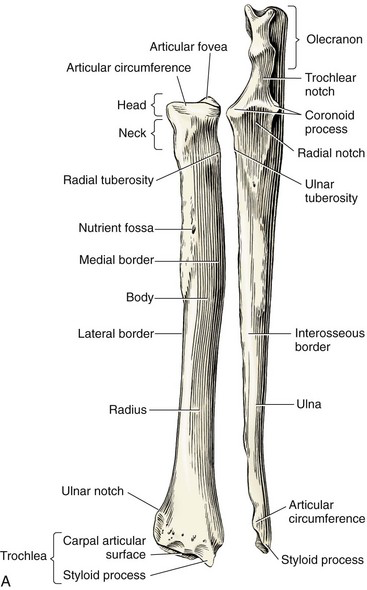
Ulna
In dogs/cats, starts medially and ends laterally
Olecranon process
Point of elbow
Attachment site of tendon of triceps brachii
Trochlear notch
Anconeal process
Fits into olecranon fossa during extension
Coronoid processes: medial and lateral
Styloid process (distal): lateral
Horse and cow
Ulna partially fused to radius (incomplete)
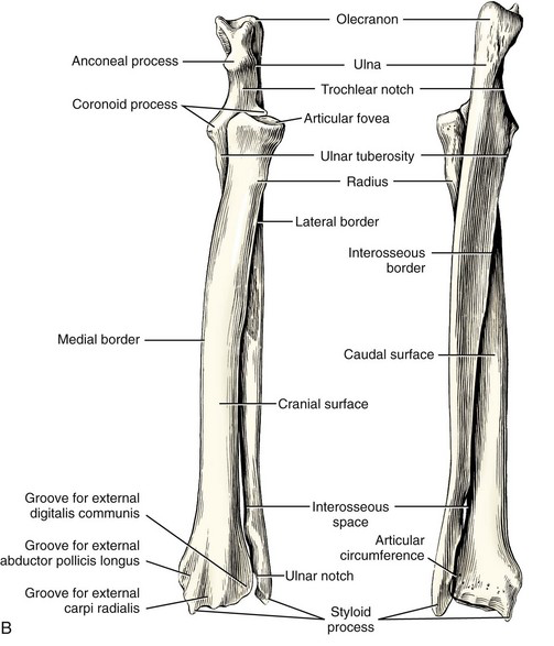
Radius
Main weight bearing bone
Head
Radial tuberosity
Medial
Styloid process: medial
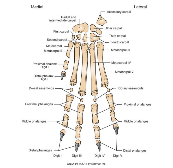
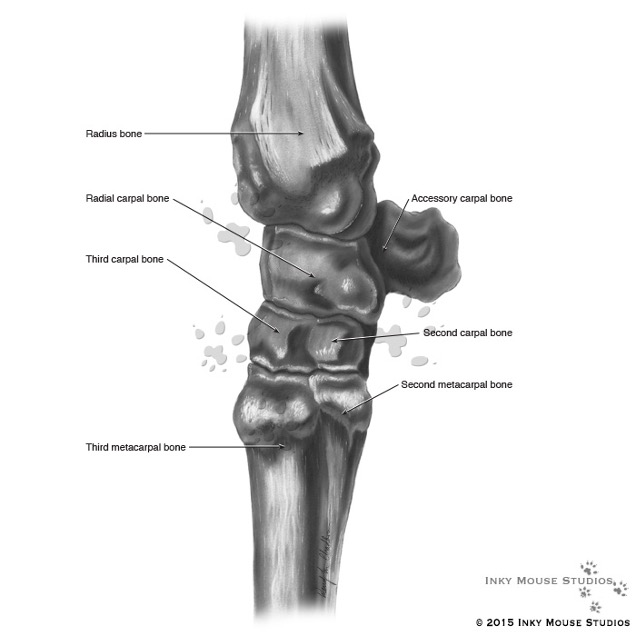
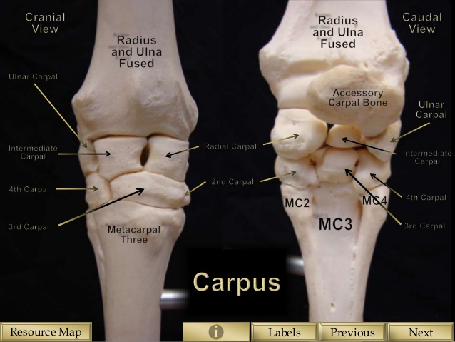
Carpal Bones
Carpus = wrist
Horse = knee
Variety among species
Humans and pigs have 8 bones
2 rows of bones
Proximal
Radial carpal bone: medial
Ulnar carpal bone: lateral
Accessory carpal bone: lateral
Distal
Numbered, starting at medial side
(I – IV)
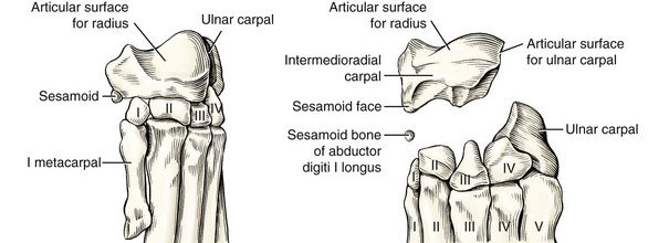
Metacarpals
Numbered from medial (MC I = dewclaw) to lateral (MC V)
Horse: one toed
Only one metacarpal is weight bearing
MC III = canon bone
Bracketed by vestigial metacarpal bones MC II and MC IV (splint bone; incomplete)
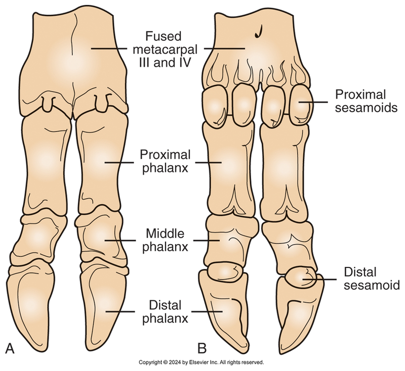
Cattle: two toed
Weight bearing on fused MC III and MC IV
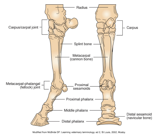
Phalanges
Digits
Each composed of 2-3 bones (phalanges)
Proximal, middle, distal (P1, P2, P3)
Distal ends in ungual process (claw, nail)
Sesamoid bones (inside of tendons)
Location and number vary among species
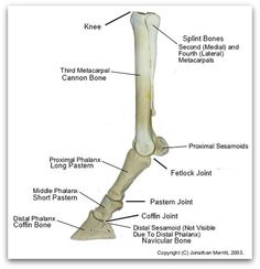
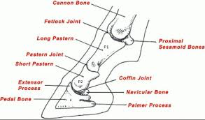
Horses – only 1 digit
Long pastern (P1) (proximal)
Short pastern (P2) (middle)
Coffin bone (P3) (distal)
Sesamoids:
proximal at fetlock joint
distal inside of coffin bone (navicular bone)
Cattle: 4 digits
3rd and 4th support body weight
2nd and 5th vestigial = dewclaws
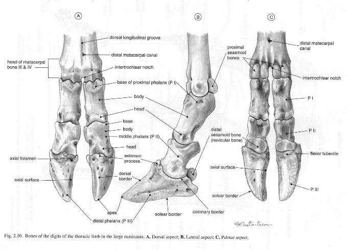
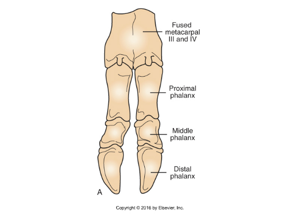
Thoracic Limb Bones of Cattle
Two fused metacarpal bones
Metacarpal III
Metacarpal IV
Four digits
2 weight-bearing
2 dewclaws
Sesamoid bones
2 proximal
1 distal
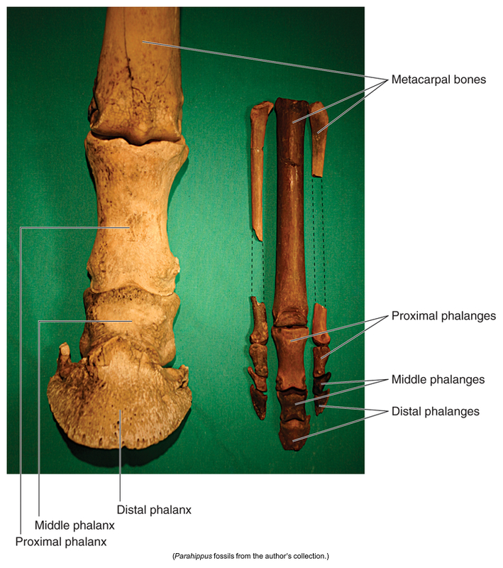
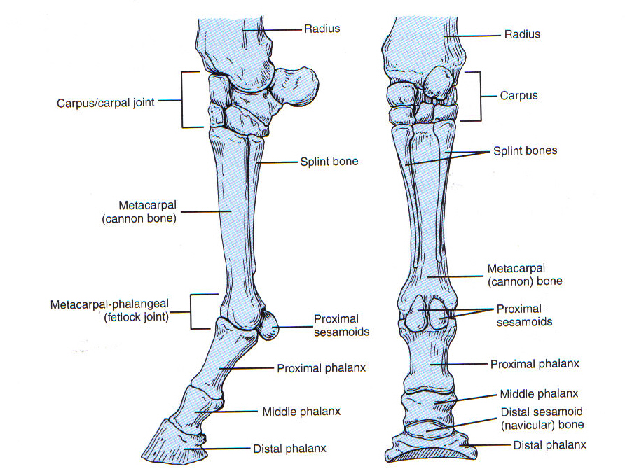
Equine Thoracic Limb Bones
1 digit with 3 phalanges
Long pastern bone (proximal phalanx)
Short pastern bone (middle phalanx)
Coffin bone (distal phalanx)
3 sesamoid bones
2 proximal at the fetlock
1 distal = navicular
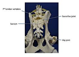
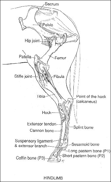
Pelvic Limb
Directly connected to axial skeleton via sacroiliac joint
Ilium of pelvis with sacrum of spinal column
Consists of:
Pelvis
Femur
Patella
Tibia
Fibula
Tarsal bones
Phalanges
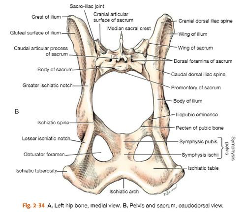
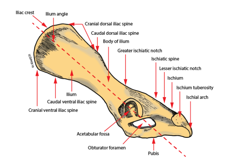
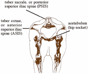
Pelvic Girdle
Pelvis (os coxae) is actually 3 bones on each side that are fused
Ilium
Wing
Cattle/horse: processes
Medial: tuber sacrale – part of sacroiliac joint
Lateral: tuber coxae – point of hip
Ischium
Caudal most part of pelvis
Ischial tuberosities = Sitz bones
Pubis
Medial
Obturator foramen
2 sides of pelvis joined with fibrocartilage ventrally by pelvic symphysis
Union between ilium and sacrum is a
fibrocartilaginous joint – sacroiliac joint
3 bones come together at acetabulum
Encloses head of femur to make coxofemoral (hip) joint
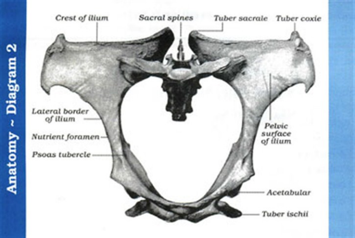
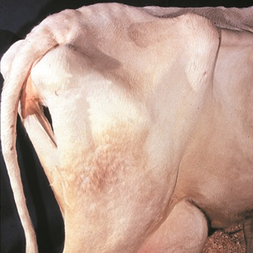
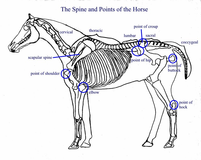
Horse Pelvis
Tuber sacrale
Point of croup
Tuber ischii
Point of buttock
cow: pin
Tuber coxae
Point of hip
cow: hook
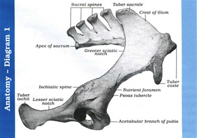
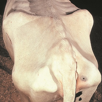
Cattle Pelvis
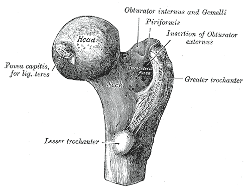
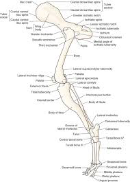
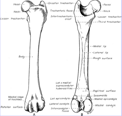
Femur
Head of femur
Ball-and-socket coxofemoral (hip) joint
Attached to shaft by neck
Greater and lesser trochanter
Fovea capitis
pit in head of femur where ligamentum teres (round ligament) inserts
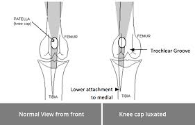
Distal:
Trochlea (patellar groove)
Surface which patella glides against
Medial and lateral condyles
Medial and lateral epicondyles
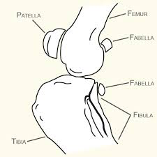
Patella and Fabellae
Sesamoid Bones
Largest sesamoid:
Patella
In distal tendon of quadriceps femoris muscle
Helps to protect tendon
Fabellae (“little beans”)
Caudal to joint: 2 in proximal gastrocnemius (calf muscle) muscle tendons behind femoral condyles (lateral > medial)
Caudal to joint: 1 in tendon of popliteus (near lateral condyle of tibia)
Present in dogs and cats
10-30% humans
Not present in cattle or horses
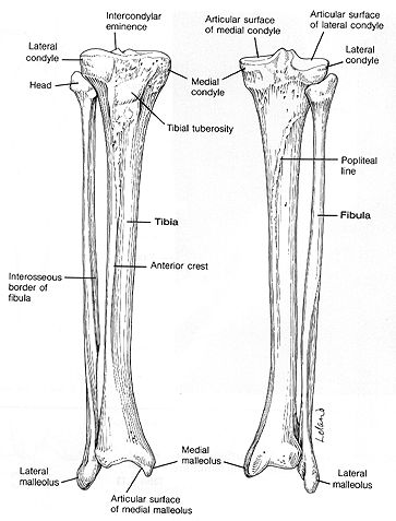
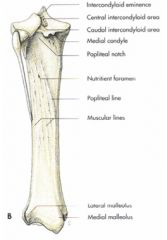
Tibia
Main weight bearing bone of lower leg
Stifle joint - tibia - hock joint
Condyles
Intercondylar eminence
Tibial tuberosity
Attachment for patellar ligament
Tibial ridge/(anterior)crest
Medial malleolus
Medial ankle “knob”
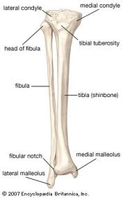
Fibula
Lateral structure
Proximal extremity
Fibular head
Shaft
Distal extremity
Lateral malleolus
Lateral “knob” of ankle
Shaft not present in horse and cattle
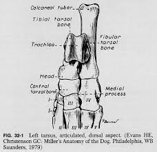
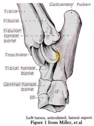
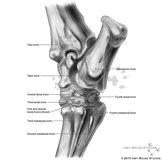
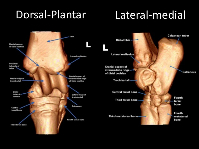
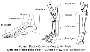
Tarsal Bones
Tarsus – dog 7 bones
Human = ankle
Domestic animals = hock
Proximal row (3 bones)
Tibial tarsal bone - medial
Large trochlea articulates with tibia
Fibular tarsal bone - lateral
Calcaneal tuberosity (human heel)
Attachment for tendon of gastrocnemius muscle
Central tarsal bone
Distal row
Numbered medial to lateral
(I – IV)
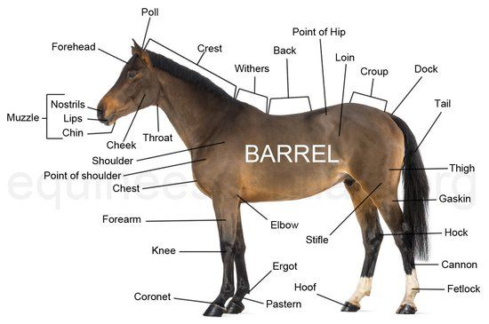
The Horse
Labeled Horse
The Horse: Carpus
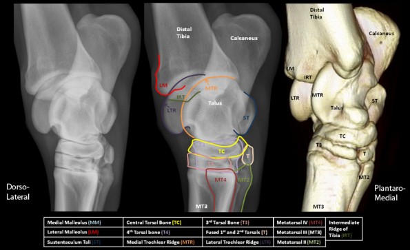
The Horse: Tarsus
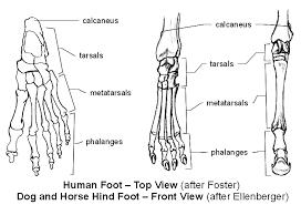
Phalanges
Similar to forelimb
Dogs/cats usually only have 4
Digits II, III, IV, V
The Horse
Equine Terminology
Forelimb:
Knee: carpus
Fetlock: metacarpal-phalangeal joint
Pastern: the part of the foot extending from the fetlock to the top of the hoof
Pastern joint: P1-P2
Coffin joint: P2-P3
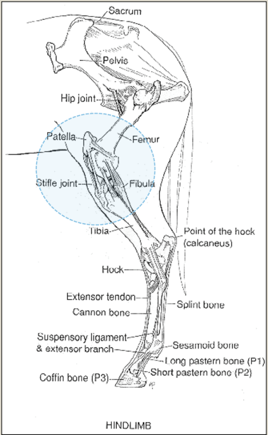
Hindlimb:
Stifle: the femoral-tibial joint (aka your knee)
Hock: the tarsus
Fetlock: metatarsal-phalangeal joint
Pastern: the part of the foot extending from the fetlock to the top of the hoof
Pastern joint: P1-P2
Coffin joint: P2-P3
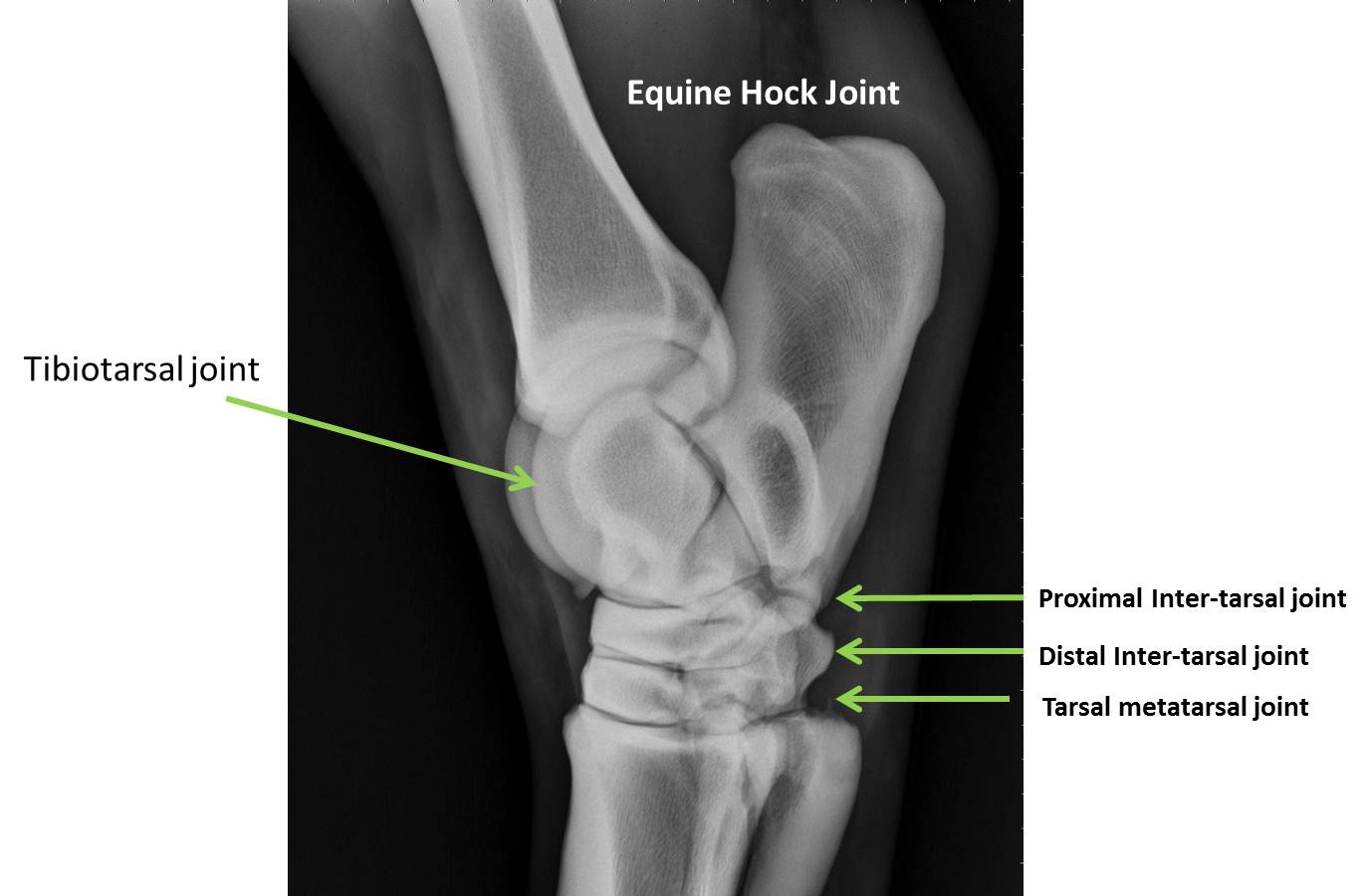
Equine Hock Joint
Horse Hindlimb
Visceral Skeleton (random bones)
Bones that form in soft organs
Os cordis
Ruminants
Supports valves of heart
Os penis
In penis of dog, beaver, walrus, chimps
Os rostri
Swine
Strengthens the snout: rooting
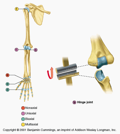
Hinge Joint
Pulley shaped connection fits into concave space
Works exactly like a hinge
Flexion/extension
Ex: elbow
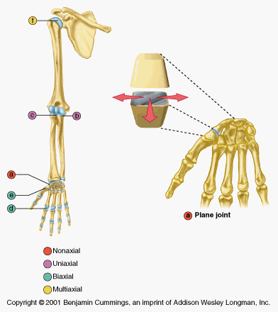
Gliding/Plane Joint
Flat articular surfaces
Sliding movements
Range of motion is limited in these joints and does not involve rotation
Ex: carpal bones
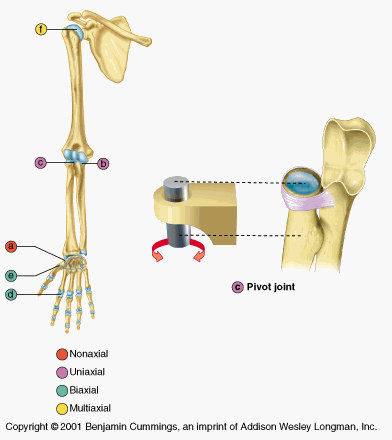
Pivot Joint
Rounded end of one bone fits into a “sleeve” of either bone or ligament
Rotation movement
Ex: atlantoaxial joint
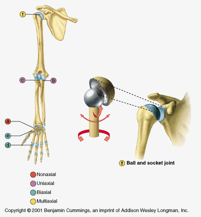
Ball and Socket Joint
Spherical head of one bone fits into cup of another.
Movement in many planes
Greatest range of motion, as all movement types are possible in all directions
Extreme freedom of movement
Ex: coxofemoral joint; scapulohumeral joint
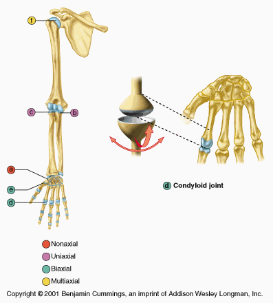
Condyloid Joint
Also called ellipsoidal joint
Oval surface fits into depression (fossa) of another bone
Allows angular movement along two axes
Ex: metocarpophalangeal joint
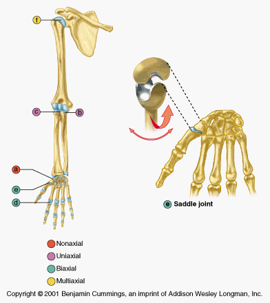
Saddle Joint
Both ends have concave and convex surfaces for increased freedom of movement
Saddle joints allow angular movements similar to condyloid joints but with a greater range of motion
Ex: carpometacarpal joint (thumb)