Embryology 2 Head and Neck - Basic Science Core
 Call Kai
 Learn
Learn Practice Test
 Spaced Repetition
 Match
Match Flashcards
 Knowt Play
1/43

Earn XP
Description and Tags
Basic Science Core
Name | Mastery | Learn | Test | Matching | Spaced | Call with Kai | Chat |
|---|
No analytics yet
Send a link to your students to track their progress
44 Terms
Gastrulation week three
Cells of the epiblast begin to invaginate
1st wave of cells migrating through the streak interdigitate with the hypoblast layer → ENDODERM
2nd wave → MESODERM
Remaining cells in the epiblast layer for the → ECTODERM
The Notochord
Originates from mesoderm
Some mesoderm cells condense and form the notochord
The notochord is essential for guiding adjacent cell differentiation
Rod like structure
Formed on the dorsal side
Present during Embryonic development
Know as the back bone
Mesoderm cells differentiate into specialized types (4)
Paraxial mesoderm
• Intermediate mesoderm
• Lateral plate mesoderm
• Extraembryonic mesoderm
Paraxial mesoderm
Closest to the notochord
Forms structures of the head and somites
3 pairs form per day from
cranial to caudal direction
• 42-44 pairs total
• Resulting structures
Vertebrae, ribs, rib tendons
Muscles of the back, ribs, and
limbs
Parts of the dermis
How does the Notocord Effect Endoderm?
Notochord signals neural tube formation and results in the ectoderm dividing into 3 sets of cells:
Neural tube
• Neural crest cells
HIGHLIGHT
Cranial neural crest cartilage, bone, cranial ganglia &
connective tissue of the face
• Epidermis
Main event during head and neck development:
FORMATION OF PHARYNGEAL APPARATUS
PHARYNGEAL APPARATUS
Consists of Pharyngeal (Branchial) ARCHES, POUCHES, and CLEFTS
Appear in the 4th-5 th week of development
Consist of 5 paired prominences with 3 layers:
an outer covering of ectoderm
an inner covering of endoderm
A middle core of mesenchyme
Pharyngeal Apparatus Morphology
1) Pharyngeal clefts derive from the ectoderm
2) Pharyngeal pouches arise from endoderm cells
3) Pharyngeal arches arises from mesoderm +
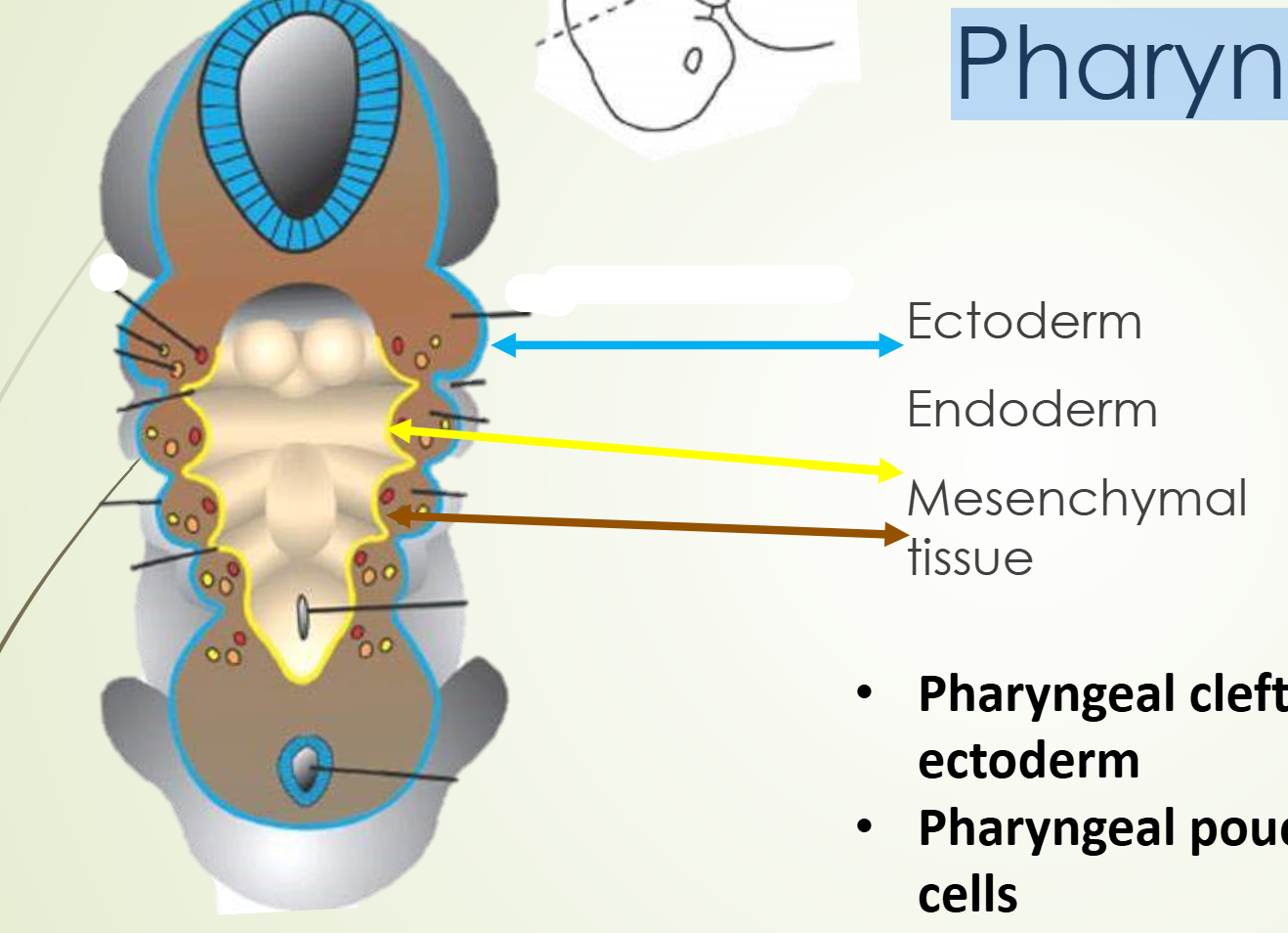
Pharyngeal arches (6)
Mesoderm tissue
No “5th” arch!/Fifth arch is transient
Involutes and produces no adult structures
Each arch has core of mesenchyme that develops into arterial supply, nerve supply, cartilage, musculature & bone
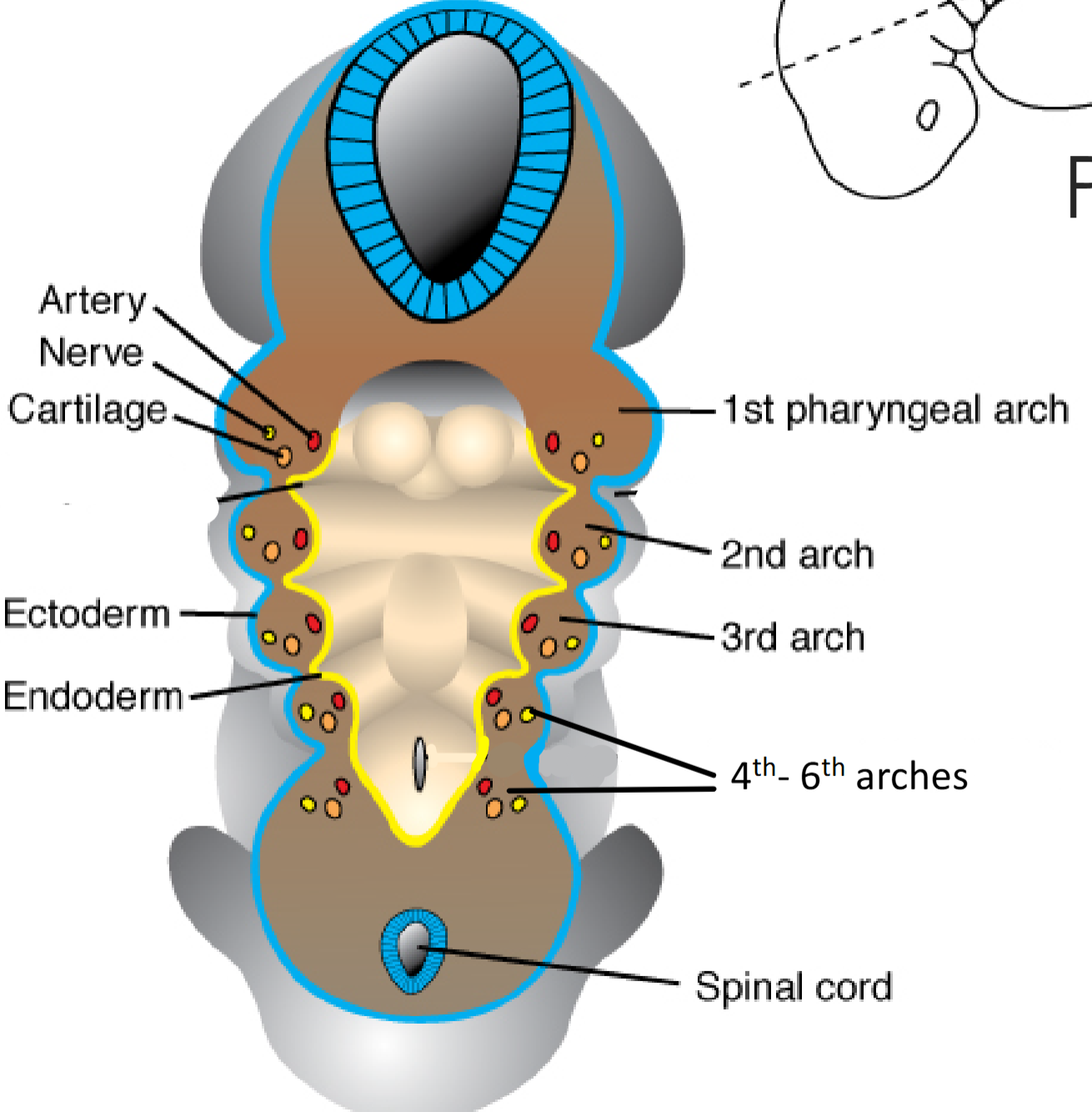
Mesenchyme
Mesenchyme is an embryonic precursor tissue that generates a range of structures in vertebrates including cartilage, bone, muscle, and the erythropoietic system
Major cell contributors to the mesenchyme of head and neck development:
Praxial and lateral plate mesoderm
Neural crest cells
Thickened regions of ectoderm called ectodermal placodes
Cranial neural crest cells
Migrate in the pharyngeal arches and face
The face is initially made up of five prominences (5)
1 central frontonasal prominence
2 paired maxillary prominences
2 paired mandibular prominences
Stomodeum
Structures surround the primitive mouth
Stomodeum is initially sealed by the oropharyngeal membrane which breaks down ~day 30
The 1st pharyngeal arch
Mandibular Arch
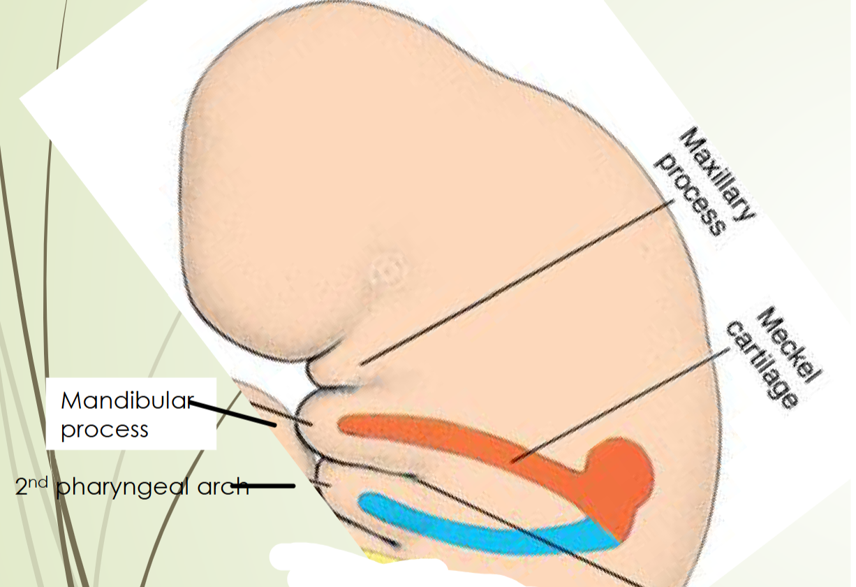
Maxillary and Mandibular prominences arise from the 1st pharyngeal arch
The 1st pharyngeal arch (2 Processes)
1) Maxillary process
2) Mandibular process
Maxillary process
Mesenchyme here → premaxilla, maxilla, zygomatic bone and part of the temporal bone
Mandibular process
Contains Meckel cartilage
Mesenchyme here → mandibular bone, part
of external ear, incus and malleus bones of the
middle ear and external auditory meatus
Meckel cartilage
In the Mandibular process
Contributes to incus and malleus and serves as a template for mandible formation
Muscles of the 1st pharyngeal:
Masseter,
Temporalis, Lateral
pterygoid, Medial pterygoid], anterior belly of the
digastric, mylohyoid, tensor tympani, and tensor
palatini
![<ul><li><p>Masseter, </p></li><li><p>Temporalis, Lateral</p></li></ul><p>pterygoid, Medial pterygoid], anterior belly of the</p><p>digastric, mylohyoid, tensor tympani, and tensor</p><p>palatini</p>](https://assets.knowt.com/user-attachments/832cd8fc-d4af-45a5-bcda-3100075a2cef.png)
Nerve supply of the 1st pharyngeal:
motor and sensory from branches of Trigeminal nerve (CN V)
The 2nd pharyngeal arch
The HYOID arch
Has Reichert cartilage
Precursor to the stapes, the styloid process,
the stylohyoid ligament and parts of the hyoid
bone & most of external ear
Muscles of the 2nd pharyngeal:
stapedius,
stylohyoid, posterior belly of the
digastric, auricular, and muscles of
facial expression
Nerve supply of the 2nd pharyngeal:
Nerve supply: Facial nerve (CN 7)
The 3rd pharyngeal arch
Small components of the hyoid bone
Muscles of the 3rd arch: (NOT CRITICAL TO memeroize)
stylopharyngeus muscle
Muscle of the pharynx
Functions in swallowing
Nerve supply of the 3rd pharyngeal arch
glossopharyngeal nerve (CN IX)
The 4th & 6th pharyngeal arches
Cartilaginous components of the fourth and
sixth pharyngeal arches fuse to form:
• Thyroid, cricoid, arytenoid, corniculate, and
cuneiform CARTILAGES of the larynx
Muscles of the fourth arch
cricothyroid, levato veli palatini,
and constrictors of the pharynx
Nerves of the fourth arch
Nerve: branches of Vagus (CN X)
The pharyngeal pouches
Lined by epithelial from the endoderm layer
4 pairs of pharyngeal pouches:
1) 1st pharyngeal pouches
2) 2nd pharyngeal pouches
3) 3rd pharyngeal pouches
4) 4th pharyngeal pouches
1st pharyngeal pouches
Structures arising from 1st pouch:
• Middle ear cavity
• Auditory (eustachian) tube
• Tympanic membrane (eardrum)
2nd pharyngeal pouches
Endoderm of the pouch
proliferates and forms buds that
penetrate surrounding
mesenchyme
• Mesodermal tissue enters buds and
forms palatine tonsil
• Months 3-5 tonsil is infiltrated by
lymphatic tissue
• Remnants of pouch create tonsillar
fossa
4th pharyngeal pouches
Dorsal region of the
fourth pharyngeal
pouch superior
parathyroid gland
Ventral region
Ultimobranchial body
• Ultimobranchial body later
incorporated into the thyroid
gland
• Cells of the ultimobranchial
body gives rise to
the parafollicular cells (C
cells) of the thyroid gland
that secrete calcitonin
3rd pharyngeal pouches
Structures arising from the 3rd
pouch:
• Dorsal region of the third pouch
differentiates into the inferior
parathyroid gland
• Ventral region of the third pouch
differentiates into the thymus
Pharyngeal Clefts (4)
1 st cleft external auditory meatus (No permejnt structures)
Second third and fourth clefts merge and form the
cervical sinus
Cervical Sinus
Second third and fourth clefts merge and form the
cervical sinus
• Cervical sinus normally disappears during development
Development of the TONGUE
Tongue appears in the embryo ~4 weeks
• Origins of tissue for the tongue:
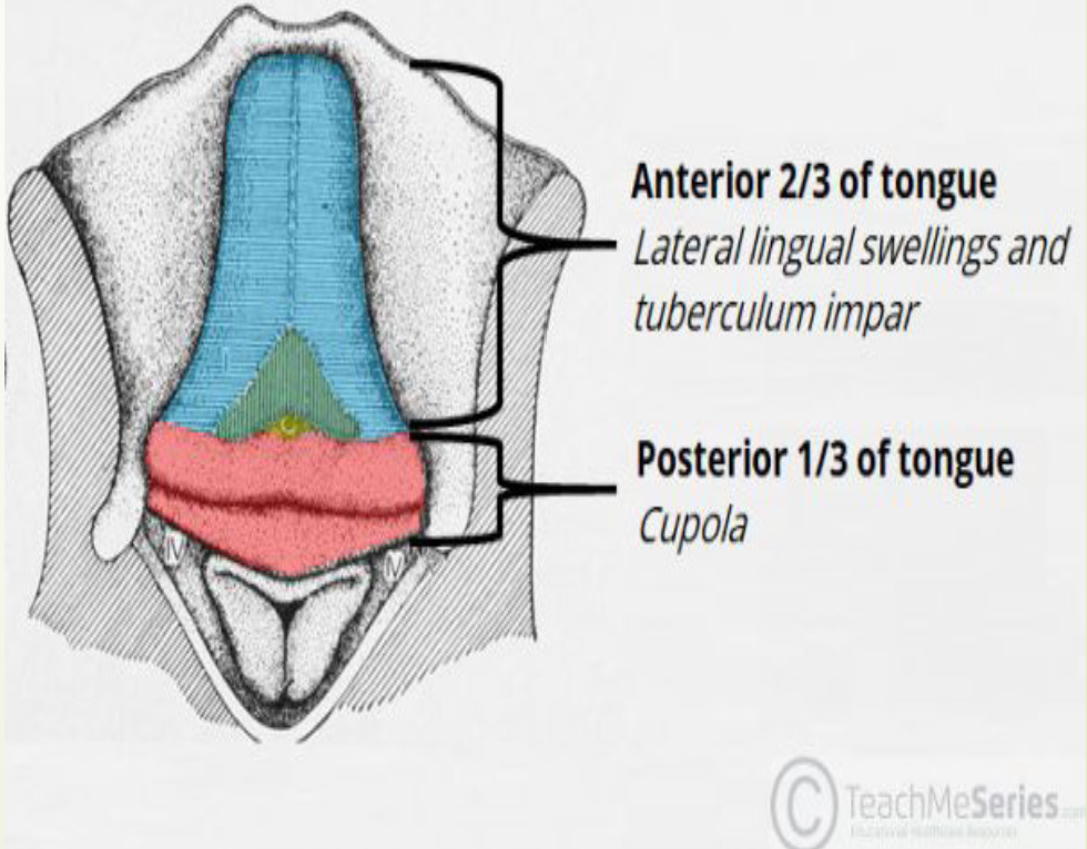
FROM FIRST ARCH: formation of anterior 2/3 of tongue aka
body of the tongue
• 2 lateral swellings: Lingual swellings
• 1 medial swelling: Tuberculum impar
FROM SECOND, THIRD, & FOURTH ARCH: posterior 1/3
tongue
• 1 medial swelling: Copula [hypobranchial
eminence
Development of the TONGUE PT 2
Anterior 2/3 of the tongue
Sensory innervation is a branch of CN V because these
structures originate from 1 st pharyngeal arch
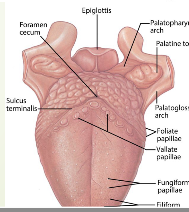
Anterior 2/3 is separated from posterior
1/3 by a V-shaped groove called
terminal sulcus
Posterior 1/3 of the tongue
• Sensory innervation by glossopharyngeal nerve,
CN IX (supplies 3rd pharyngeal arch)
Additional special sensory innervation (taste)
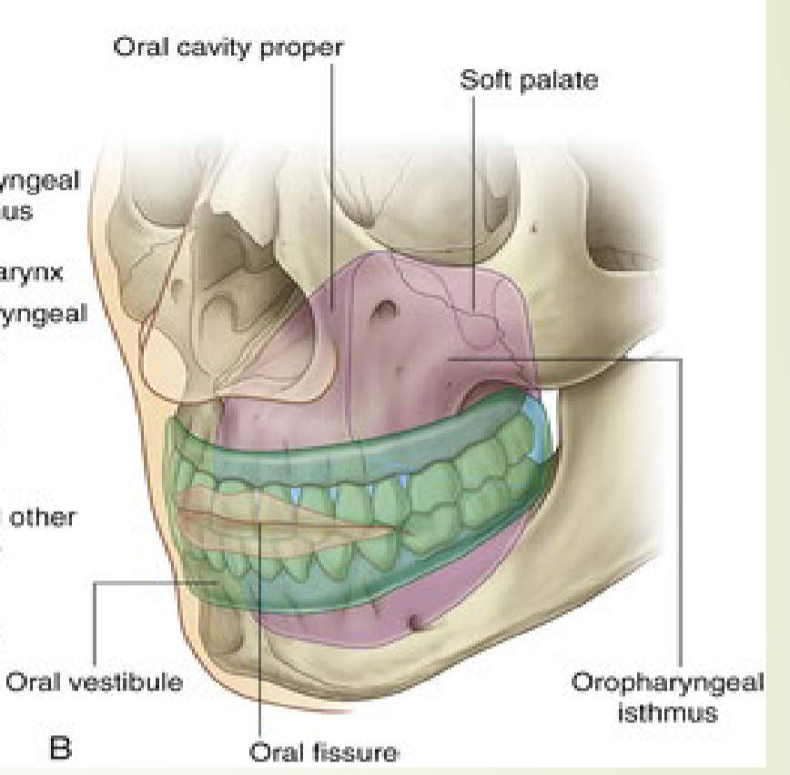
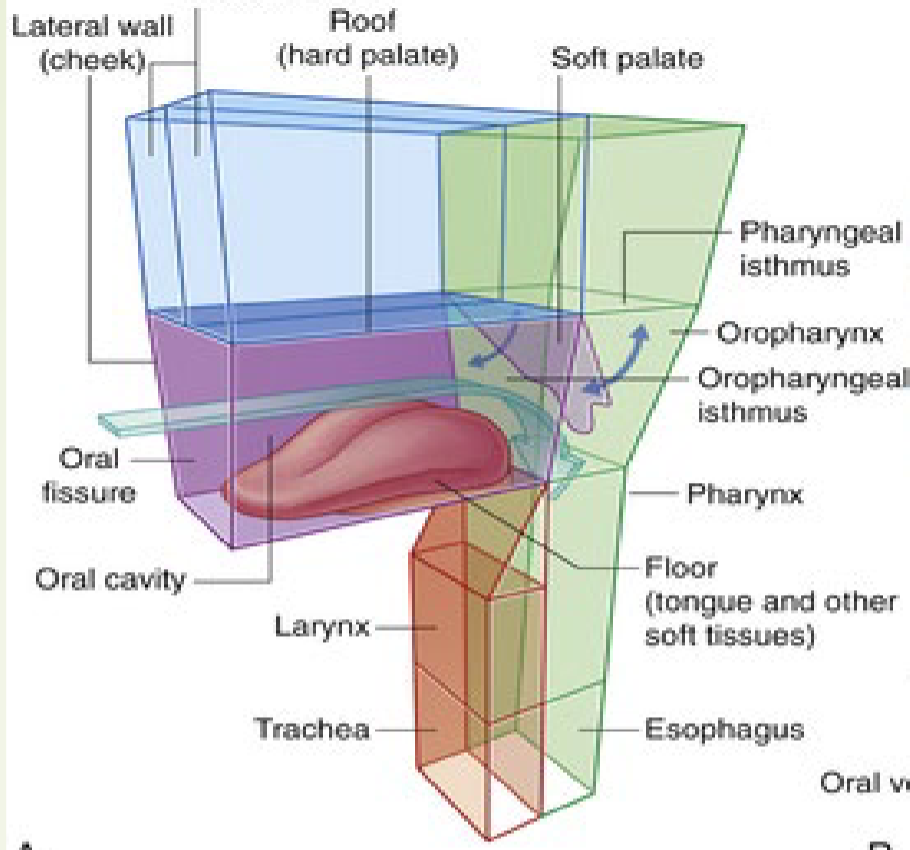
Oral cavity proper versus Oropharynx
The oral cavity is inferior to the nasal cavities ( Fig. 8.247A ). It has a roof and floor
and lateral walls, opens onto the face through the oral fissure, and is continuous with
the cavity of the pharynx at the oropharyngeal isthmus.
The roof of the oral cavity consists of the hard and soft palates. The floor is formed
mainly of soft tissues, which include a muscular diaphragm and the tongue. The lateral
walls (cheeks) are muscular and merge anteriorly with the lips surrounding the oral
fissure (the anterior opening of the oral cavity).
The posterior aperture of the oral cavity is the oropharyngeal isthmus, which opens
into the oral part of the pharynx.
The EPIGLOTTIS
Originates from tissue of 4th pharyngeal arch
Originates from Epiglottal swelling, swelling posterior to copula (posterior 1/3 of tongue)
Innervation: branch of Vagus nerve, CN X
Development of the THYROID
Tissue for thyroid gland originates from:
Endoderm between first and second pharyngeal pouches
• Development steps
Proliferation begins at junction of anterior 2/3 tongue and
posterior 1/3 of tongue
• Leaves behind a depression called foramen cecum
Thyroid descends from tongue to final position in the neck ~7th
week
Thyroid remains connected to the tongue by a narrow canal,
the thyroglossal duct which should disappear during
development
• Gland begins to function at approximately the end of the
third month (10-12 weeks)
Birth defects & the Pharyngeal Apparatus WILL NOT BE ON THE EXAM