Bio 210: Epidermis, Dermis, Hypodermis
1/20
There's no tags or description
Looks like no tags are added yet.
Name | Mastery | Learn | Test | Matching | Spaced |
|---|
No study sessions yet.
21 Terms
Integumentary System: Skin
- Body’s largest and heaviest organ
- Consists of two layers: Epidermis and Dermis
- Accessory Organs: Hair, Nails, and Glands
- Functions: protection (abrasion, dehydration, UV exposure, pathogens), sensation (pressure, touch, temperature, pain), thermoregulation, vitamin D production
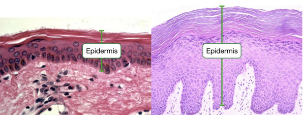
Epidermis
Superficial
4-5 layers of Stratified Squamous Epithelial tissue
Avascular and waterproof (keratin)
Resists abrasion, prevents dehydration
Stratum basale
“base layer”
Primarily basal cells (mitotic cells)
A single layer of cuboidal to columnar cells
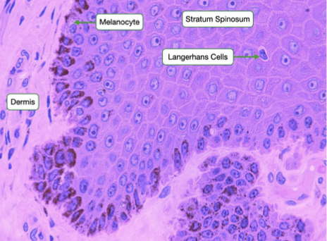
Melanocytes and Merkel cells
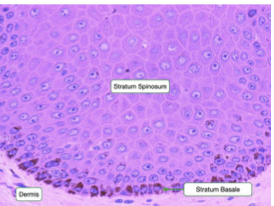
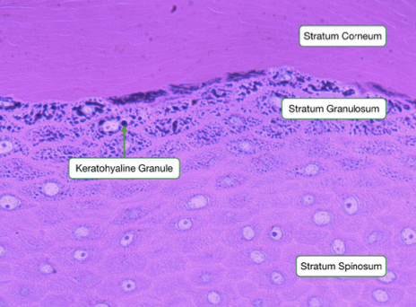
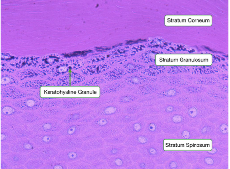
Stratum spinosum
“Spiny layer”
Thicker (8-10 layers)
Keratinocytes begin dying
Desmosome connections: keratinocytes shrink during fixation
Langerhans cells
Stratum granulosum
“Granular layer”
Thinner (3-5 layers)
Granules
Keratinocyte death: start to lose their organelles; cell membranes thicken
Stratum lucidum
“clear layer”
found only in thick skin
translucency
lack cellular components (organelles)
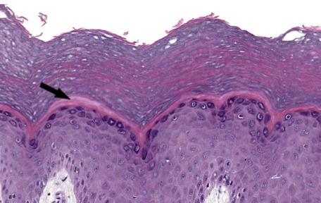
Stratum corneum
“corny layer”
corneocytes
Many layers (25+)
Brick-and-mortar architecture
bricks of keratin
lipids form the mortar (cement)
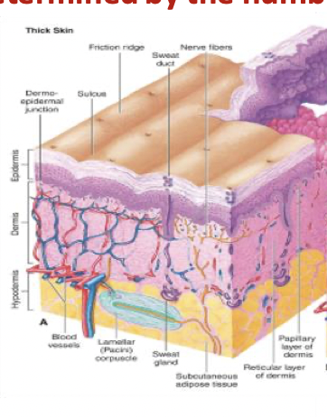
Thick Skin
All 5 epithelial strata (contains stratum lucidum)
Found in areas subject to pressure/friction (palms, fingertips, soles of feet)
Structure: contains friction (papillary) ridges; lacks hair follicles, sebaceous glands, and arrector pili
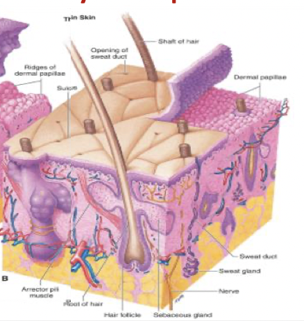
Thin Skin
Only 4 epithelial strata (no stratum lucidum)
Found everywhere that thick skin is not present
Structure: Contains hair follicles, sebaceous glands, arrector pili muscles
Keratinocytes
main type of cell in epidermis
produce keratin (fibrous protein that helps give the epidermis its protective properties)
Corneocytes
make up most of the outer layer of the skin
Anucleate, flattened, and dead keratinocytes
Melanocytes
produce melanin for skin pigmentation
Merkel cells
light touch receptors
found in stratum basale layer
Dendritic (Langerhans) cells
boosts immune functions
macrophage-like function: kills microorganisms, removes dead cells, and stimulates other immune system cells
Keratinization
Increased levels of keratinization coincide with cellular death
As cells move superficially, they become more keratinized
Function: resist abrasion, prevent dehydration
Desquamation
Skin slough/”skin peeling”
Shedding the outermost layer of tissue
Skin pigmentation
Melanin (eumelanin: black/brown pigment)(Peomelanin: red pigment)
Carotene: yellow/orange pigment
Hemoglobin: red pigment (with oxygen) and Blue pigment (without oxygen)
Dermis
- “True Skin:” core layer of cutaneous membrane
- Made up of Dense Irregular Connective Tissue
- Function: gives the skin strength and structural support
- Two layers: papillary and reticular
- Vascularized and derived from mesoderm
Papillary layer
Loose areolar connective tissue = mesh network
Dermal papillae: form friction ridges with epidermis
Extensive capillary network
Meissner’s (tactile) corpuscles: lighter touch.
Reticular
Dense irregular connective tissue allows for strength and elasticity
Thicker: 80% of the dermis
Anchor for glands and hair follicles
Pacinian (lamellar) corpuscles: strong pressure/vibrations.
Hypodermis
“Subcutaneous Membrane”
NOT part of the cutaneous membrane
Serves to connect skin to underlying fascia
Made up of loose connective tissue
High adipose content (shock absorption and insulation
Vascularized