Eye and Vision
 Call Kai
 Learn
Learn Practice Test
 Spaced Repetition
 Match
Match Flashcards
 Knowt Play
1/47

Earn XP
Description and Tags
lecture: 4/5
Name | Mastery | Learn | Test | Matching | Spaced | Call with Kai | Chat |
|---|
No analytics yet
Send a link to your students to track their progress
48 Terms
I. The Eyes & Vision — General
Approximately what percentage of the body's sensory receptors are in the eyes?
What is the approximate size of the eyeball?
Approximately 70%
Approximately 1 inch in diameter
What protects the eyeball?
Approximately how much of the cerebral cortex is used to process visual information?
A bony orbit surrounded by a layer of fat
Approximately half of the cerebral cortex
II. Accessory Structures
A. Eyebrows
Where are the eyebrows located?
What are the eyebrows?
What are two functions of the eyebrows?
On the supraorbital margins
Short, coarse hairs
They protect against sunlight by providing shade and block sweat
B. Eyelids
What is another name for the eyelids?
What separates the eyelids?
What are the two ends of the eyelids called?
What does the medial commissure contain?
Palpebrae
The palpebral fissure
The lateral and medial commissures
The lacrimal caruncle
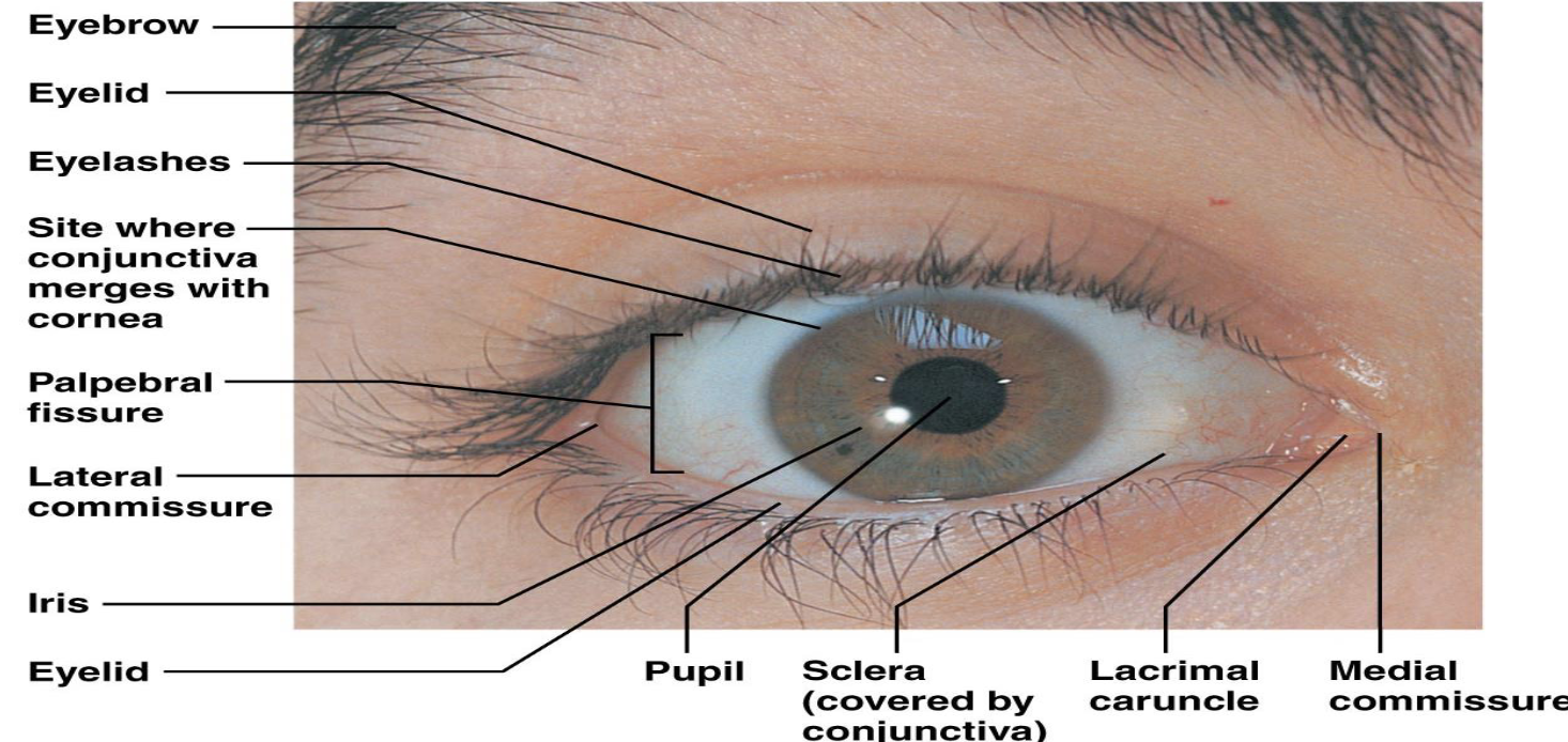
C. Eyelashes
Where are the eyelashes located?
What is special about the innervation of the eyelashes?
What does stimulation of the eyelashes trigger?
On the margins of both eyelids
They are richly innervated
Blinking

D. Conjunctiva
What is the conjunctiva?
What does the conjunctiva secrete?
A mucus membrane
Mucus

E. Lacrimal Apparatus
Where are the lacrimal glands located?
How do lacrimal glands release their secretions?
What happens when you blink according to the lecture?
Over the lateral part of the eye
Through excretory ducts
Blinking spreads the lacrimal secretions across the eye toward the medial commissure
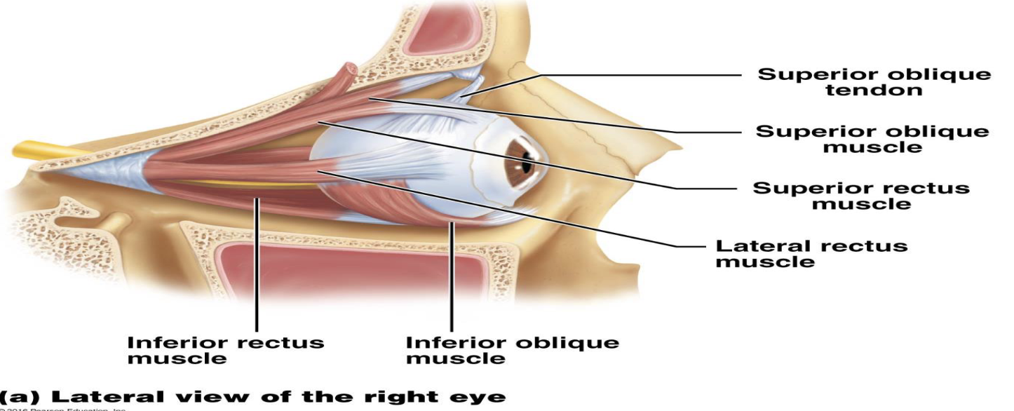
III. Extrinsic Eye Muscles
What are the functions of the extrinsic eye muscles?
What are the four extrinsic eye muscles listed in this lecture?
They allow the eye to track moving objects, maintain shape, and maintain direction of movement
Lateral rectus
medial rectus
superior rectus
inferior rectus
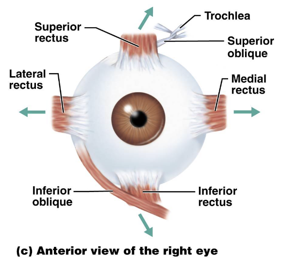
What is strabismus?
How can extrinsic eye muscles cause strabismus?
Misalignment of the eyes
Improper muscle function can cause the eyes to point in different directions

IV. The Eyeball — Three Layers
A. Fibrous Layer
What is the fibrous layer?
What are the two structures of the fibrous layer?
The outermost layer of the eyeball
The sclera and cornea

Sclera
What is the sclera?
What color is the sclera?
What is the sclera an anchoring site for?
The posterior and bulk of the fibrous layer
Opaque white
The extrinsic muscles

Cornea
Where is the cornea located?
What is a characteristic of the cornea?
What does the lecture state about immunity of the cornea?
At the anterior part of the eye, where it forms a clear bulge
It is clear
It has no immunity
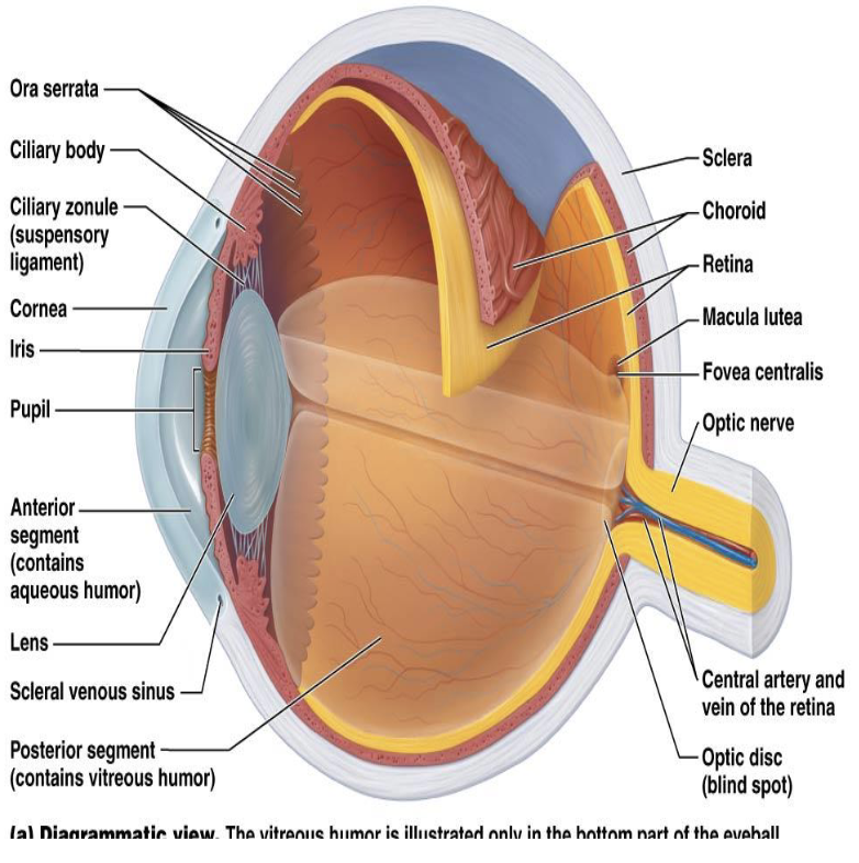
B. Vascular Layer
What is the vascular layer?
What structures are associated with the vascular layer?
The middle layer of the eyeball
The choroid, ciliary body, and iris

Choroid
What is the function of the blood vessels in the choroid?
What do melanocytes in the choroid provide?
What does the pigment in the choroid prevent?
Where is the choroid located?
They nourish the other layers
Pigment to absorb light
Light reflection
In the posterior portion of the eye
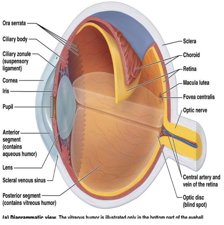
Ciliary Body
What is the ciliary body?
What controls the shape of the lens?
What holds the lens in place?
A thickened ring of tissue encircling the lens
The ciliary muscles
The ciliary zonule

Iris & Pupil
What is the iris?
What is the pupil?
The colored ring around the pupil
The opening for light

C. Sensory Layer
What is the sensory layer of the eyeball?
Where is the retina located?
The retina
It is the inner layer of the eyeball

Pigmented Layer
What is the pigmented layer of the retina?
What is the function of the pigment in the pigmented layer?
A single-cell-thick layer containing pigment like the choroid
It has the same function as the pigment in the choroid
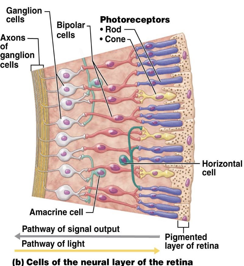
Neural Layer
What is the neural layer of the retina?
What types of cells are found in the neural layer?
Approximately how many photoreceptor cells are present?
A transparent layer containing neurons
Photoreceptor cells, bipolar cells, and ganglion cells
Millions of photoreceptor cells

What do bipolar cells do?
What do ganglion cells do?
They link photoreceptors to ganglion cells
They create action potentials, and their axons leave the eye as the optic nerve
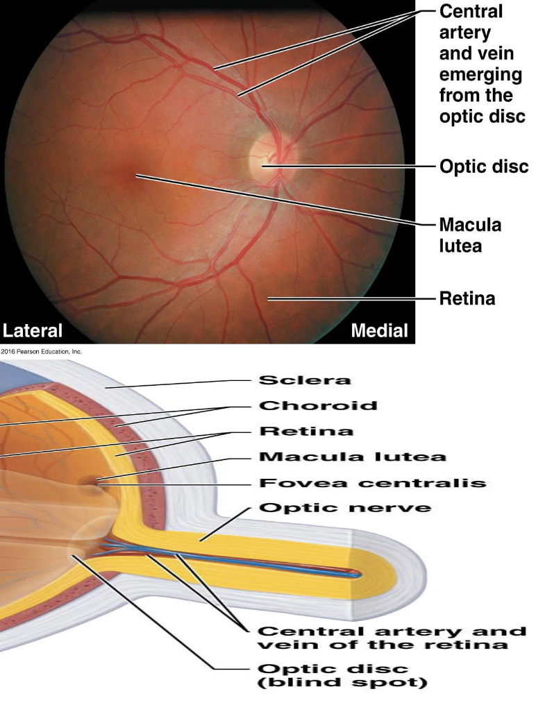
V. Important Eye Structures
Optic Disc
What is the optic disc?
What is another name for the optic disc?
The location where the optic nerve exits the eye
The blind spot

Macula Lutea
Where is the macula lutea located?
What does the macula lutea contain?
Lateral to the blind spot
A small pit containing the fovea centralis

VI. Photoreceptors — Rods vs. Cones
Rods
How do rods compare with cones in number and light sensitivity?
Do rods provide sharp images?
What type of vision do rods provide?
Where are rods located mainly?
Rods are more numerous and more light-sensitive than cones
No
Peripheral vision
Mainly in the periphery of the eye

Cones
What type of light do cones respond to?
What type of vision do cones provide?
Where are cones highly concentrated?
Bright light
Color vision and detail
In the fovea centralis

VII. Lens
What is the function of the lens?
What is the shape of the lens?
What are two physical characteristics of the lens?
It precisely focuses light on the retina
Biconcave
It is flexible and elastic

VIII. Cataracts
What are cataracts?
What factors listed in the lecture may result in cataracts?
Clouding of the lens
Aging
diabetes
heavy smoking
exposure to intense light
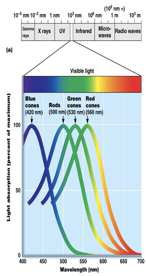
IX. Light
What is light?
What are packets of light energy called?
What do rods and cones respond to?
Electromagnetic radiation
Photons
Different wavelengths of light

X. Light Refraction
What is light refraction?
Where is light refracted as it passes through the eye?
What does the lens do to refracted light?
The bending of light as it passes from one medium to another
When entering the cornea, entering the lens, and leaving the lens
It refracts light so that the focal point is on the retina

XI. Focusing
Distance Vision
What are light rays like during distant vision?
How much lens adjustment is needed for distant vision?
What is the far point of vision?
They are parallel
Minimal lens adjustment is needed for focus
The distance that does not require the lens to focus

Near Vision
What is accommodation?
What is the maximum point of accommodation called?
A process in which the ciliary muscles change the shape of the lens
The near point

What happens to the pupil during near vision?
What is convergence?
The pupil constricts through the pupillary reflex
Both eyes rotate medially
XII. Refraction Problems
Myopia
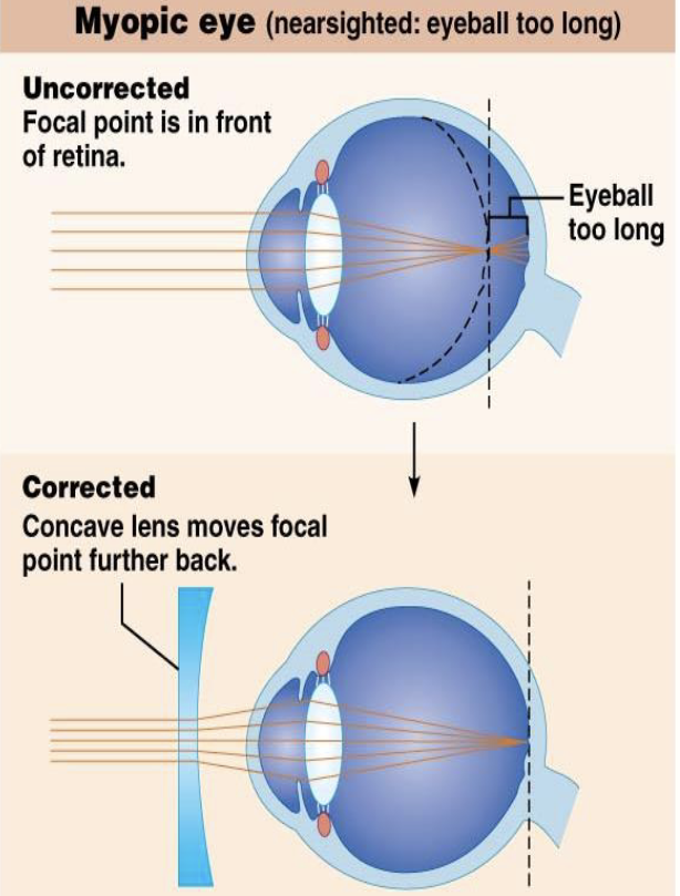
What is myopia?
What causes myopia according to the lecture?
Where is the focal point in myopia?
What type of lens corrects myopia?
Nearsightedness
A long eyeball
In front of the retina
A concave lens

Hyperopia
What is hyperopia?
What causes hyperopia according to the lecture?
Where is the focal point in hyperopia?
What type of lens corrects hyperopia?
Farsightedness
A short eyeball
Behind the retina
A convex lens
Astigmatism
What causes astigmatism?
How may astigmatism be corrected?
Uneven curvature of the cornea or lens
Glasses/contacts or eye surgery
XIII. Phototransduction
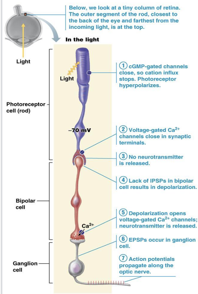
What is the sequence of cells involved in phototransduction according to the lecture?
Where are photoreceptor cells located?
Photoreceptor cell → bipolar cell → ganglion cell (→ Optic nerve)
Embedded in the retina
What does the bipolar cell do?
What happens to the ganglion cell axon?
It connects the photoreceptor to the ganglion cell
It forms the optic nerve

XIV. Visual Pathway Through the Brain
What forms the optic nerve?
Where does the optic nerve exit the eye?
Axons of ganglion cells
Through a weak spot in the sclera

What happens at the optic chiasm?
What do the optic tracts carry?
The medial optic nerve fibers cross
Information from the same half of the visual field

Where are the lateral geniculate nuclei located?
What happens after the lateral geniculate nuclei?
In the thalamus
Axons from the thalamic neurons connect to the primary visual cortex

Where is the primary visual cortex located?
What does the primary visual cortex do?
Visual Pathway?
In the occipital lobe
It interprets visual stimuli into perceived images
Optic nerve → Optic chiasm → Optic tracts → Lateral geniculate nuclei → Primary visual cortex
XV. Depth Perception
What contributes to depth perception?
What does the visual cortex do with the two visual fields?
Overlap of the two visual fields
It fuses the two fields together
Eyeball Layers
Fibrous → ??
Vascular → Sensory/Retina
Retina
Photoreceptor → ??
Bipolar → Ganglion → Optic nerve
Rods vs. Cones
Rods → ??
Cones → ??
peripheral vision + more light sensitive
color + detail + fovea
Focusing
Myopia → ??
Hyperopia → ??
Astigmatism → ??
long eyeball → focal point in front → concave lens
short eyeball → focal point behind → convex lens
uneven cornea/lens curvature
Visual Pathway
Optic nerve → ???
optic chiasm → optic tracts → lateral geniculate nuclei → primary visual cortex
Fovea
Small pit in macula lutea → ?
highly concentrated cones
Conditions
Cataracts → ?
clouding of lens → aging, diabetes, heavy smoking, intense light