Embryo Development
5.0(1)
Card Sorting
1/45
Earn XP
Description and Tags
Last updated 2:01 AM on 3/3/23
Name | Mastery | Learn | Test | Matching | Spaced | Call with Kai |
|---|
No analytics yet
Send a link to your students to track their progress
46 Terms
1
New cards
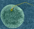
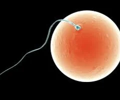
fertilization
Begins with contact between egg and sperm, ends with transfer of paternal chromosomes into the egg to form a zygote
2
New cards
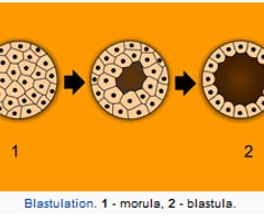
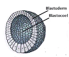
cleavage
the process ofd vision, single-cell zygote begins a series of organized, successive mitosis to produce a multicellular, hollow structured called the blastula; no growth of embryo
3
New cards
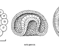
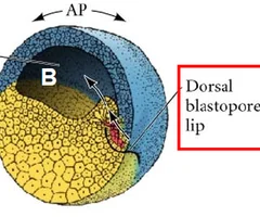
gastrulation
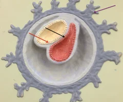
the period of cell movement; starts with the formation of a curved groove (blastopore) in the region formally occupied by the gray crescent
4
New cards
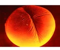
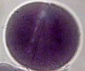
unfertilized egg
stage 1; appears as a large cell with a clear nucleus and nucleolus
5
New cards
gray crescent
stage 2; a region located opposite the site of sperm penetration, becomes the dorsal side, where gastrulation will begin
6
New cards
2 cell stage
stage 3; The result of the first cleavage event after fertilization
7
New cards
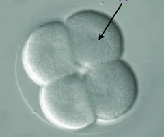
4-cell stage
stage 4; The result of a second cleavage event

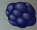
8
New cards
16-cell stage
stage 6
9
New cards
mid cleavage
stage 8; characterized by continued irregular cleavage and intrusion of pigmented area over pale area
10
New cards
late cleavage
stage 9; cells in animal hemisphere are small and pigmented and extend and well down toward the vegetal pole
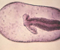
11
New cards
dorsal lip
stage 10; The region above the blastopore on the dorsal side of the amphibian embryo.

12
New cards
mid gastrula
stage 11; the groove of the blastopore and the dorsal lip have spread to form a circle around the small area of the remaining yolk
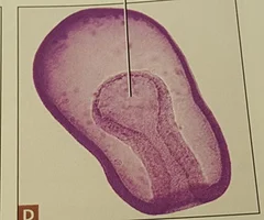
13
New cards
late gastrula
stage 12; Blastoporeformed, surrounding yolk plug, blastopore elevated to point of posterior axis of embryo.
14
New cards
zygote
fertilized egg
15
New cards
yolk
made of proteins and lipids
16
New cards
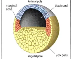
vegetable pole
yolky side
17
New cards
animal pole
dark colored

18
New cards
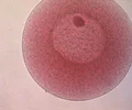
blastula
a multicellular, hollow structure produced after a series of organized successive mitoses
19
New cards
blastomere
each cell of the blastula
20
New cards
blastocoel
the central cavity of the blastula
21
New cards
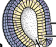
blastopore
the opening though which the cells on the surface of the embryo begin migrating to the Indies (into the blastocoel)
22
New cards
dorsal lip
The region above the blastopore on the dorsal side of the amphibian embryo
23
New cards
yolk plug
the remaining yolk in which the dorsal lips forms a circle around
24
New cards
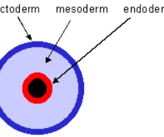
ectoderm
outer germ layer, the brain and spinal cord
25
New cards
mesoderm
middle germ layer, muscles, skeleton, urogenital organs, muscle layer, and outer walls of digestive and respiratory tracts

26
New cards
endoderm
outer germ layer; inner lining of digestive and respiratory tracts
27
New cards
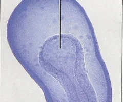
archenteron
the cavity formed by the endoderm; primitive gut; replaces the blastocoel and will eventually form the lumen of the digestive tract
28
New cards
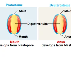
dueterostomes
blastopore will form anus opening first
29
New cards
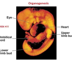
organogenesis
processes by which the cells of the three primary germ layers form actual organ systems in the embryo
30
New cards
differentiation
cells express their different morphological and functional specializations
31
New cards
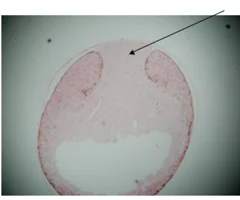
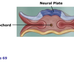
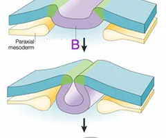
neural plate
the flattening of the neural ectoderm
32
New cards
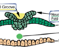
neural folds
forms when the neural plate rolls up at the edges
33
New cards
neural groove
area between the neural folds
34
New cards
neural tube
formed when the neural folds eventually fuse together to form an enclosed, hollow structure, ultimately forms the brain and spinal cord
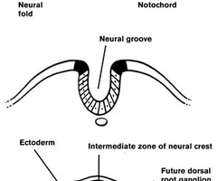
35
New cards
notochord
A flexible rod that supports a chordate's back

36
New cards
yolk plug
A group of large, nutrient-laden endodermal cells surrounded by the completed blastopore in an amphibian gastrula. These cells will be covered by ectoderm and end up inside the embryo.
37
New cards
stage 22
tail fin circulation
38
New cards
stage 21
mouth open, cornea transparent
39
New cards
stage 20
gill circulation
40
New cards
stage 19
heart beat
41
New cards
stage 18
muscular response
42
New cards
stage 17
tail bud
43
New cards
stage 16
neural tube
44
New cards
stage 15
rotation
45
New cards
stage 14
neural folds
46
New cards
stage 13
neural plate, dorsal flattening