Nursing Review Summer 2025
1/94
There's no tags or description
Looks like no tags are added yet.
Name | Mastery | Learn | Test | Matching | Spaced | Call with Kai |
|---|
No analytics yet
Send a link to your students to track their progress
95 Terms
Integumentary system
skin, hair, nails
Functions of integumentary system
1. Protection
2. Body Temperature Regulation
3. Works with immune system
4. Sensation (Mechanoreceptors)
five stratums of epidermis
1. Stratum corneum
2. Stratum lucidum
3. Stratum granulosum
4. Stratum spinosum
5. Basale stratum
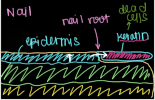
Basale stratum (deepest layer)
This layer:
1. Contains keratinocytes (give tough outer layer)
2. Contains Melanocytes (more melanocytes=more pigment=darker skin).
3. Cells rapidly divide in this layer and move upward to other layers.
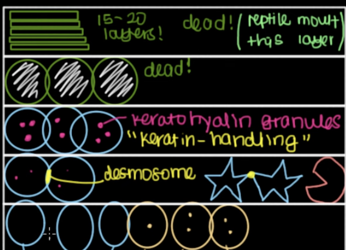
stratum spinosum (spiny layer)
In this layer:
1. Desmosomes connect the keratinocytes that have come up from the last layer.
2. Keratinocytes look star shaped because those cells under microscopes have lost water and look pointy because they are connected by these desmosomes but are shrivelled.
3. Langerhan cells: help immune system
stratum granulosum
1. Keratinocytes in this layer have a lot of granules containing keratin moving proteins.
2. keratinocytes also secrete lamellar bodies which form lipid layer that is impermeable layer at top of skin.
Stratum lucidum
Died! clear layer, hold the zombie keratincytes which have died after working in those other layers, they have lost their nuclei and organelles which give the cell color under a microscope.
Stratum corneum
Top layer, second layer of dead skin which holds stacked layers of flat keratinocytes (15-20) which died in stratum lucidum which will randomly fall off which makes way for newer keratinocytes from lower layer.

Dermis contains
Two layers that contain: blood vessels, nerves, and connective tissue (which is just proteins)

Papillary layer of dermis
The uppermost layer of the dermis, characterized by loose connective tissue, it contains capillaries (branch off from arteries) and sensory neurons.
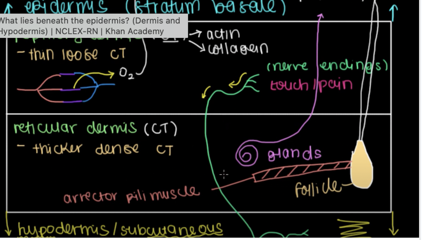
Reticular dermis
The thicker, deeper layer of the dermis, composed of dense connective tissue containing glands (i.e sweat glands), hair follicles, and arrestor pili muscles connected to those follicles so hair stands up.
Hypodermis
Layers of fat that absorb shock and insulate the tissues and bone below.
Types of burns
Burns are classified by which layers they reach.
First degree=epidermis
Second=dermis when nerve will be killed off and pain will recede
Third=past hyperdermis into fat/muscle/bone below, burn off nerve endings.
Types of sweat glands
Holocene, apocrine, merocrine
Holocrine sweat glands
face, chest, back)
secretes by disintegrating the whole cell to release sebum (oily substance) which lubricates skin and slows bacterial growth
Apocrine sweat glands
armpits, groin, around nipples
release clear substance into hair follicle that combines with bacteria to make odor: top of cell buds off and releases proteins/lipids/steroids.
Don’t work until after puberty, because these release things like pheromones in animals. In us, they do emotional sweating, which activates in times of emotional stress.
Merocrine sweat glands
everywhere else: palms, soles of feet, etc
release sweat by exocytosis, secret mostly water, help us cool down due to evaporative cooling, and get ride of waste products (like nitrogen, and extra electrolytes)
also release antibodies that help tag bacteria and lysozymes.
Afferent vs efferent nerve cells
Going to/going from CNS
Stimulus makes signal which goes to central nervous system vs nervous system makes stimulus (Like when somethings itchy which cause you to feel stimulus and move hand for example)
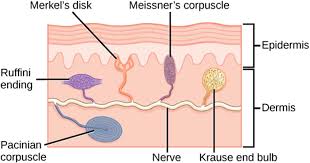
Mechanoreceptors
These cells and the nerve endings form structures that detect deformation of the skin and other tissues, opening ion gated channels, allowing sodium into the nerve fibers that send the signal to the CNS.
Types of mechanoreceptors
Meissner corpuscle
Pacinian corpuscle
Merkel’s disc
Ruffinis corpuscle
Hair follicle receptor
Pain and temperature (nociception/thermoception)
TrpV1 receptor:
Within cells on your hand for example are these receptors, they will send a signal to a nerve that will eventually reach the brain.
Whenever there’s a change in temperature or molecule signals pain, it causes a conformational change in the protein
When a cell gets poked, thousands of cells get broken up, which releases molecules that travel around and bind to TrpV1 receptor etc etc activating the cell and sending signal to the brain
Thermoregulation
Hypothalamus (front=fire, respond to heat vs back=burr, responds to cold) sends signals to muscles that respond
Thermoregulation In heat
Movement of blood cells in bloodstream hold energy, to get rid of heat we increase blood flow towards skin.
Vasodilation: arterioles are dilating since smooth muscle has been told to relax which wides blood vessels.
Skeletal muscles dont do anything when its hot
Thermoregulation In cold
Smooth contracts and causes vasoconstriction that decreases how much the cells in bloodstream are bouncing around, so more energy is moved inward.
Shivering: Skeletal muscles also contracts when it’s cold, taking ATP and making ADP by snapping off a phosphate group, which produces energy/heat.
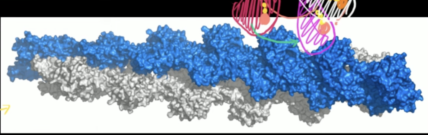
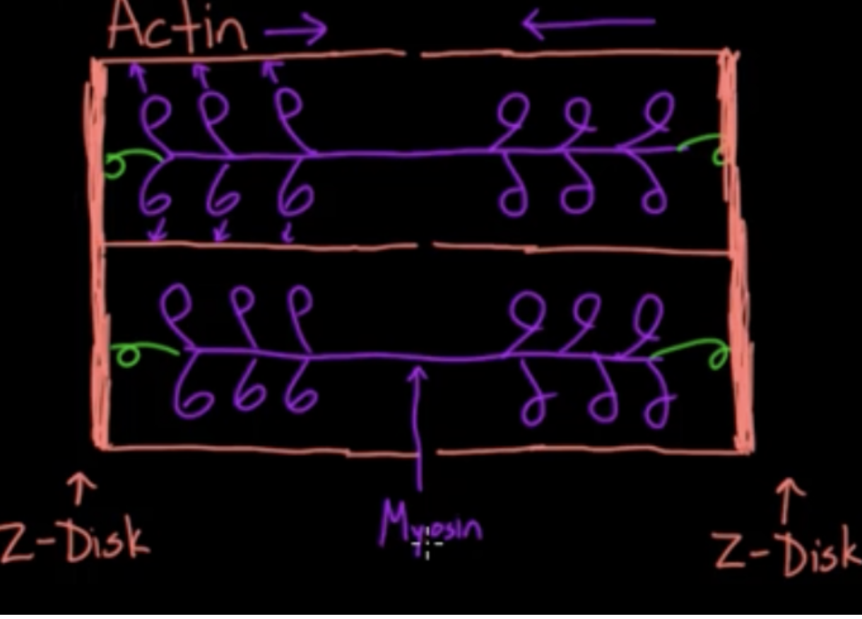
How myosin and actin make movement in muscle cells
Basically theres and ATP cycle that continually cocks and releases the gun that is continually pushing on Actin
When ATP binds to myosin, Myosin releases Actin
Then ATP hydrolizes, which releases energy to energize the myosin protein to high energy conformation with ADP and P still attached into a diff area of the actin
Phosphate is released from the Myosin, which causes Myosin in a mechanical movement to push the Actin
ADP released from Myosin, back to beginning but were one rung to the left
This is basically how we move, but multiple Myosins are acting on Actin at once
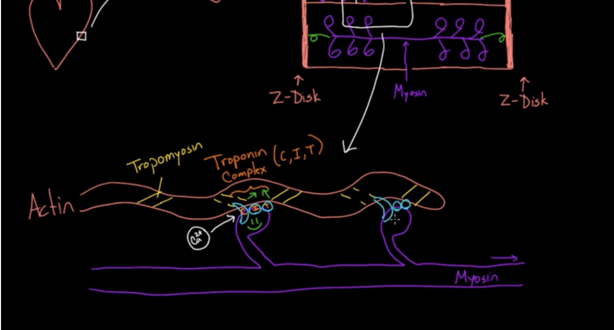
How to tropomyosin and troponin affect muscle movement
Tropomyosin is coiled around the Actin, Troponin attaches the tropomyosin to the actin
When a muscle is not contracting, the tropomyosin is blocking the myosin from attaching or from sliding to diff rungs, blocking it from working
Only way for it to unblock is for the troponin to change shape, which only occurs when there is a high calcium ion concentration. They will bind to troponin and change their shape, which moves the tropomyosin out of the way. If low concentration then the troponin blocks it once again.
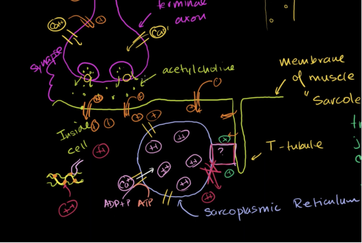
How sarcoplasmic reticulum affects calcium concentration in cells
Muscle cell has a sarcoplasmic reticulum, which has calcium ion pumps on its membrane, which use ATP to pump calcium into it, so resting muscles have high concentrations of calcium inside.When muscle needs to contract these get dumped out into rest of cell, that then bind to troponin to allow tropomyosin to move so myosin can bind.
How does cell know when to open SR?
An action potential will travel across the cell through voltage gated ion channels and into the T-tubule in muscle cells which activates a protein complex to release the calcium into the cell. When the signal leaves the voltage gated channels close and calcium is actively transported back into SR.
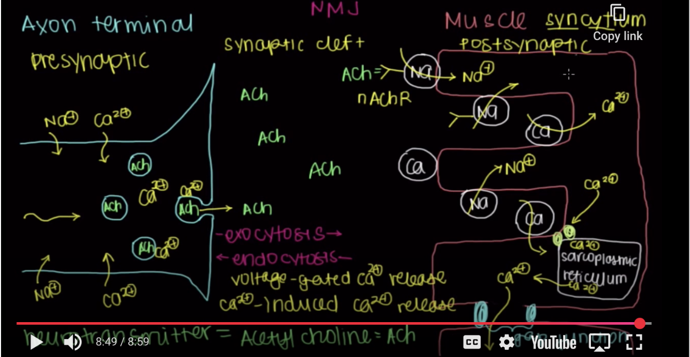
Neuromuscular Junction: Where our neurons talk to our muscles
When an action potential reaches the axon terminal it signals calcium ion channels to open. This calcium will bind to vesicles holding neurotransmitters (acetylcholine) that then binds with the neuron membrane through exocytosis deposit the signal into the synaptic cleft. Sodium channels in the muscle membrane have receptors that when binded with the ACH signal open the channels and cause an influx of sodium into the muscle cell. (insert myosin/actin intercellular response here).
Types of muscle: skeletal
Attached to tendons (except when they’re not I.e. external obliques attached to aponeurosis (like a flat tendon) or bones
Voluntary
Fastest (movement must happen almost instantaneously)
Many nuclei (located on periphary)
Types of muscle: cardiac
Specialized muscle cells in the heart only
Help your heart beat
Involuntary
Middle speed
Branched: sometimes have two nuclei
Striated under microscope
Types of muscle cell: smooth
Found in hollow organs (like stomach or bowels) or blood vessels
Contracts and helps things move through
Involuntary
Slow (vasodilation/constriction happens slowly)
Look like an eye/almond shaped, striated under microscope
Heart cells
Myosin is pulling Actin inward from both sides of the cell, so it contracts inward
When calcium is around, the myosin can attach to actin and do work
Inotropy (strength of contraction): either increase amount of calcium available to bind to troponin or increase calcium ability to bind to troponinT
Type 1 fibers (M1tochondr1a more prevalent (Golden rule))-fiber=cell
Red (hearts have more oxygen than our veins do, why theyre red instead of blue)
Slow contraction speed (lot of steps in oxidative phosphorylation)
Slow twitch (same reason)
Aerobic respiration
Long duration contractions (more energy made with mitochondria)- back, arms, glutes)
Fatigue resistant (enough energy to contract for a long time)
Strong muscle groups (more energy to contract, more muscle cells contracting at same time=stronger contraction overall)
Store energy in triglycerides
Type 2 fibers (fiber=cell)
White
Fast contraction speed
Fast twitch
Anerobic respiration (don’t have as much mitochondria)-you dont stand on your hands
Short duration contractions (less energy)
Easily Fatigue (less energy)
Weak contractions (not enough mitochondria=not enough energy=not enough power)
Stored in ATP (energy used right away), or creatine phosphate
Voluntary control is located in
cortex/spinal cord
Involuntary control is located in
Beyond me=brain stem/besides
ganglia (PNS)
Bundle of neurons’ somas
automatic vs somatic
Somatic controls voluntary movement of skeletal muscles (acetylcholine) while automatic controls involuntary processes such as digestion, breathing, heart rate.
Automatic NS: sympathetic vs parasympathetic
Sympathetic: Fight or flight response (norepinephrine)
parasympathetic: rest/digest, such as slowing heart beat when asleep/telling stomach to digest after big meal (norepinephrine)
Motor neuron
One motor neuron and all the skeletal muscles it contacts and controls along with the neuromuscular junction
Lower motor neuron vs Upper Motor neuron
Upper Motor Neuron sends signal to lower motor neuron while LMN is messenger that tells muscle to contract
UMN sends that message to LMN but also tells LMN when to stop
LMN keep their somas in the brain or in the spinal cord and send their axons (through nerves) to synapse on and control skeletal muscle cells or other cells.
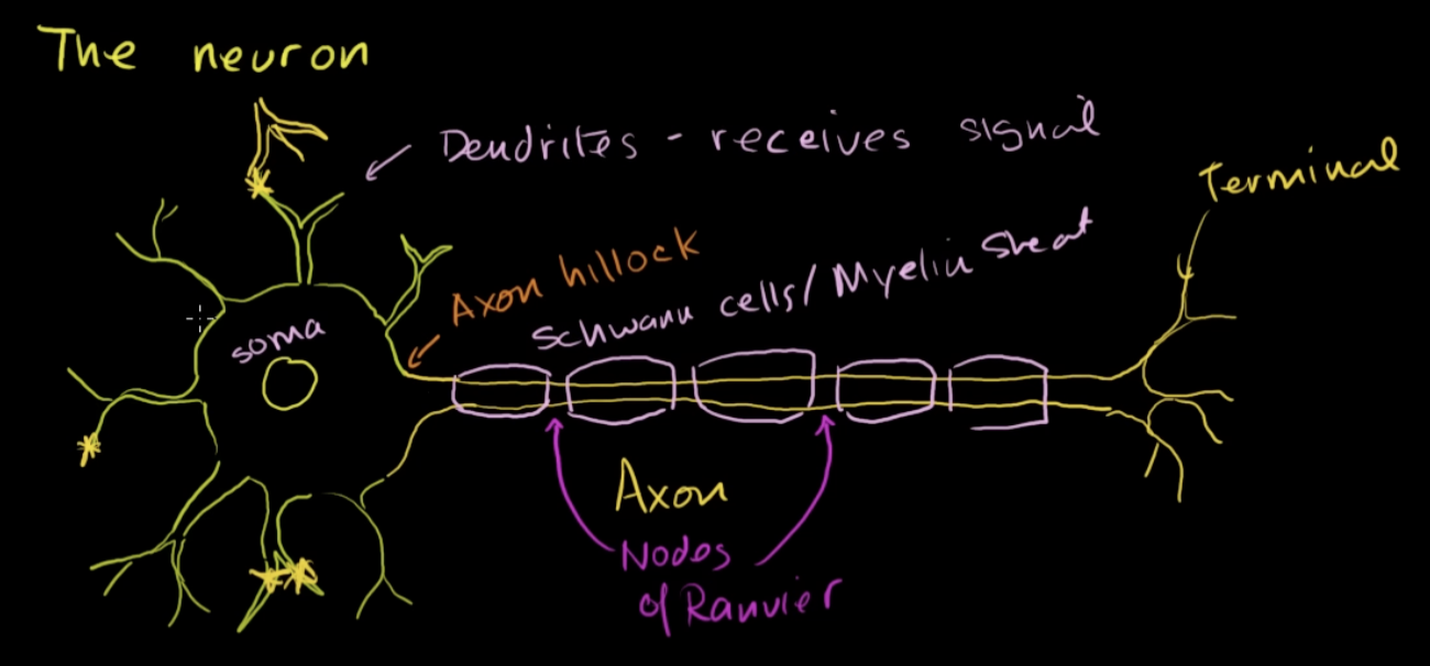
Neuron structure
Soma with its processes (dendrites/axons)
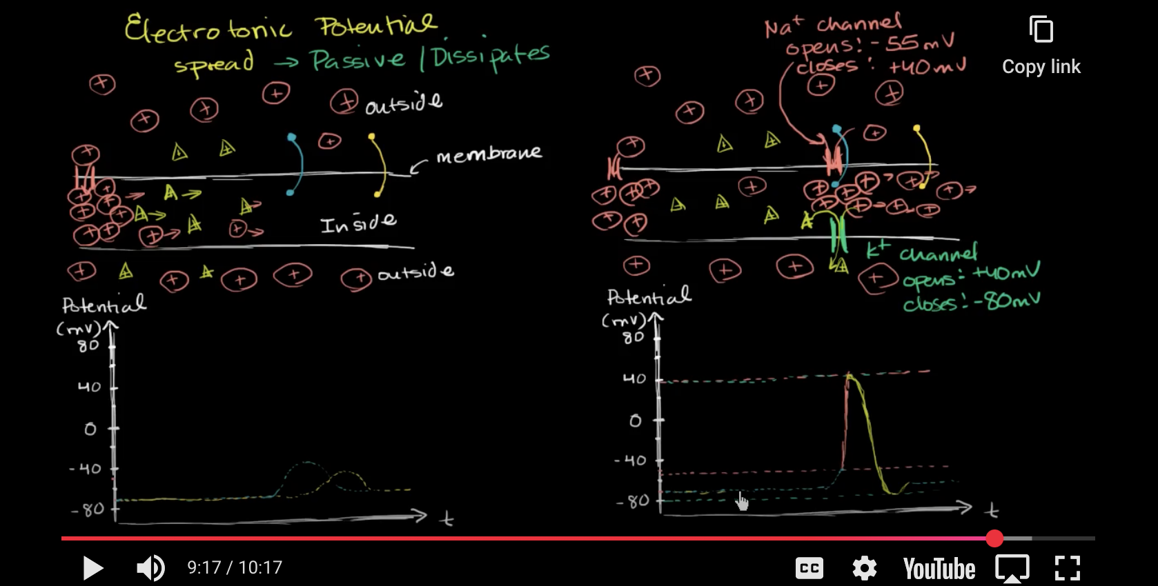
How does an action potential work?
a sodium ion channel will open in the trigger zone depolarizing the membrane potential.
Electrotonic spread/potential: as positive charge moves out other areas get more positive but to a lesser extent since less charge is available to spread out as you get further away, aka signal lessons
However, this depolarization will cause voltage gated ions to open, allowing sodium to flood in (because more sodium outside cell).
Basically as ions enter and are repelled by each other , causing them to move down the axon and depolarize those areas, causing more voltage gated ions to open, the cell is boosting the signal.
Once enough sodium is in the cell (lets say 40mv) then the ion gated channel closes and the potassium ion channel opens causing hyperpolarization (moving back to baseline)
Skeletal system main functions:
Support
Protection of organs
Storage of calcium
Hematopoiesis
Blood cells, white blood cells and platelets are all formed within bone marrow
Axial vs Appendicular skeleton
Axia (center)l= cranium, rib cage, vertebral column
Appendicular= four appendages, pelvis
Flat bones vs long bones
Both have Inner spongy bone with outer shell of compact bone, both do hematopoiesis
flat=skull, ribs, pelvis
Protect organs
long=humerus, femur, etc
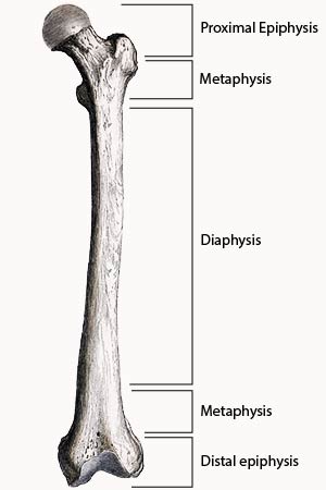
Long bones structure
Diaphysis (middle stretch of bone) vs epiphysis (the ends) vs metaphysis (in between which holds growth plates in children)
Red bone marrow vs yellow bone marrow
red=hematopoiesis (production of red blood cells=red bone marrow)- in flat bones and epiphyses of long bones
yellow = fat storage, fat cells called adepocytes -diaphases of long bones
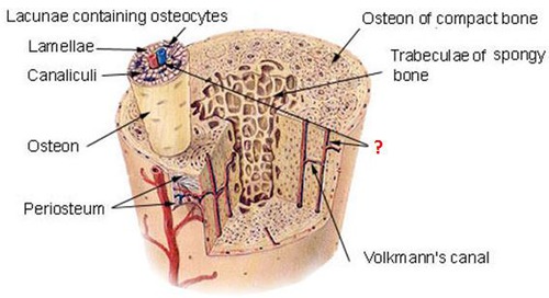
Haversian system
Sheets of bone called lamella that wrap around eachother
In center is the haversian canal wher blood vessels, nerves, and lymph nodes
Canaliculi branch out from the canal into the empty spaces called lacunae (empty space) for osteocytes (bone cells) which function to communicate about nutrient availability and such.
Different types of skeletal cells
Osteoprogenitors
Precursor of osteoblasts, differentiate in presence of growth factors
Osteoblasts
Synthesize collagen and proteins: osteocalcin and osteopontin
Alkaline phosphatase
Mature to osteocyte
Osteocytes
In lacunae (like lake: empty space)
Star like appearance: communicate with other cells that help maintain bone
Osteoclasts
Bone resorption: Break bone down again
Osteoclasts vs osteoblasts
Osteoclasts break/crash down and Osteoblasts build up.
How does skeletal system regulate amount of calcium In blood stream?
Hormones alter ratio of osteoclast activity to osteoblast activity
Increase osteoclast activity=more calcium in blood stream
Increase osteoblast activity= less calcium in blood stream and more in bone
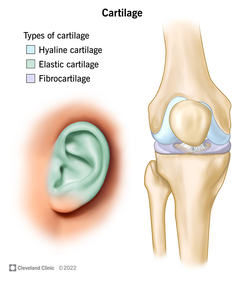
What is cartilage?
a strong, flexible (Chondrocytes: make collagen and elastin) connective tissue that protects your joints and bones

Types of cartilage
Hyaline: Joints, reduce friction, absorb shock
Elastic: outer ear/epiglottis, provide shape and support
Fibrous: inter vertebral discs (in between vertebrae)/pubic (where two halves of pubic bone come together), provides rigidity and absorb shock
cartilage vs ligaments vs tendons
cartilage is the shock absorber at the end of bones and ligaments connect bones to other bones while tendons connect bones to muscles
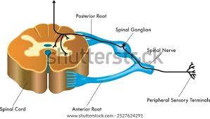
What is in the central nervous system?
Brain and Spinal cord
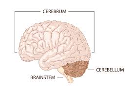
What is the cerebrum?
Top part of the brain (two hemispheres)
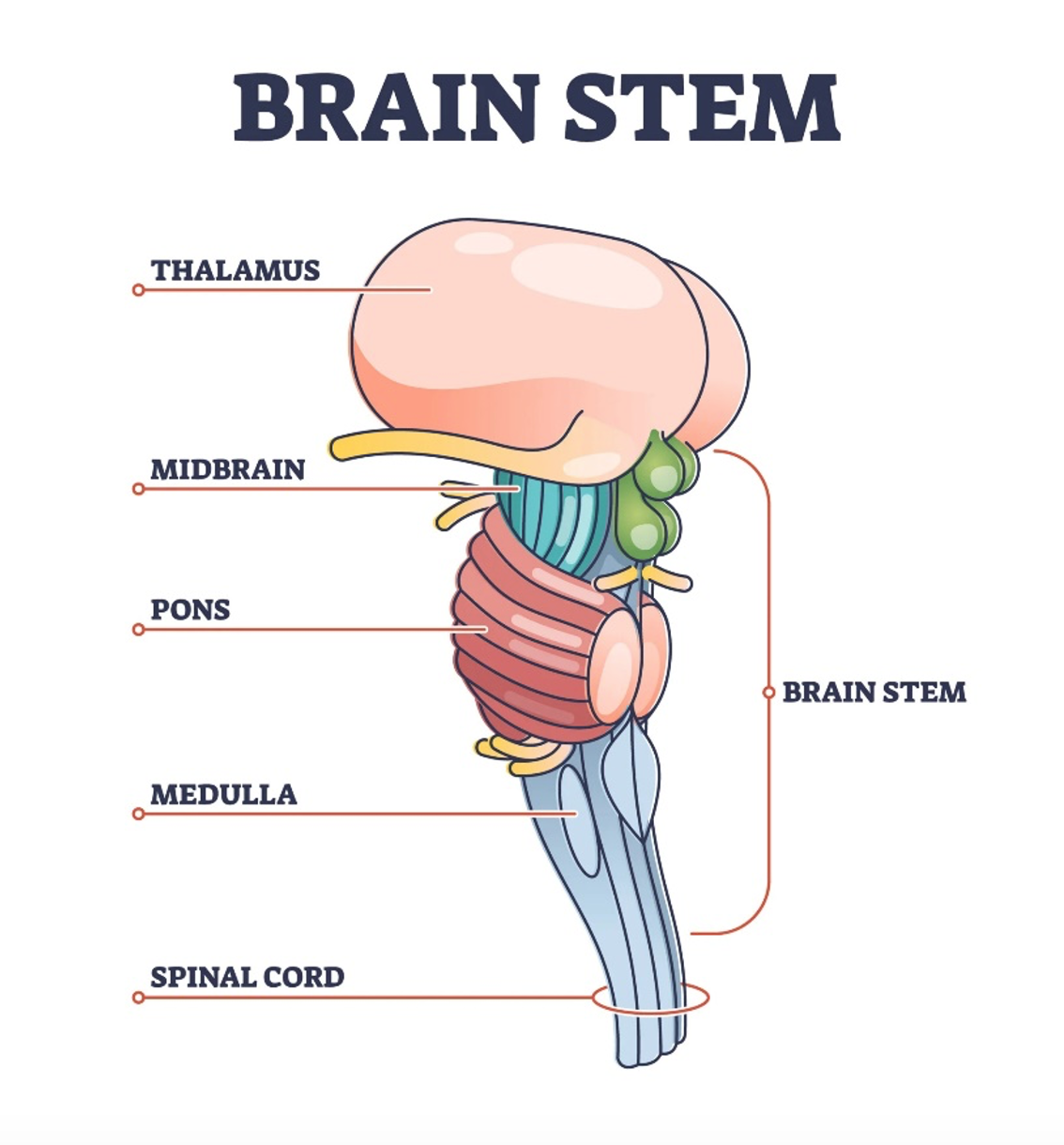
What is the brain stem
Connects brain with the spinal cord, three parts:
Midbrain
Pons
Medulla
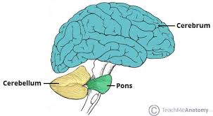
What is the cerebellum?
“little brain” holds more than half of neurons in the brain and does a lot of stuff
What is in the Peripheral Nervous System?
All the nerves not in the brain/spinal cord
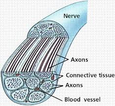
What are nerves
Bundle of nerve fibers (nerve fiber=axon of neuron, neuron=nerve cell)
cranial vs spinal nerves
Exit the brain out of skull vs come out of spinal cord.
Functions of NS
Basic functions
Motor (control of skeletal muscle=movement, tone, posture)
Sensory (senses=more than five, detection)
Automatic (reflexes
Higher functions
Cognition (thinking, learning, memory, language, executive functions: creating goals and organizing to achieve them)
Emotions
Consciousness: awareness of being alive/controlling actions
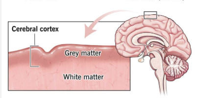
What is the Cerebral Cortex?
Grey outer layer of cerebrum
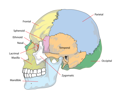
Sections of cerebral cortex (names for part of cranium they cover)
Frontal lobe
Parietal lobe
Temporal lobe
Occipital lobe
Left vs Right
Language is most often a function occurring in the cortex of the left hemisphere
Information like visuals and somatosensory input gained on the right side of the brain is processed on the other side of the brain and vice versa. The frontal lobe has a motor cortex kinda thing where signals from right brain will cross spinal cord and affect left side of movement,
Everything else is processed on both sides
Corticospinal Tract
UMN axon travels down through the brain stem from the cerebral cortex, where the brain stem meets the spinal cord most of the axons will cross to the other side until they synapse on a LMN.
Corticobulbar tract
lots of UMN axons come down and synapse with cranial LMN on both sides
Mechanoreceptors function for these somatosensations:
Position awareness (certain mechanoreceptors are attached directly to muscle, and send signal back to CNS about things like stretch/movement which allow awareness of limbs)
Vibrations
Touch
all above will:
Use mechanoreceptors (Respond to physical forces) that:
Connect to axons coming into the skin
Carried in large diameter axons with thick myelin sheaths=fast
Somatosensation: Pain
Nociceptors that:
Connect to axons coming into the skin
Don’t have structure at end of axon, usually axon just ends at an uncovered terminal (bare nerve endings)
short diameter axons=longer response time
Somatosensations: temperature
Thermoreceptors:
Connect to axons coming into the skin
Don’t have structure at end of axon, usually axon just ends at an uncovered terminal (bare nerve endings)
short diameter axons=longer response time.
What is Automatic nervous system split in to?
Sympathetic Nervous system VS Parasympathetic Nervous system. Both basically decide where blood is flowing.
Sympathetic nervous system
“Fight or Flight” (i.e. blood flow: in “fight” the sympathetic nervous system will decrease blood flow to intestines and increase to skeletal muscles or increase sweat glands production (cool us down=move faster and farther)
Parasympathetic nervous system
Rest and Digest (i.e. heart output goes down when PNS is activated because less blood flow is needed or salivary glands are stimulated to increase digestion)
Why is a neural cell/a neuron sometimes called a nerve cell?
cause in ye old days that’s what they were called but now we know nerves are really made up of only a part of a neuron, so its antiquated.
Neural cell types?
Glia and Neurons
Types of Glia: Astrocytes
Structure: Soma with processes that are highly branched
Function:
Scaffold for CNS, form structural support and form glial scar when injury occurs (surround and wall off injured area)
Homeostasis (ion concentration, interstitial fluid levels, lactate concentration for instant energy for neurons when needed)
Blood brain barrier (large molecules can’t go to CNS)
Clear out synapses between neurons
Types of glia: microglia
Structure Small soma and lot of long highly branched processes
Resting vs active
Active are larger and more blob shaped
resting= monitoring fluid, if detected they turn into active microglia
Looking for inflammation/ bacteria or virus that has entered CNS
Functions:
If finds foreign cell will secrete cytotoxic factors to kill bacteria
Phagocytosis: Microglia then eat debris/other dead cells of CNS
Antigen presentation: Sticks tags on the outside of that debris which causes more inflammation
types of glia: Ependymal cells
Barrier between Cerebral spinal fluid Interstitial Fluid (very leaky).
Types of glia cells: Oligodendrocytes
Structure: a soma with a few processes,
Functions:
These cells form the myelin sheaths that cover neuron axons with their long membranes in layers like a wrapped wire.
form around multiple neurons at a time.
types of glia cells: Schwann cells
Form protective myelin sheath around axons: membrane wrapped around the axons with a little bump on the outside where its nucleus etc is.
How do neurons function?
Membrane potential Neurons are often more negative on inside of membrane than on outside.
Info usually is first through the dendrites and moves through the soma to the axons through a graded potential.
If summation of graded potentials is enough the trigger zone will then fire an action potential down the axon.
Neurons don’t just send information but also receive it when axon terminals form their own synapses with the dendrites of another neuron.
Neurotransmitter removal
Enzymes present will break some down
Re-uptake pumps in axon terminal membrane, to reuse signal
Astrocytes pump neurotransmitters into to break down/pump back into axon terminal
types of neurotransmitters
Amino acids
Most functions
Peptides
I.e. opioids like endorphins
Pain
Monoamines
attention/cognition/emotion
Serotonin, histamine, dopamine, epinephrine, norepinephrine
Other
I.e. acetylcholine
How do excitatory vs inhibitory synapses work to fine tune a rx?
In neuron: Ligand binds so receptor opens channel and causes either sodium and calcium to enter (excitatory) or chloride or potassium to enter (inhibitory) which causes a graded potential (more localized action potential)
In target cell: When ligand binds it causes creation of second messenger that can do various excitatory/inhibitory things such as opening channels/affecting proteins/genes etc
Neuroplasticity
Potentiation vs Depression
Strength of flow of information increases/decreases and amount of response or strength of response increases/decreases due to how often this neuron is used causing a decrease/increase in dendrites/length of axon etc.

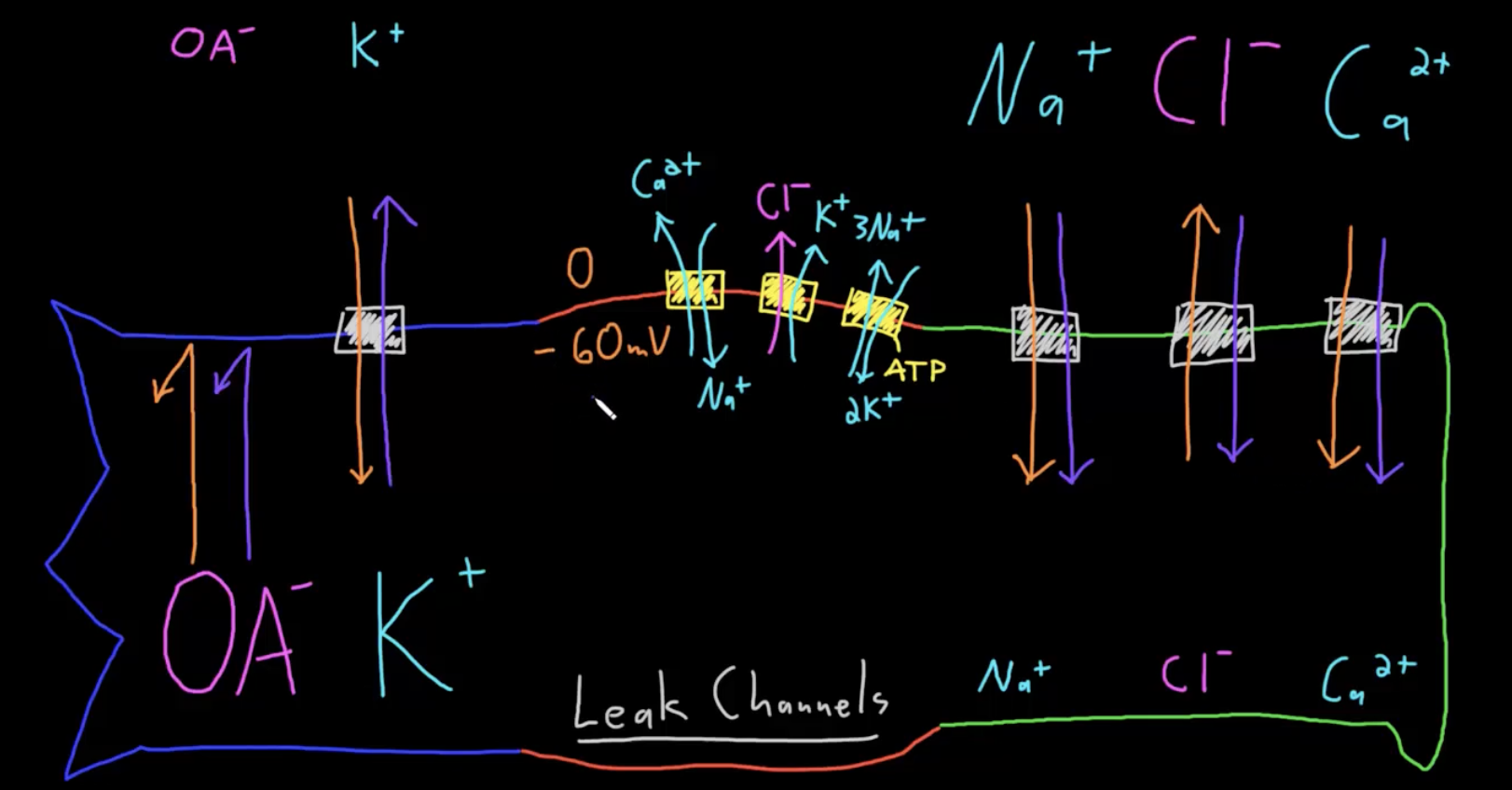
Why does a neuron have a membrane potential?
Basically various ions and organic anions are being pushed by a diffusion force and by their electrical force. Add this along with membrane permeability and various pumps in the cell and you get the concentration shown in the video.
Graded potential (what is it?)
Through excitatory or inhibitory inputs they increase/decrease the likelihood of an active potential by changing the membrane potential. Multiple graded potentials will build so it reached trigger point.
Graded potential mechanism (depolarizing/hyperpolarization of neuron)
The neurotransmitter receptor opens channels and allows ions to move into neurons.
Strength of potential depends on how many neurotransmitters are released, how long the neurotransmitters remain in synapse, which channels are opened and which ions they allow through, and how many channels are opened.
Depolarization usually occurs when a sodium or calcium channel is opened bringing positive charges into the negative inside of the neuron.
Hyperpolarization usually occurs when a chloride channel is opened and chloride is driven into the inside of a cell (diffusion is greater than electrical force) or when a potassium channel opens because of its diffusion force and causes outside to be more positive 9same thing as inside being more negative)
Basically its important to note that just because its more positive on outside does not mean cations will not naturally move out of cell because of their diffusion force.
Depolarization vs hyperpolarization
Decreasing/increasing likelihood of threshold of difference in charge being met
Neuron action potential mechanism (why does potassium enter cell)
Voltage gated ion channels: open when membrane potential crosses threshold value. Open and sodium pours in because of both electrical and diffusion forces
Trigger zone: has highest amount of voltage gated ion channels
Sodium will enter cell, trying to reach its equilibrium potential
At a certain point, inside the cell is more positive than outside, causing potassium to leave the cell because of its electrical force and diffusion force. Voltage gated potassium channels also open, contributing to the quick fall of charge to even more negative than resting potential.
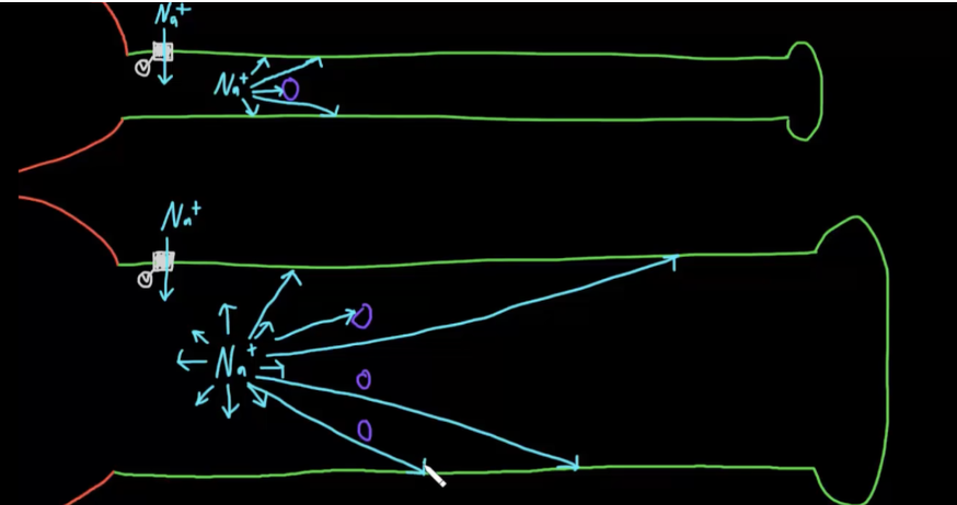
Small vs large diameter axon
larger=ions hit the membrane less=less resistance and quicker to move action potential down the axon. Also more room to move around various tubules/stuff in the axon.
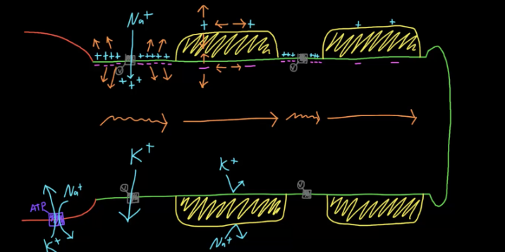
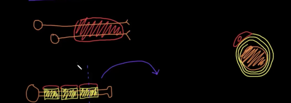
Why is conduction of action potentials faster in myelinated axons?
In nodes of ranvier, the short membrane causes cations who are more strongly attracted to the anions than repelled from each other to congregate together.
However Myelin sheath makes the membrane much thicker, meaning the strength of attraction between ions across the membrane is much less, which means the attraction between anions and cations does not overcome the repelling force between like charged ions, so ions are spread out from each other.
Effect: myelinated parts of the axon will take less time to depolarize because there are less cations that need to move away from the axon and less anions that need to move inward.

Saltatory conduction
Signal dissipates slightly across myelin sheaths, but is reboosted across nodes of Ranvier because of the voltage gated channels located there.
Saltatory conduction: saltar=latin for hop around, explaining how it jumps around across the axon.