Bacterial CNS
 Call Kai
 Learn
Learn Practice Test
 Spaced Repetition
 Match
Match Flashcards
 Knowt Play
1/27

There's no tags or description
Looks like no tags are added yet.
Name | Mastery | Learn | Test | Matching | Spaced | Call with Kai | Chat |
|---|
No analytics yet
Send a link to your students to track their progress
28 Terms
List the Aerobic, gram-negative bacilli that can infect the CNS
Other CNS Bacterial PAthogens
Aerobic, gram-negative bacilli
Escherichia coli
Main virulence factor is the K1 (capsular) antigen
Acinetobacter baumanii
Pseudomonas aeruginosa
Serratia sp.
Salmonella sp.
NOTE:
In addition to neonates, and older adults other at risk (for above listed pathogens) individuals are those who
Experienced head trauma
Undergone neurosurgical procedures
Other CNS bacterial pathogens
Staphylococcus aureus
Coagulase-negative, Staphylococcus sp.
High risk individuals are those whO have undergone neurosurgical procedures
Or those with CSF shunts
Anaerobic streptococci, Bacteroides sp., Fusobacterium sp.
Rare
Associated with brain abscess, hematogenous spread
What are the symptoms of Bacterial Meningitis
>2 yrs old
newborns, infants
Bacterial Meningitis Symptoms
Symptoms
Greater than 2 years of age
Sudden high fever
Stiff neck
Severe headache
Seizures
Sleepiness/difficulty waking
Sensitivity to light
Skin rash (meningococcal meningitis)
Newborns, infants
High fever
Constant crying
Excessive sleepiness/irritability
Poor feeding
Bulge in the soft spot on top of baby’s head (fontanel)
Stiffness in baby’s body and neck
Pathogenesis
nasopharyngeal colonization -> mucosal barrier invasion -> blood stream -> meninges -> Subarachnoid space -> bacteria replicates
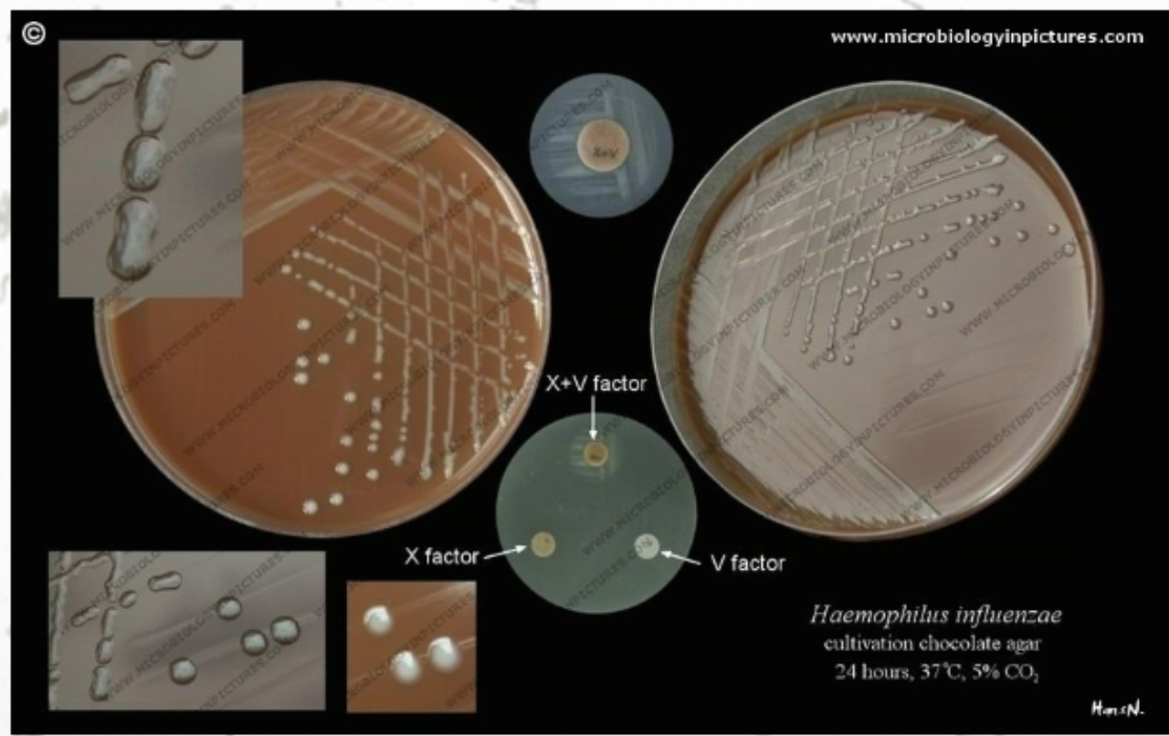
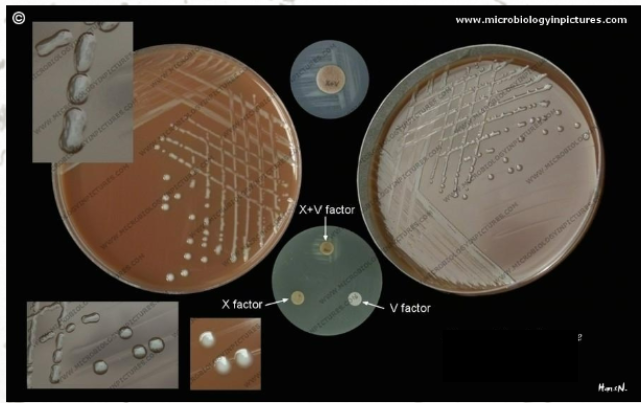
Describe Haemophilus influenzae
Characteristics
Haemophilus influenzae
Characteristics:
Gram-negative rods
Non-motile
Facultative anaerobes
Oxidase positive
Catalase positive
Reduce nitrates to nitrites
Require preformed growth factors:
X (hemin), and V (NAD)
Describe Meningitis by H. influenzae type b
Pts affected
Symptoms
Virulence Factor
Pts Affected:
children 3 months to 6 years of age
Generally, infants 1 – 23 months old
Symptoms:
Mild respiratory disease w/
– Headache
– Stiff neck
– Brudzinski sign
Involuntary flexion of the hip and knee when the neck is bent forward
Kernig sign
Inability to extend the leg while the hip is flexed to 90 degree
Virulence factors
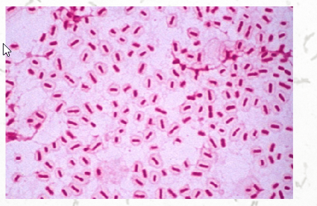
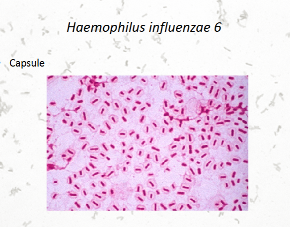
Capsule (most imp.)
a, b, c, d, e, f subtypes (det. By polysaccharide)
Immunoglobulin A proteases
Adherence by fimbriae
Outermembrane proteins and LPS
NOTE:
Fimbriae have not been found on isolates of H. influenzae from CSF or blood of patients with invasive form of this disease
Invade bloodstream by a breakdown in the tight junctions between epithelial cells
Besides meningitis, what other diseases does H. Infl. cause?
Haemophilus influenzae Clinical Manifestations
Septicemia
Arthritis
Epiglottitis
Intense edema -> airway obstruction
Children age 2 —4 years
Tracheitis
Usually arises after an acute viral respiratory infection
Thick secretions -> occlude
Must be differentiated from epiglottitis
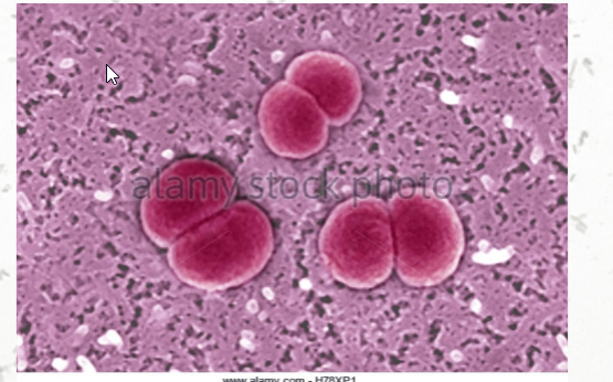
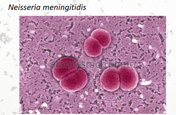
List the characteristics of Neisseria meningitidis
Characteristics:
Aerobic, nonmotile, non-spore forming
Gram-negative, diplococci
Cytochrome oxidase positive
Catalase positive
Capnophilic
Requires CO2
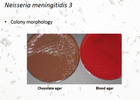
Culture
Chocolate agar
Requires iron for growth
Competes with human host by binding human transferrin
Possible reason for being strict human pathogens
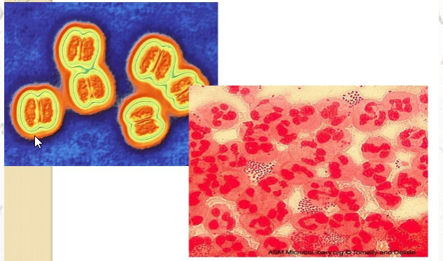
Neisseria meningitidis: microscopy

Describe the Neisseria meningitidis
Virulence Factors
MOA
STATs
Incubation
List the Meningococcemia Symptoms
List the Meningitis Symptoms
Neisseria meningitidis – Virulence Factors
Pili (fimbriae)
mediate adherence to nasopharyngeal
epithelial cells
Capsule
Receptors for human transferrin
Endotoxin
Immunoglobulin A protease
Neisseria meningitidis
MOA:
respiratory droplets -> mucosal surfaces of nasopharynx and oropharynx -> blood stream
STATs
30% of the population
Military recruits and college students have increased risk of exposure
@ risk groups:
Infants 1-23 months
Children greater than 2 years, adults
Older adults greater than 65 years
Incubation 1 – 10 days
Meningococcemia Symptoms
Purpura of skin and mucous membranes
Petechial rash
Tachycardia, hypotension
DIC (disseminated intravascular coagulation)
Meningitis Symptoms
Headache
Fever
Meningismus
Inflammation of meninges, or subarachnoid hemorrhaging
Nuchal rigidity
Kernig’s sign, Brudzinski’s sign
Altered sensorium
Inability to think clearly or concentrate
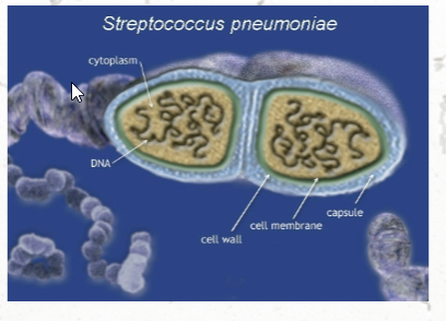
Streptococcus pneumoniae
Streptococcus pneumoniae Characteristics
Virulence Factors
STATs
Pneumococcal meningitis symptoms
Gram-positive diplococci
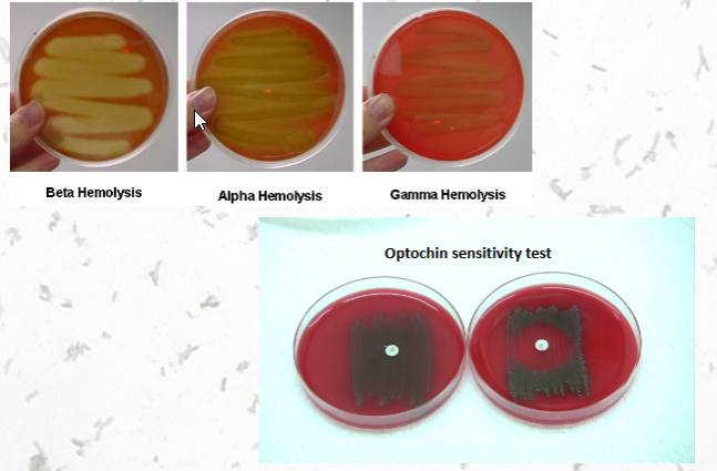
Alpha-hemolytic on blood agar
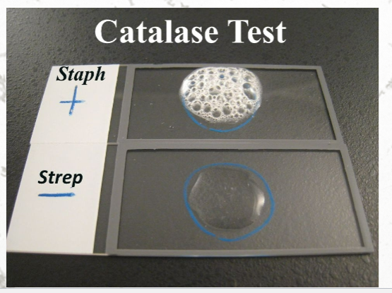
Catalase negative
Optochin sensitive
Oxidase negative
Urease negative
Ferments
Glucose
Lactose
Virulence Factor:
Polysaccharide capsule
Abs can work against this
STATs
Most common cause of meningitis in adults
Second most common cause of meningitis in children
Pneumococcal meningitis symptoms
Onset is usually rapid
Chest pain, chills
Cough, fever
Confusion, headache
Stiff neck
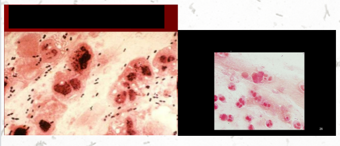
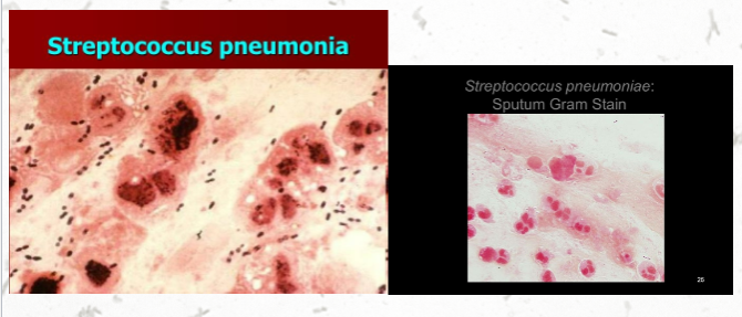
Streptococcus pneumoniae
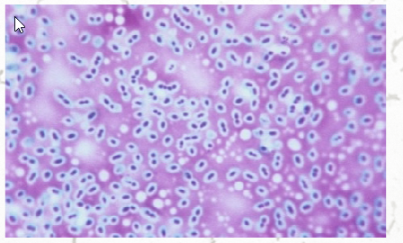
Streptococcus pneumoniae
Describe Haemophilus influenzae, Neisseria meningitidis, Streptococcus pneumoniae
Immune Defense vs Evasion
Diagnostic
Immune Defense vs Evasion:
Defense:
Ability to adhere to mucosal surfaces can be inhibited by IgA
Evasion:
Produce IgA proteases -> cleaves hinge region
Diagnostics:
Multiplex bacterial PCR diagnostic assay
For All mentioned
can detect low numbers of bacteria
Can detect nonviable etiologic agents in pts receiving antimicrobial therapy
Specificity 99.1 – 100% predictive
Describe Listeria monocytogenes
Characteristics
Pathogenesis
STATs
High Risk Groups
Clinical Symptoms
Characteristic
Gram-positive rods
Facultative anaerobe
Catalase positive
Oxidase negative
Tumbling motility at 25 degC
Growth on blood agar
Exposure from raw vegetables, raw milk, cheese, meats (deli)
Pathogenesis
Ingestion of contaminated foods
Consumption of Dairy and processed meats
Incubation 11-90 days
Endocytosis across mucosal barrier -> BBB
Hematogenous dissemination
predilection for the CNS and placenta
CNS invasion by 3 methods
Invasion of endothelial cells of BBB
Transportation -> CNS w/in circulating leukocytes
(phagocyte facilitated)
Bacteria inoculated into oral tissues -> macrophage phagocytosis -> invade CNs
STATs
Fifth most common cause of meningitis
High risk groups
Neonates (less than 1 month)
Older adults (greater than 65 years)
Clinical symptoms
Stiff neck
Movement disorders (ataxia, tremors)
Seizures
Fluctuating mental status
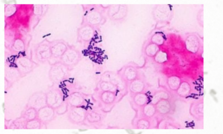
Listeria monocytogenes
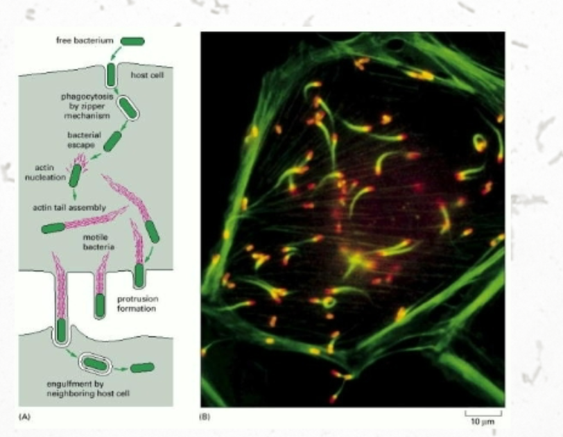
Listeria monocytogenes
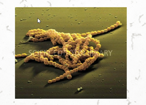
Streptococcus agalactiae
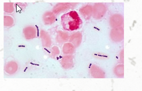
Streptococcus agalactiae
Describe Streptococcus agalactiae
Characteristics
Virulence Factors
Risk Groups
Characteristics
Gram-positive cocci
Normal bacterial flora of the GI/GU
Lancefield group – B
Beta hemolysis
Bacitracin resistant
Catalase negative
Virulence Factors
Capsule (imp.)
Inhibits
complement deposition
phagocytosis
Pore forming beta-hemolysin/cytolysin
Risk groups based on age
Neonates less than 1 month old
vertical transmission: mother to infant
Infants from 1-23 months old
Adults >60
Underlying risk factors: diabetes mellitus, malignancy
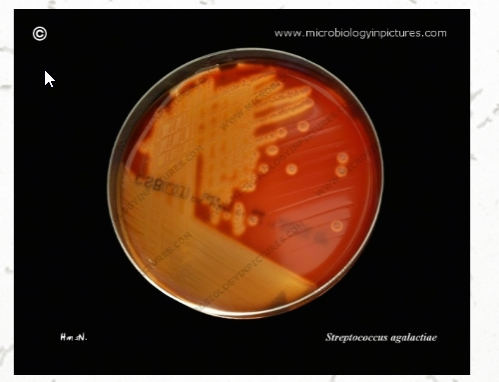
Streptococcus agalactiae
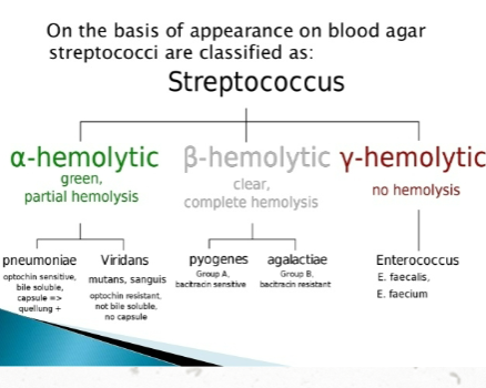
Describe the different hemolytic presentation on agar
Describe CSF Analysis
Procedure
What is the normal CSF cell count
What can cause an abnormal CSF cell count
What is the normal glucose levels?
These measurements determine?
What can cause abnormal Glucose levels?
What is the normal protein levels?
What can cause elevated levels?
What diseases can it indicate
Describe microbial analysis
Types
Procedures
Procedure:
Specimen collection
Up to 4 samples collected
2-3 mL each
Each sample is labeled based on order of draw
Tube #1 – chemistry/serology
Tube #2 – microbiology
Tube #3 – hematology
Tube #4 – special immunologic tests, protein electrophoresis
CSF cell counts
Normal – no cells
0-5 wbc/uL
Differential
Obtained via cytospin prep
Tube #3 – hematology = specimen of choice
CSF Cell Counts abnormal:
Neutrophilia
Bacterial meningitis
Tubercular meningitis
Fungal meningitis
Increased lymphocytes
Viral meningitis
Glucose
60% of the blood glucose level
Measurement is useful to determine
Impaired transport of glucose from plasma to CSF
Utilization by leukocytes and microbes
Abnormal Glucose Determinates
Increased WBC w/ predominant neutrophils + decreased CSF glucose
bacterial meningitis
Increased WBC w/ predominant lymphocytes + decreased CSF glucose
tubercular meningitis
Increased WBC w/ predominant lymphocytes + normal CSF glucose
viral meningitis
Protein:
Normal: 15-45 mg/dL
Usage of Protein Levels:
Nonspecific indicator of some type of degenerative process
Meningitis, brain abscess, MS
Elevated Levels ->
increased permeability of BBB
Increased immunoglobulins in CNS
Decreased clearance of normal protein from CSF
Degeneration of neuronal tissue
Traumatic tap
Microbial analysis
Types:
Gram stain,
Acid-fast stain,
fluorescent antibody stains
Procedure:
CSF Centrifuged
Slide or cytospin prep, and cultures are prepared
blood cultures
India Ink
detect fungal meningitis