osteology and arthology of head and neck
1/177
There's no tags or description
Looks like no tags are added yet.
Name | Mastery | Learn | Test | Matching | Spaced | Call with Kai |
|---|
No analytics yet
Send a link to your students to track their progress
178 Terms
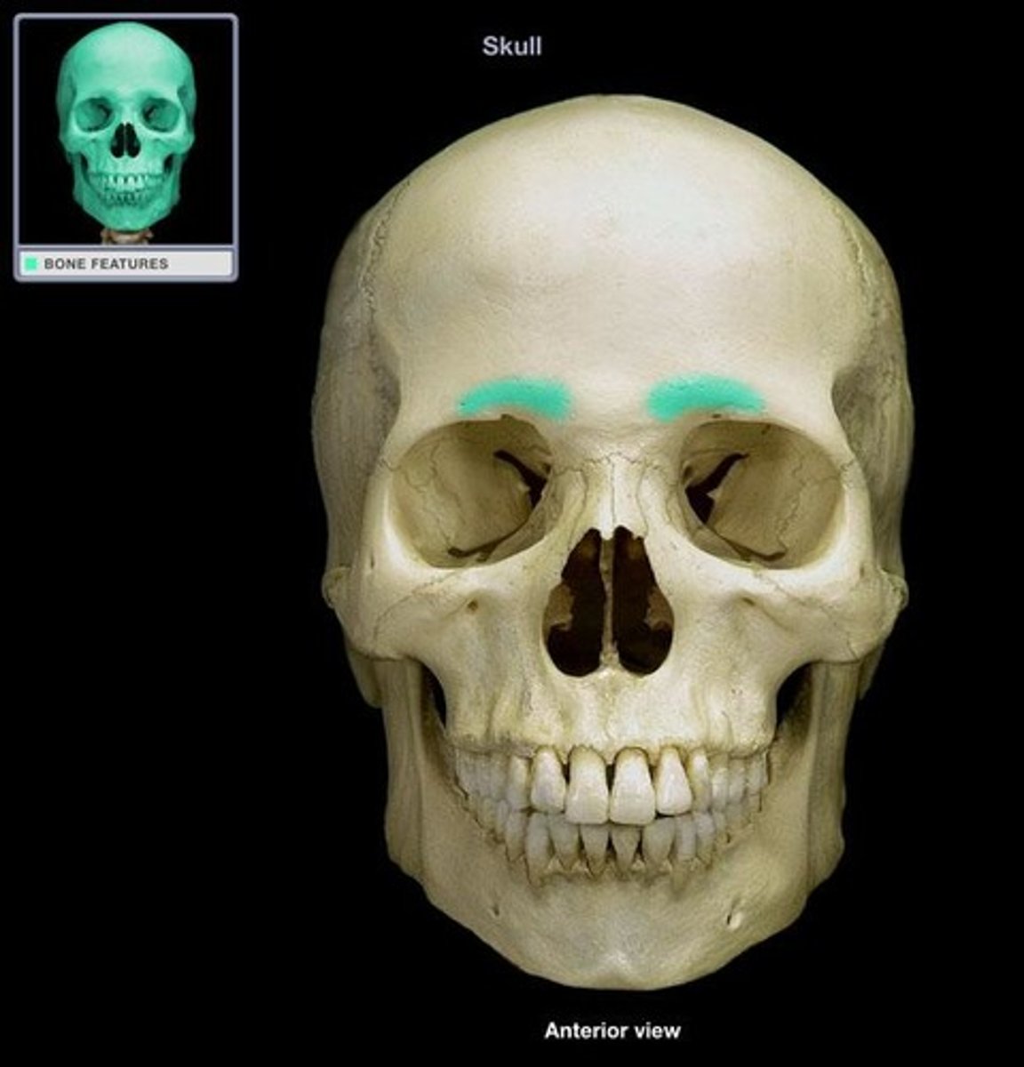
glabella
smooth, slightly depressed area between the superciliary arches (red)

supraorbital margin
angular boundary between squamous (flat) and orbital parts
supraorbital foramen
located in the supraorbital margin (orange)

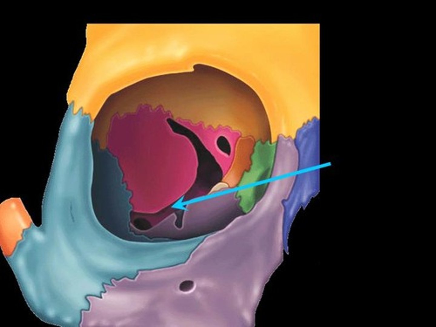
orbital plate of ethmoid bone
posterior to the lacrimal bone, very thin bone, contributes to medial wall
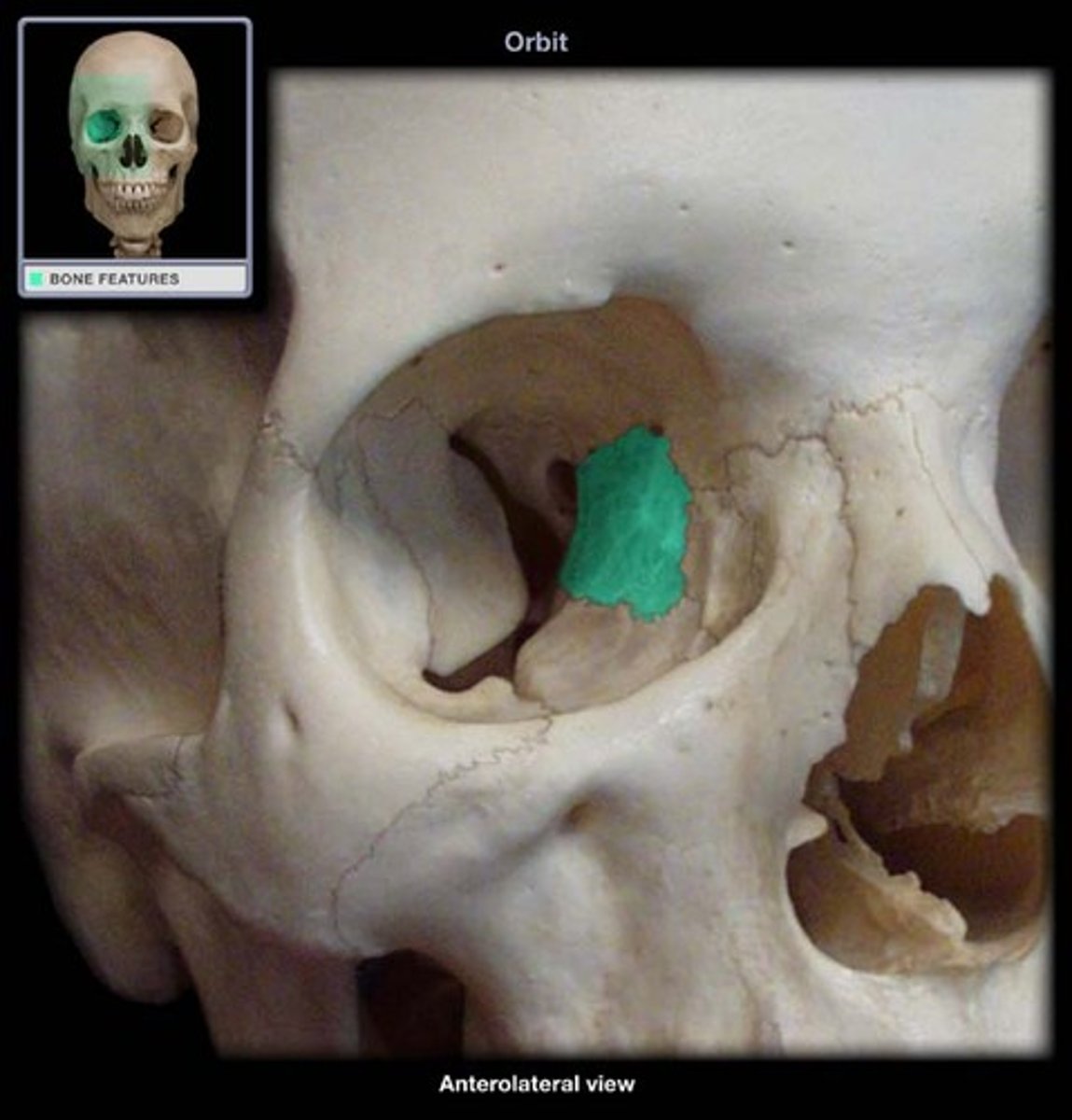
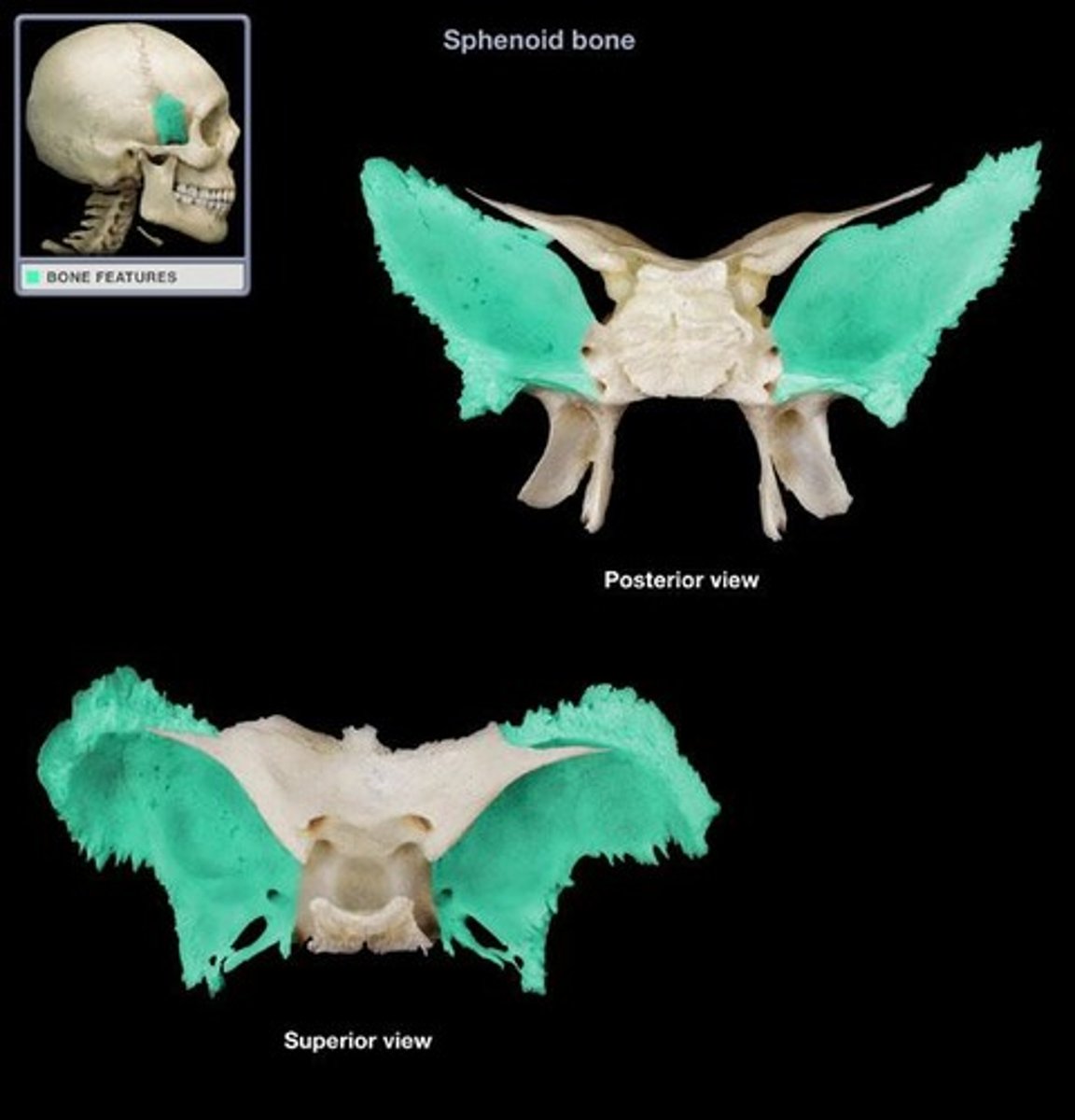
sphenoid bone
greater wing contribute to lateral wall, lesser wing contribute to superior wall near apex
where does the optic nerve go through?
is through the lesser wing of the sphenoid bone
orbital process of palatine bone
contribute slightly to apex

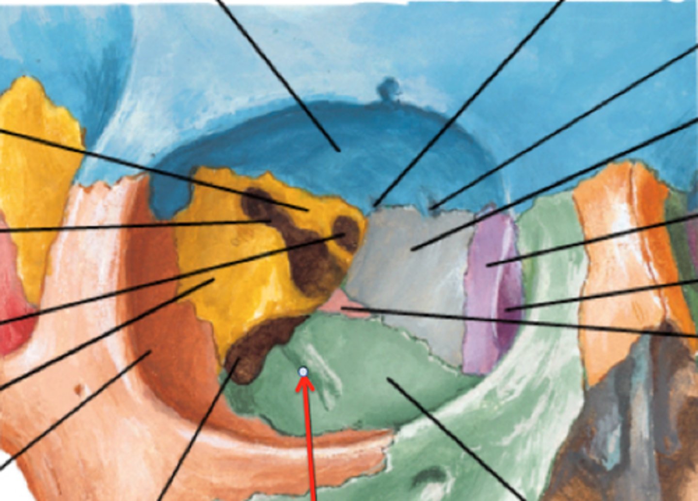
superior orbital fissue
located between the greater and lesser wings of sphenoid
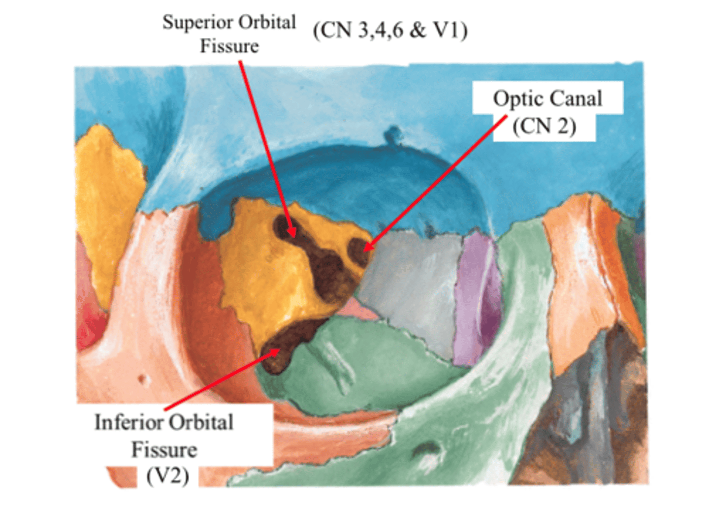
inferior orbital fissure
between maxilla, zygomatic and greater wing of sphenoid
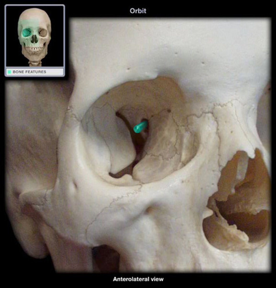
infraorbital groove
depression on the orbital surface of maxilla
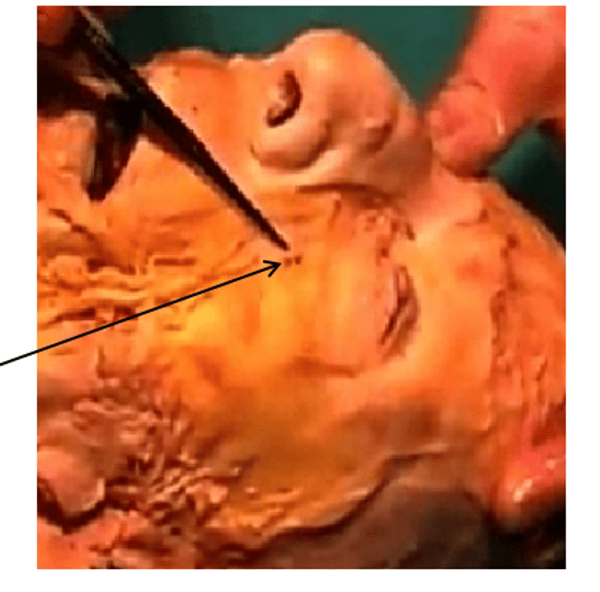
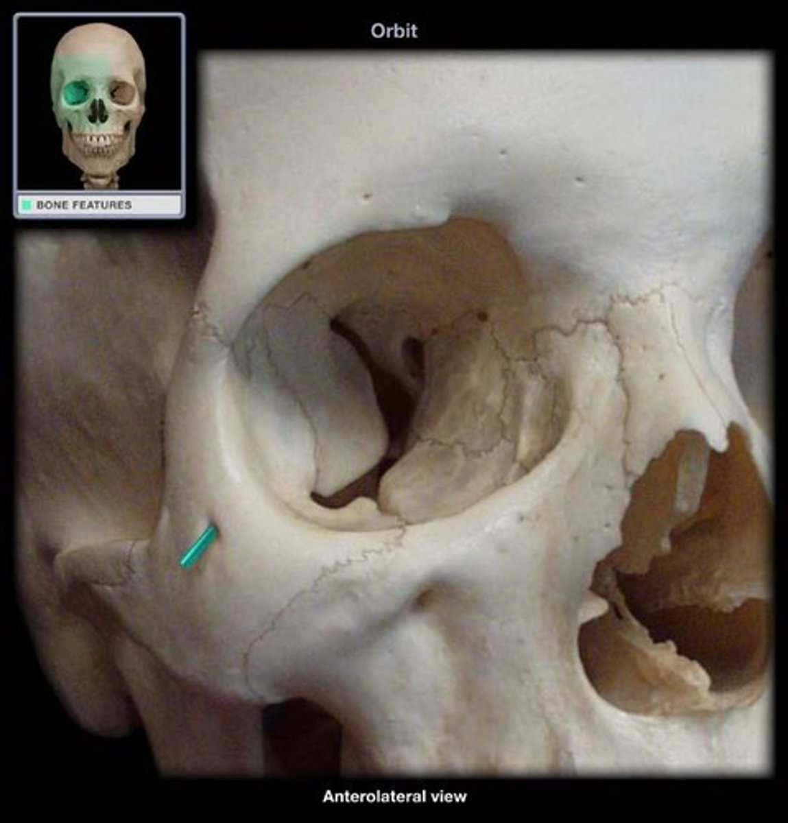
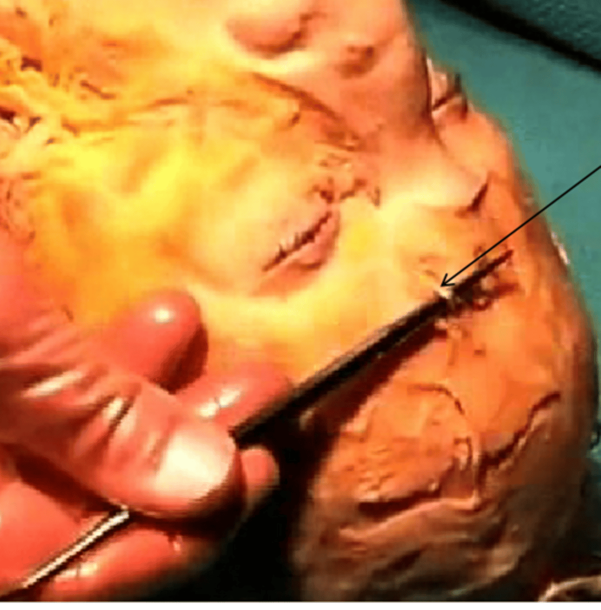
cadaver correlate -> infraorbital
infraorbital nerve on top of probe after locating the infraorbital foramen
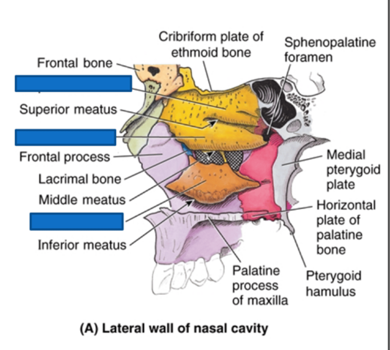
what are the four parts of the nasal cavity?
nasal bones, piriform, nasal septum, nasal conchae
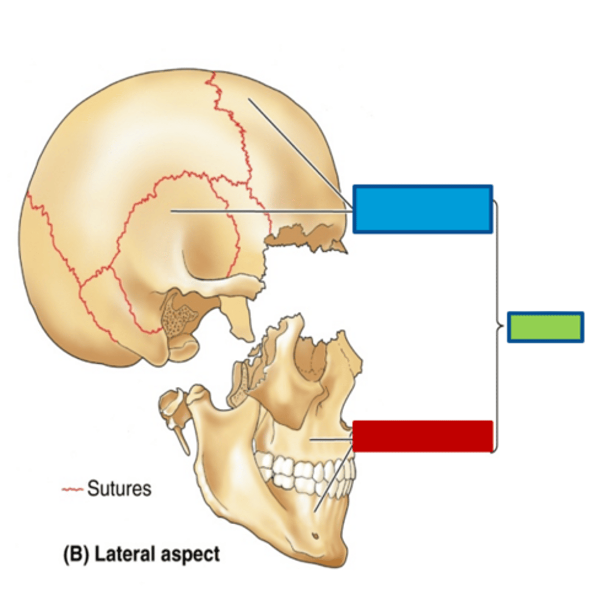
cranium
skeleton of head
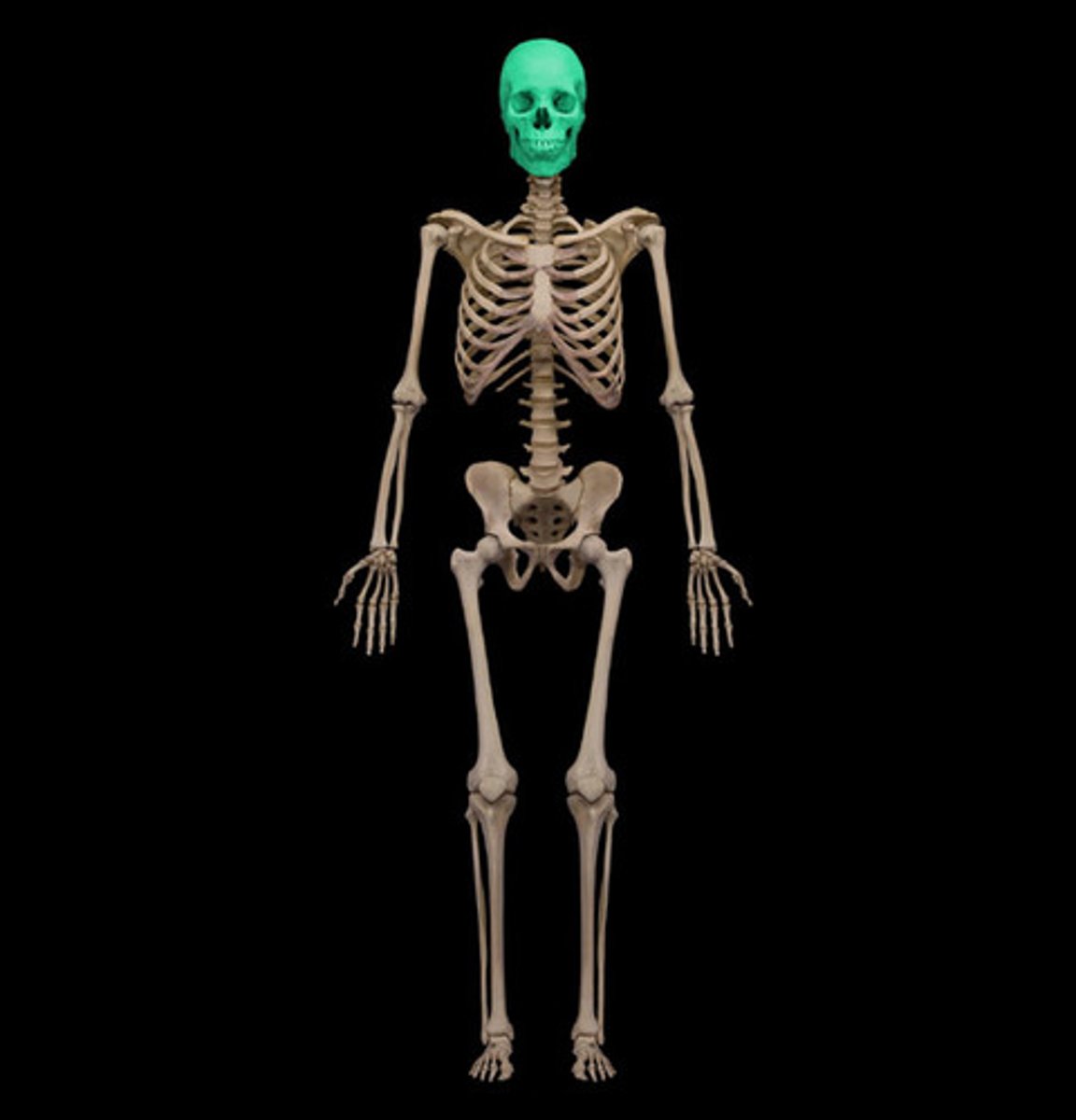
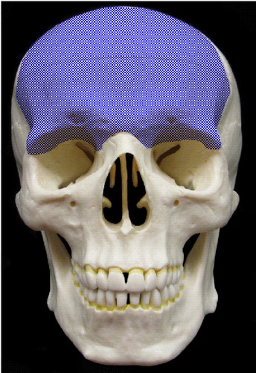
neurocranium
boney case of head, including cranial meninges (blue)
which bones are singular in the neurocranium?
frontal, ethmoid, sphenoidal, occipital
which bones are paired in the neurocranium?
temporal and parietal
calvaria
dome-like roof of the neurocranium (skullcap)
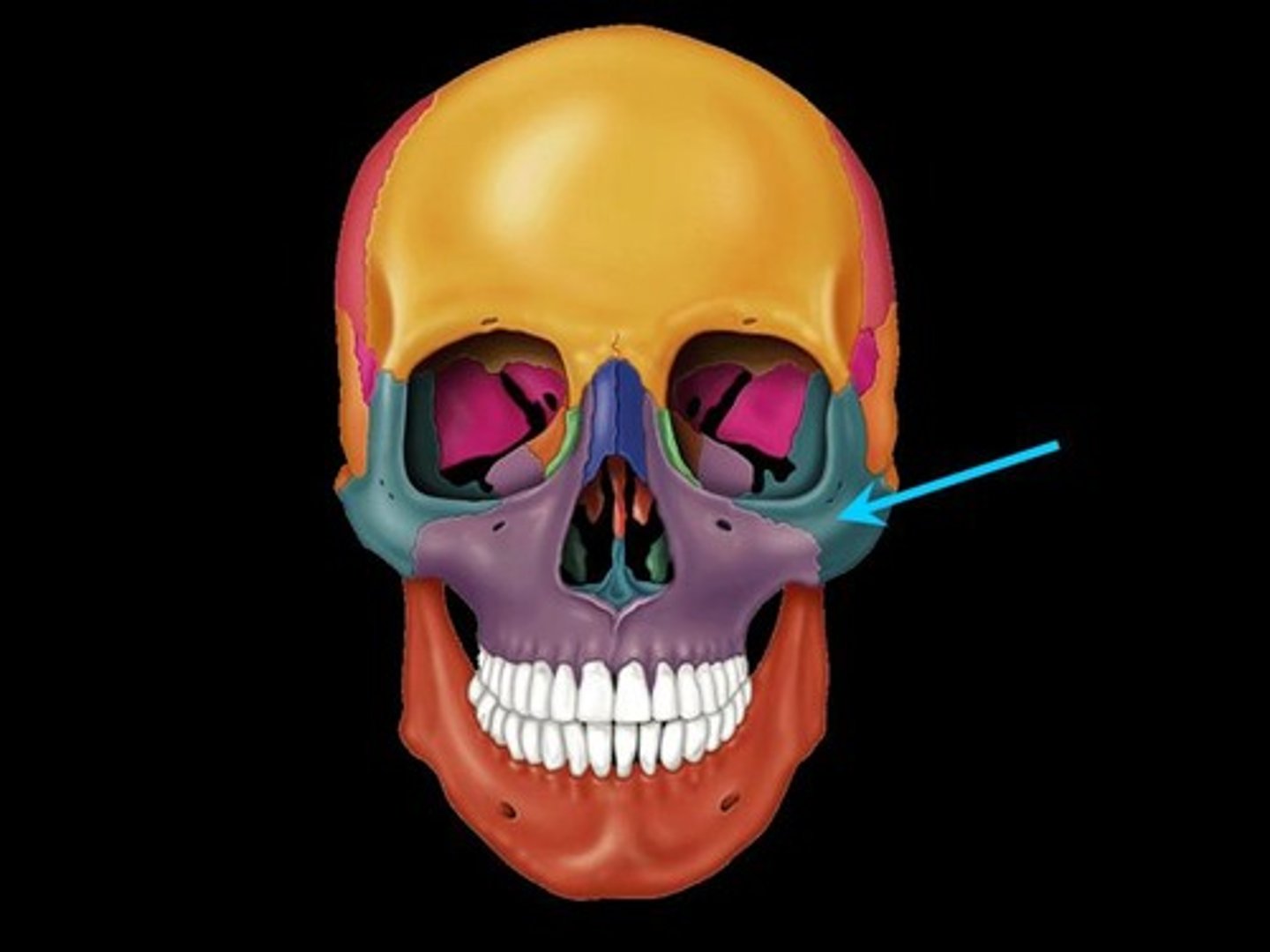
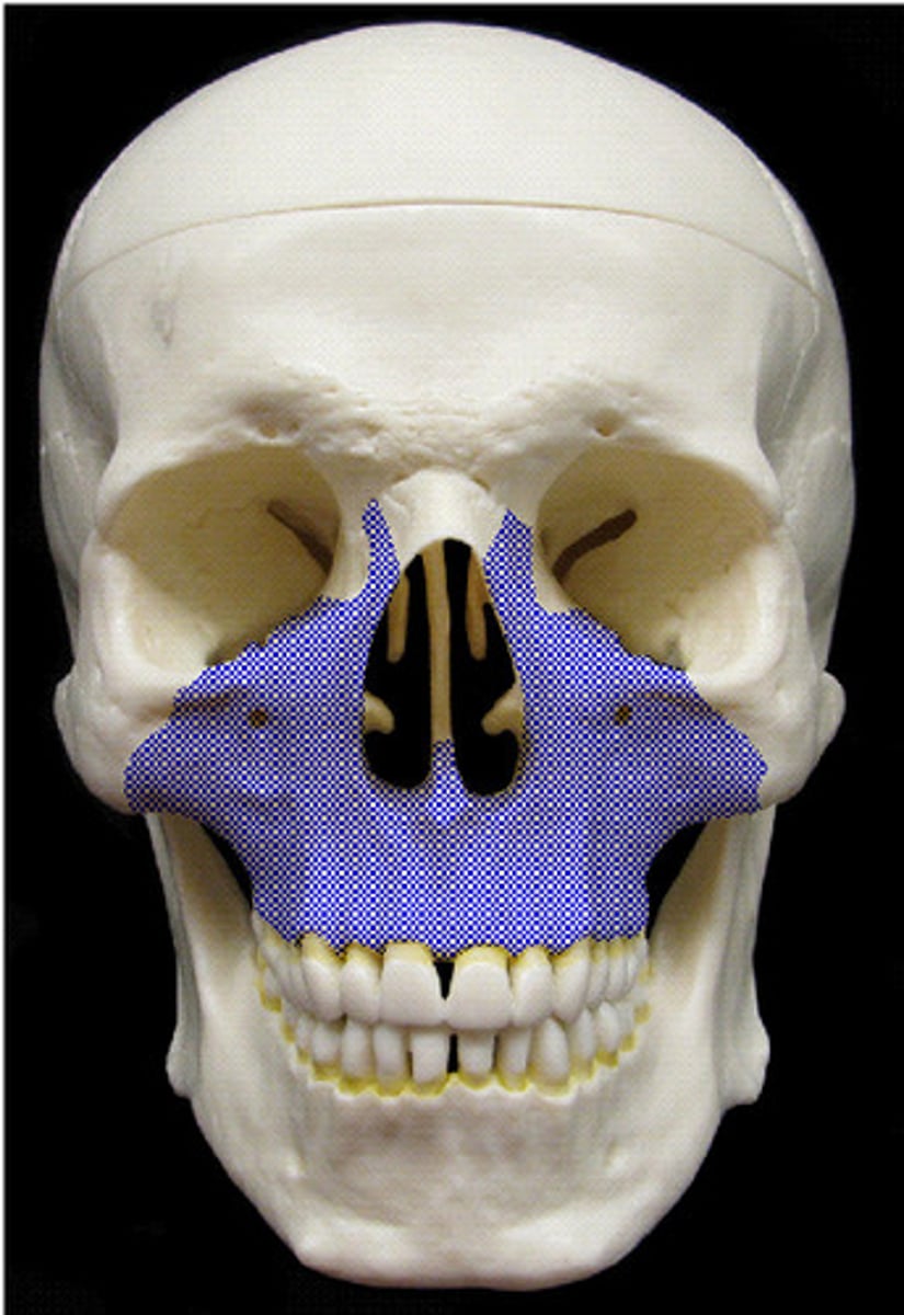
viscerocranium
anterior part of cranium (red)

what are singular bones of the viscerocranium?
mandible, ethmoid, vomer
what are the paired bones of the viscerocranium?
maxillae, inferior nasal conchae, zygomatic, palatine, nasal, lacrimal
fontanel
unossified area in the infant cranium
what are air sinuses?
pneumatized bones that contain air spaces that increase with age
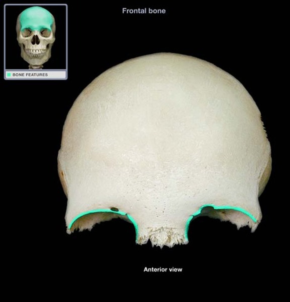
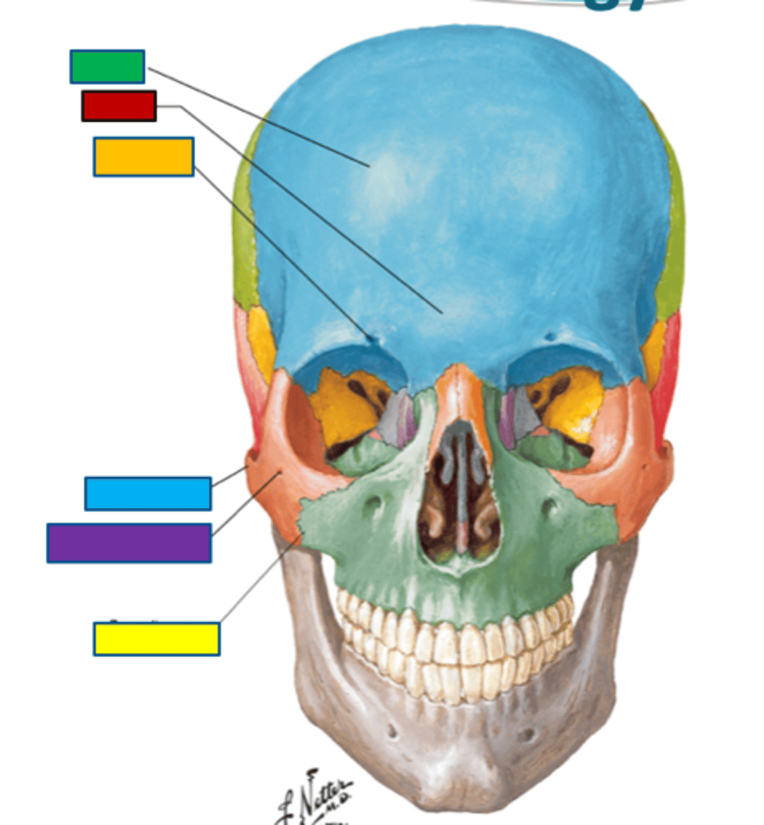
frontal bone
skeleton of forehead and superior margin of the roof of the orbit (green)
superciliary arches
ridge extending on each side of the labella typically larger in males
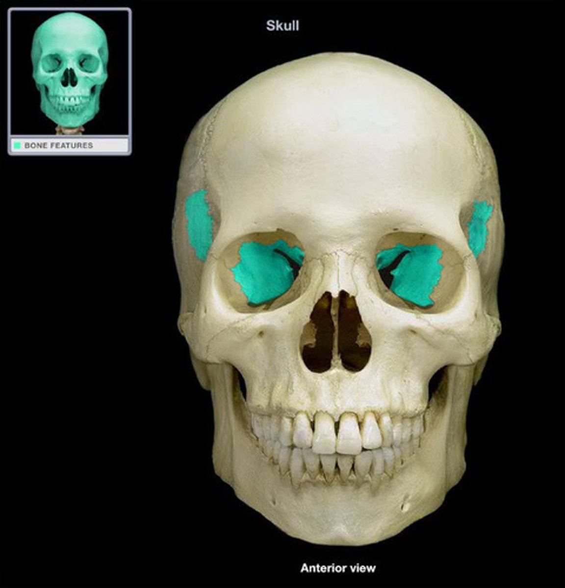
zygomatic bones
form prominence of cheeks, lie inferolateral to orbits and rests on maxillae
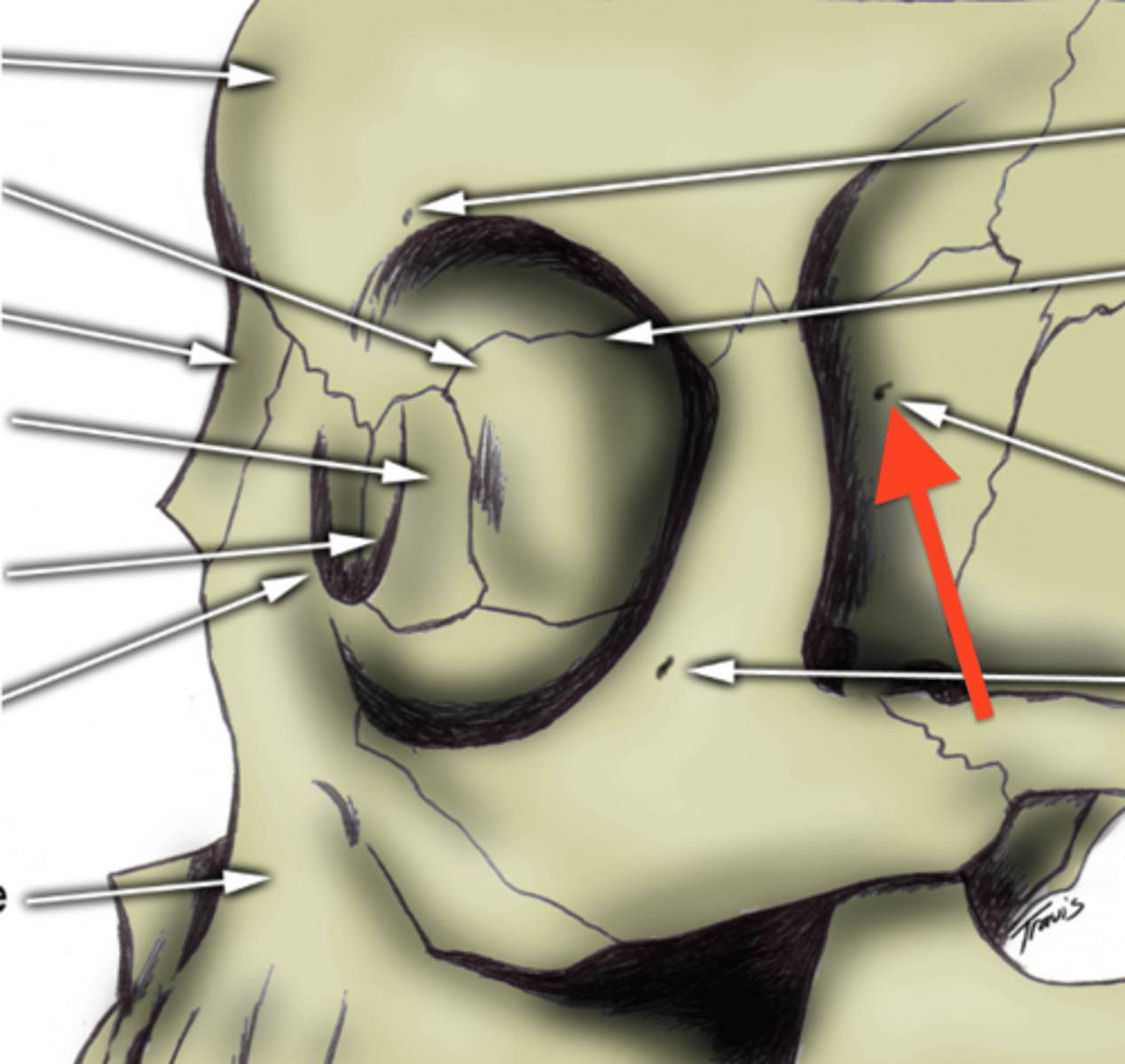
zygomaticofacial foramen
zygomatic bone on facial side
zygomaticotemporal foramen
on zygomatic bone temporal side
cadaver correlate
supraorbital nerve on top of probe after locating supraorbital foramen or notch
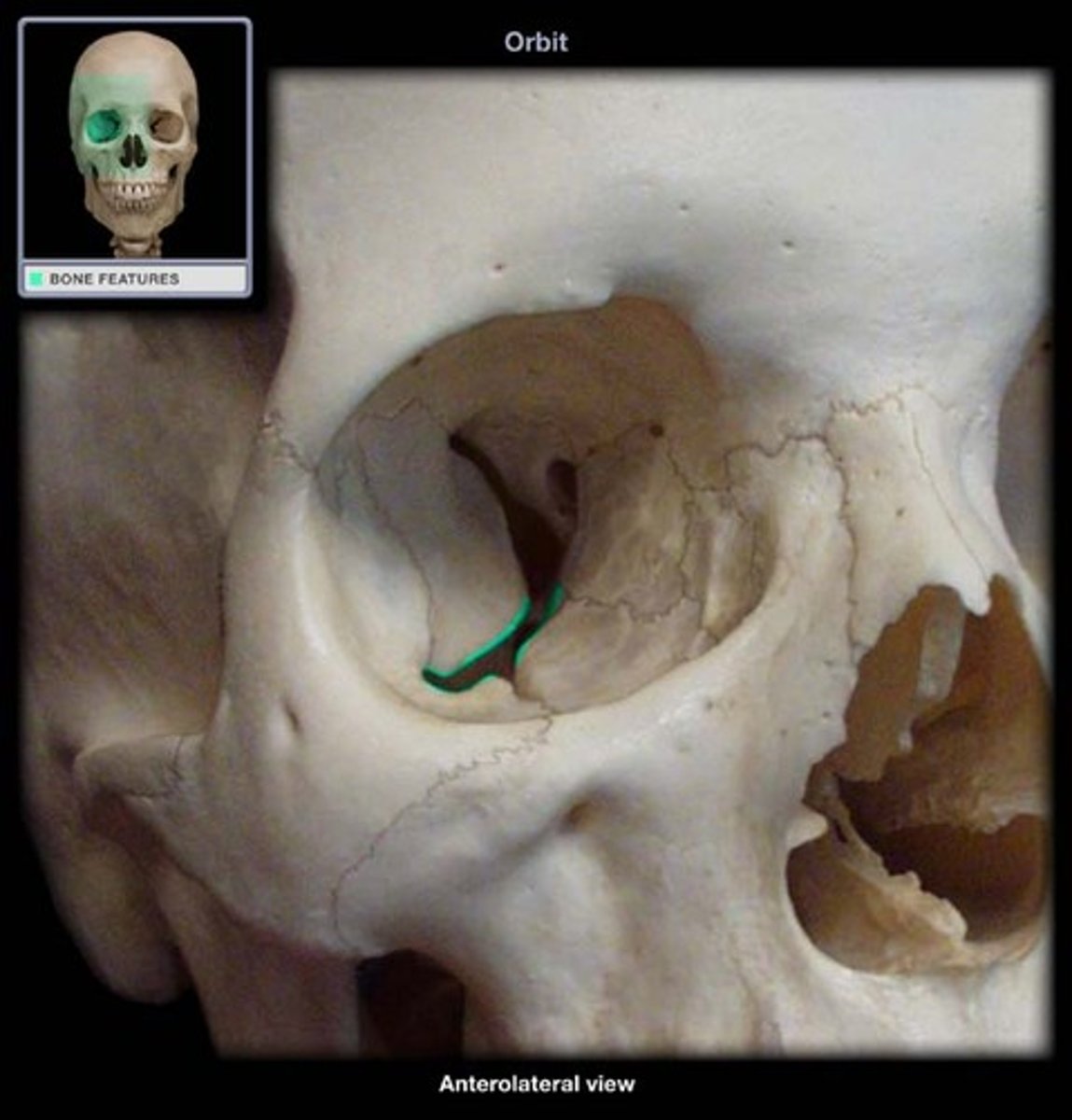
orbit
quadrangular pyramid, base anteriorly, apex posteriorly and 4 walls
lacrimal bone
contribute to medial wall, has fossa for lacrimal sac
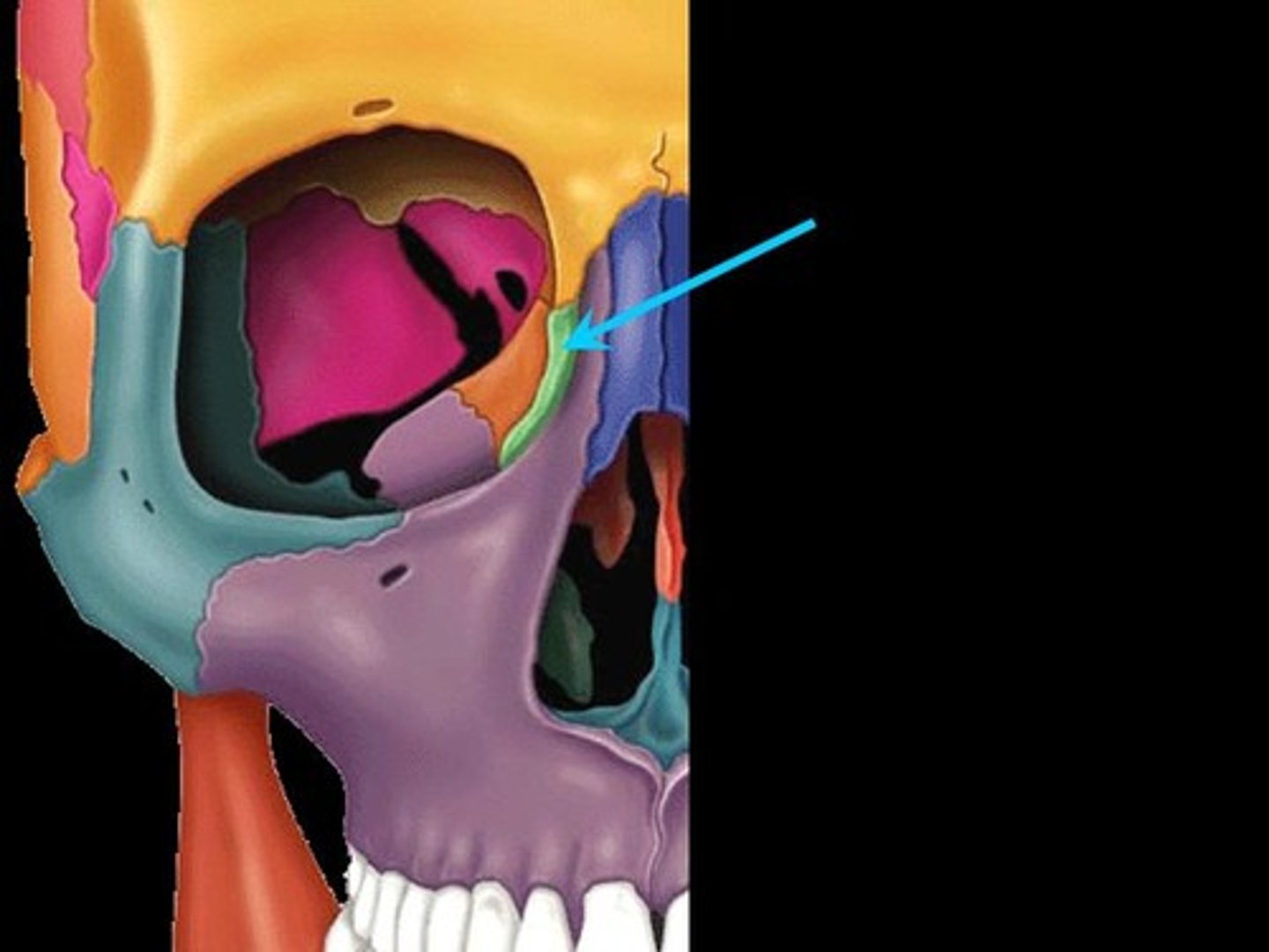
frontal bone (frontal view)
contribute to superior wall
zygomatic bone (frontal view)
contribute significantly to lateral wall

maxilla (frontal view)
contribute significantly to inferior wall
suture
fibrous joint between cranial bones
nasal bones
lies over ridge of nose, make up base of the nose
piriform aperture
pear-shaped anterior opening of the nose in cranium
nasal septum
made of perpendicular plate of ethmoid, vomer
nasal conchae
curvy bony plates on the lateral wall of each nasal cavity
what is the inferior, middle and superior parts of the nasal conchae located?
superior, middle, inferior
what are the two parts of the nasal conchae
inferior (individual bone) and middle + superior (part of ethmoid bone)
what part of the nasal conchae is an individual bone?
inferior aspect
how many inferior nasal conch are there?
2, right left
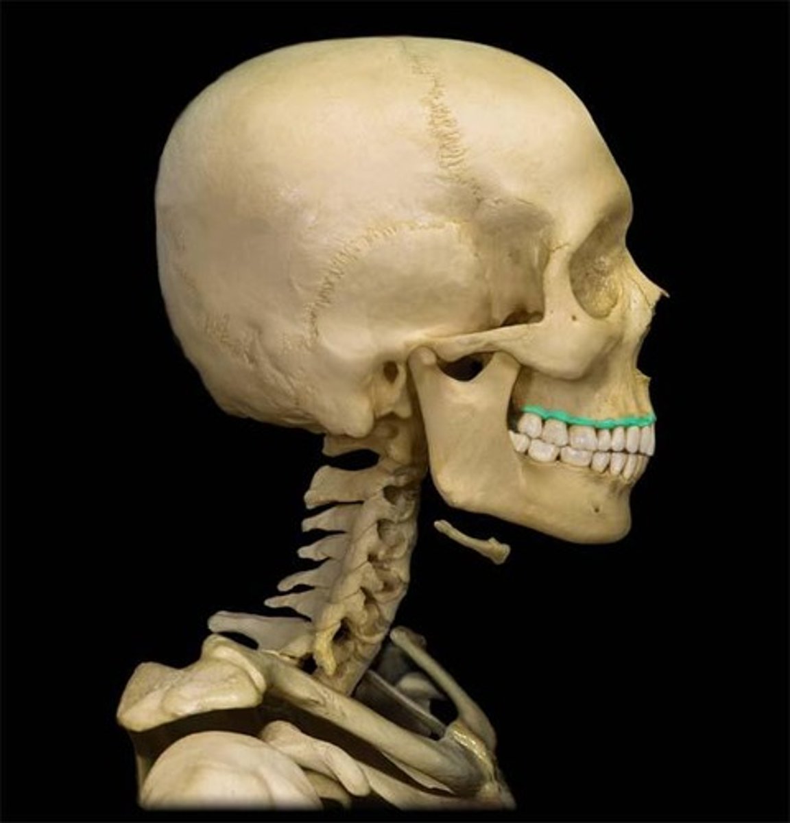
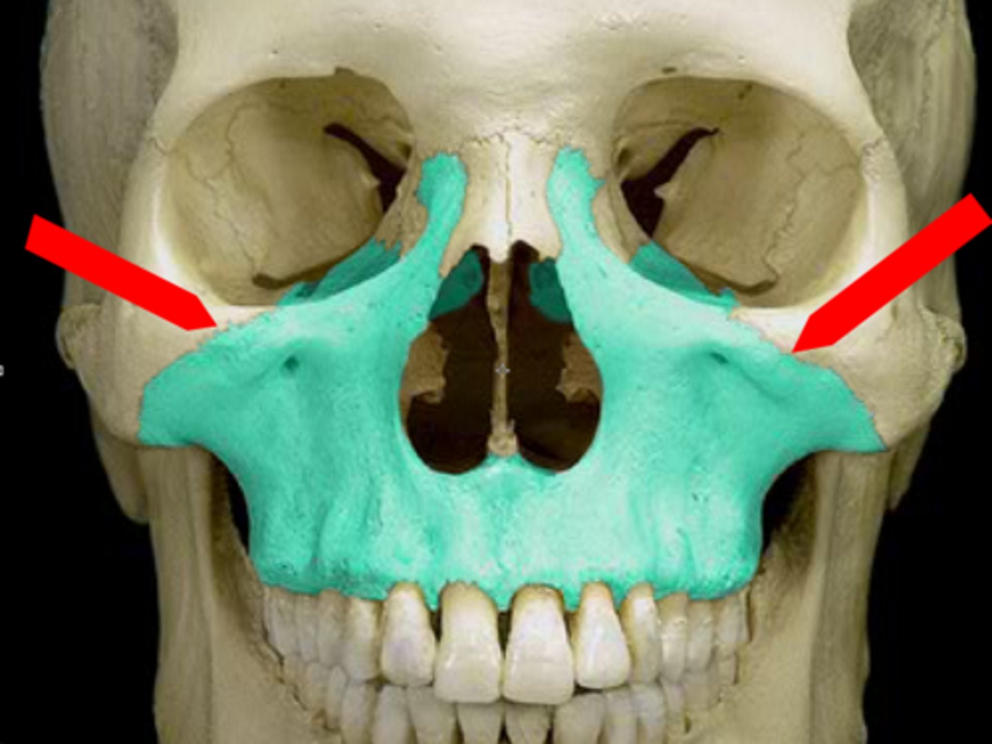
maxillae (2 right left)
form upper jaw and supporting for maxillary teeth
alveolar processes
tooth sockets (alveoli)
infraorbital foramen
inferior to each each orbit
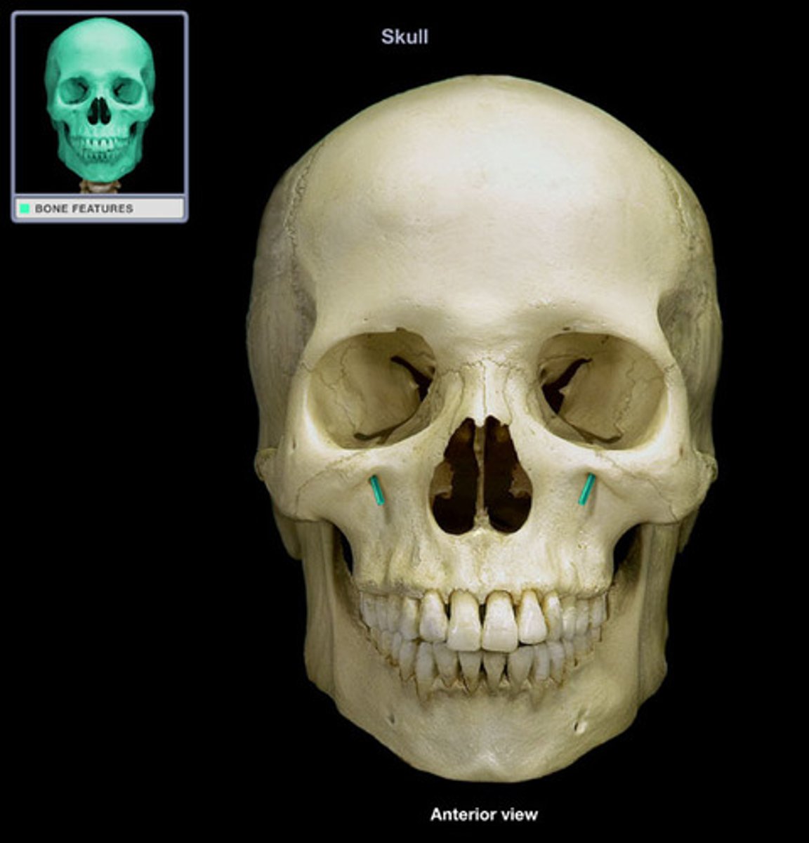
what exits the infraorbital foramen?
infraorbital nerve
zygomatic process of maxilla
articulates with zygomatic bone
maxillary tuberosity
posterior on maxilla behind last tooth, most prominent after growth of wisdom tooth
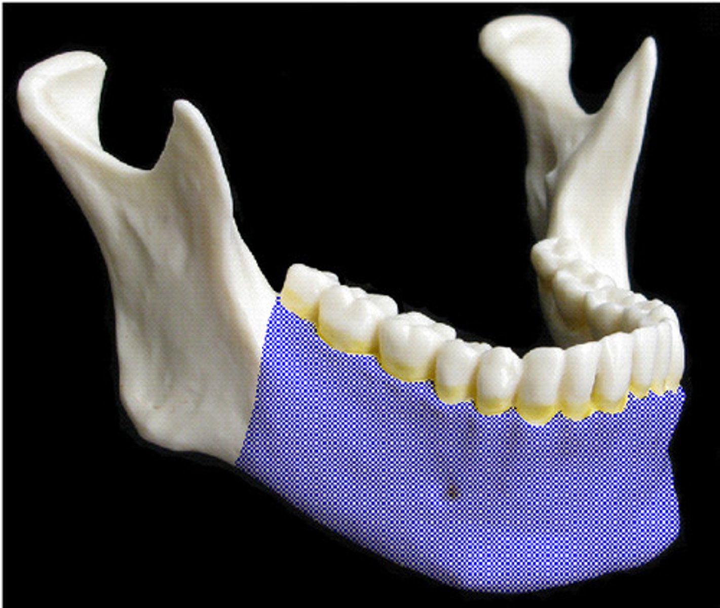
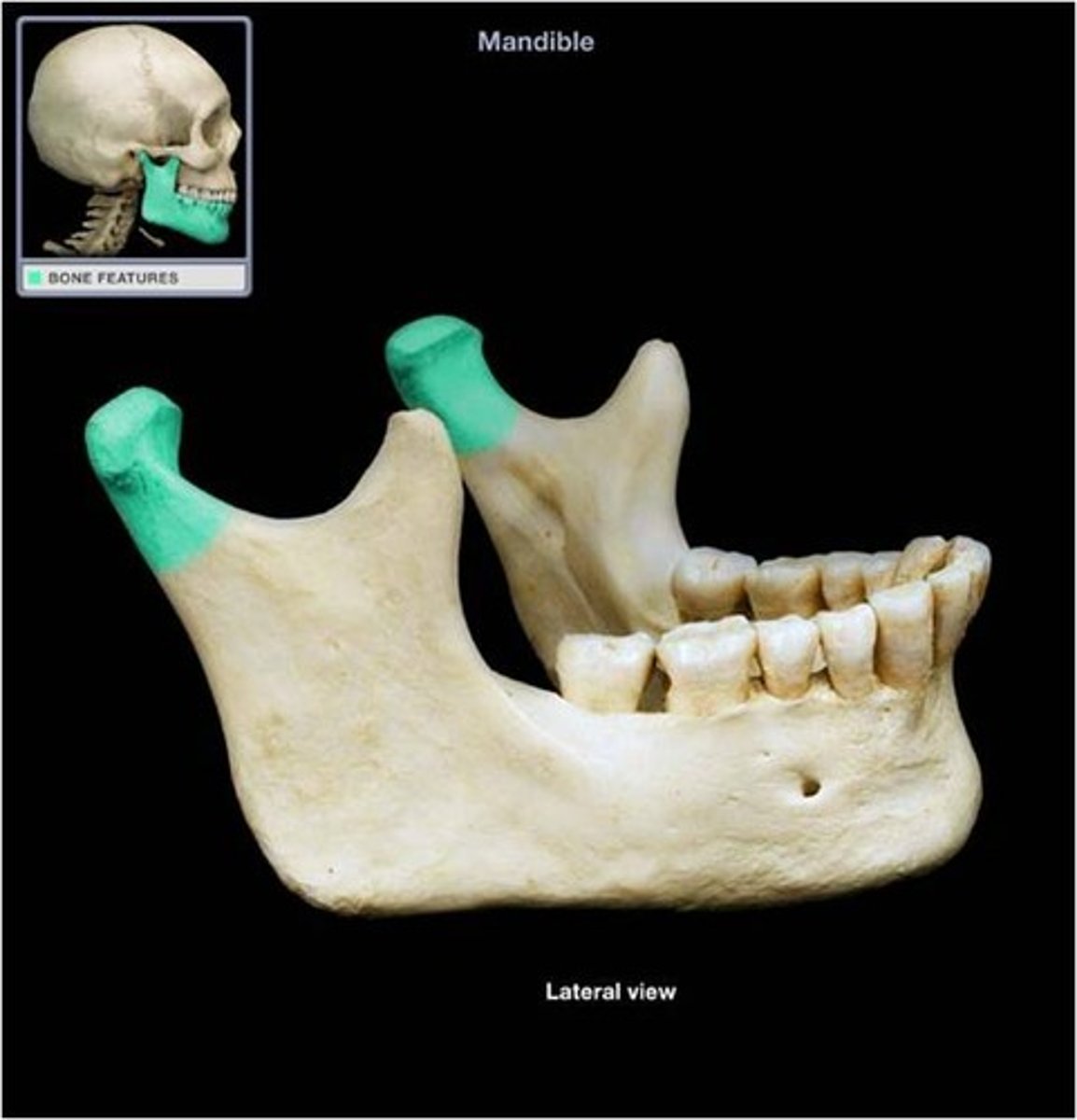
mandible
u-shaped that supports the mandibular teeth
body of mandible
horizontal part of mandible
ramus of mandible
vertical part of mandible
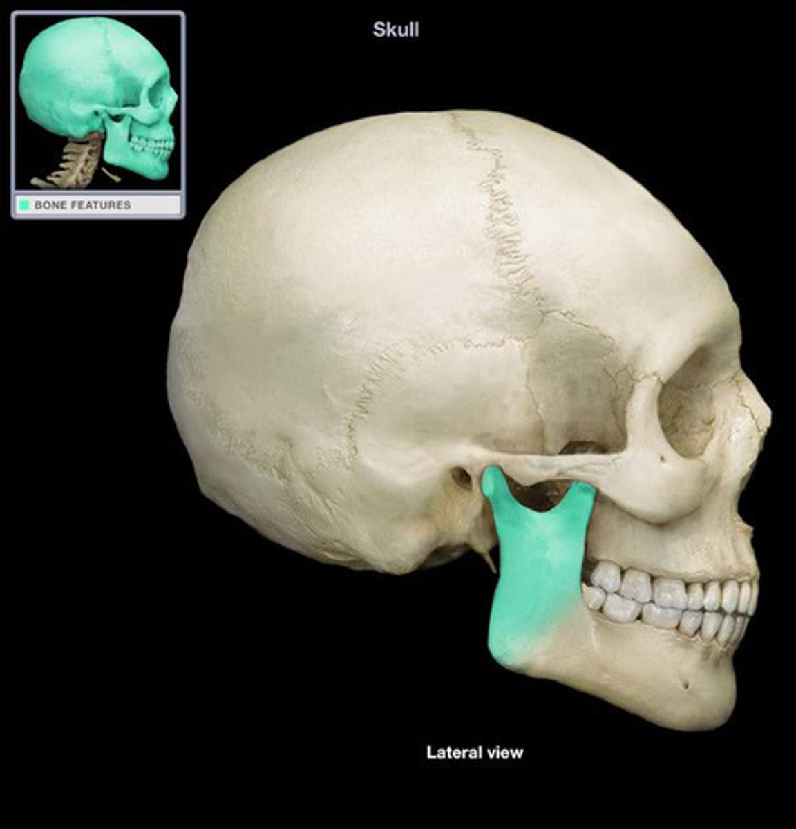
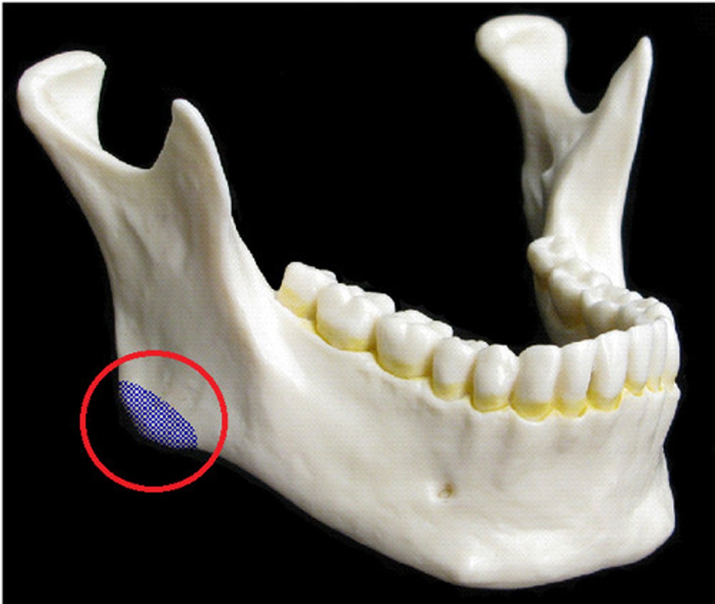
angle of mandible
union of body and ramus of mandible
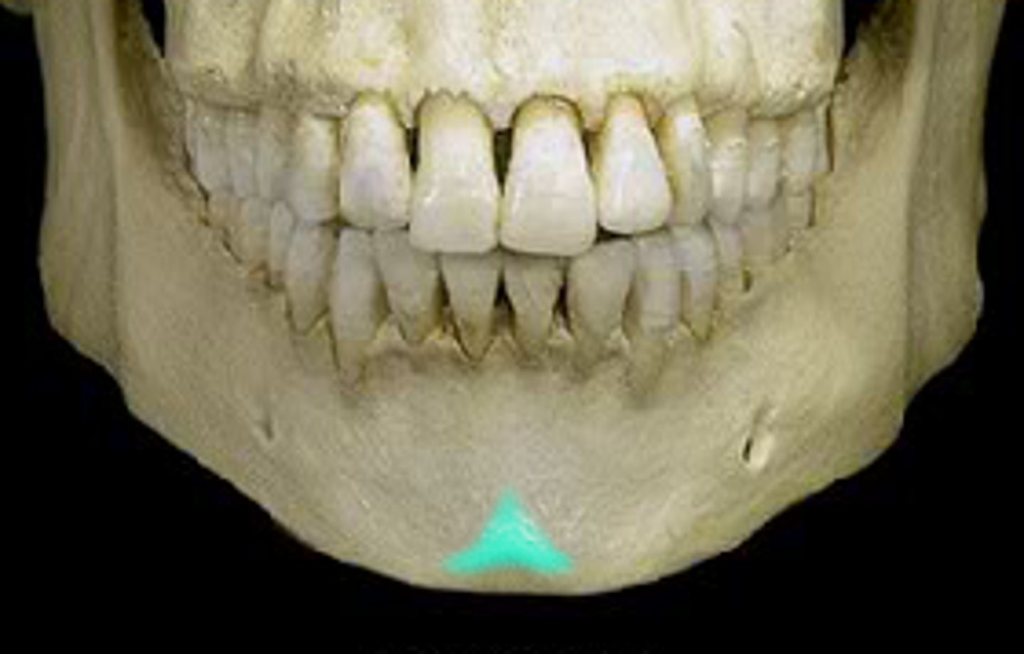
mental protuberance of mandible
forms prominence of chin
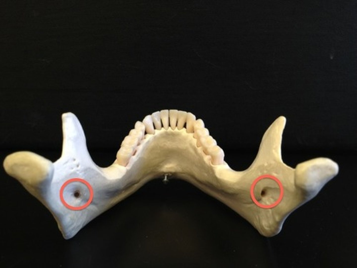
mental foramen of mandible
on the outside of body of mandible
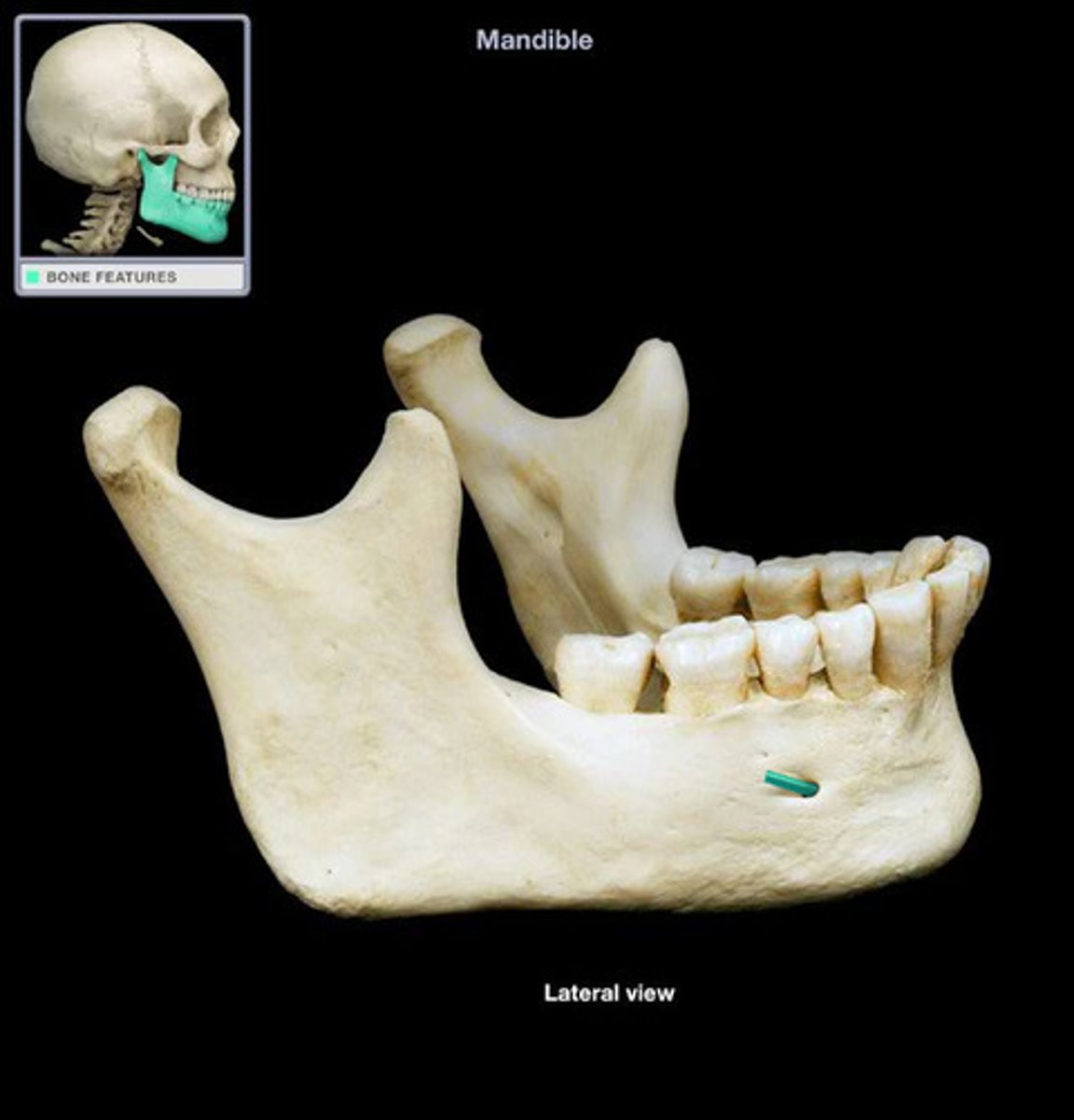
what exists the mental foramen?
mental nerve (started off as interior nerve in mandibular foramen)
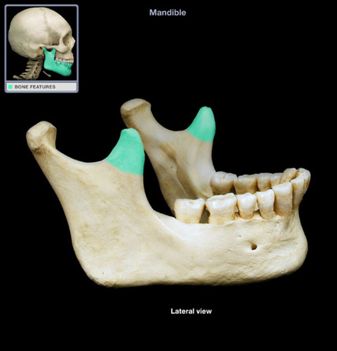
coronoid process of mandible
anterior to condylar process of mandible
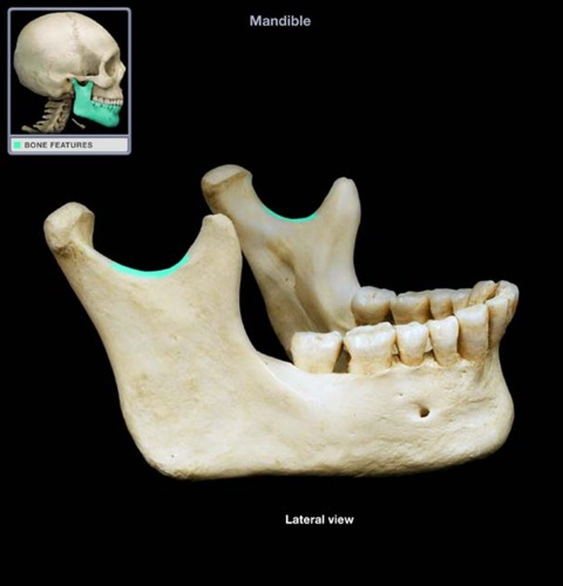
mandibular notch of mandible
between coronoid and condylar process of mandible
condylar process
inserts into mandibular fossa of temporal bone to form the TMJ
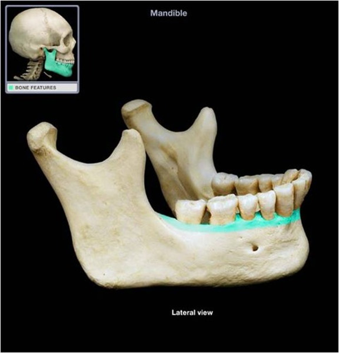
alveolar process of mandible
house the lower teeth
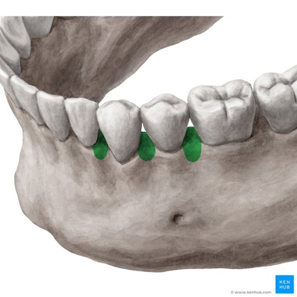
interalveolar septa
bony partitions separating tooth sockets
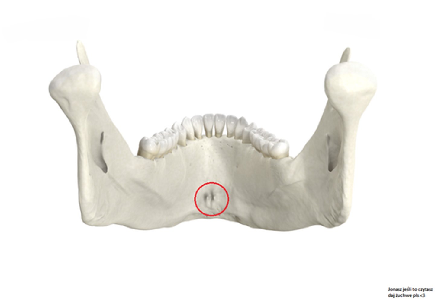
mental spine of mandible
site of attachment for muscles
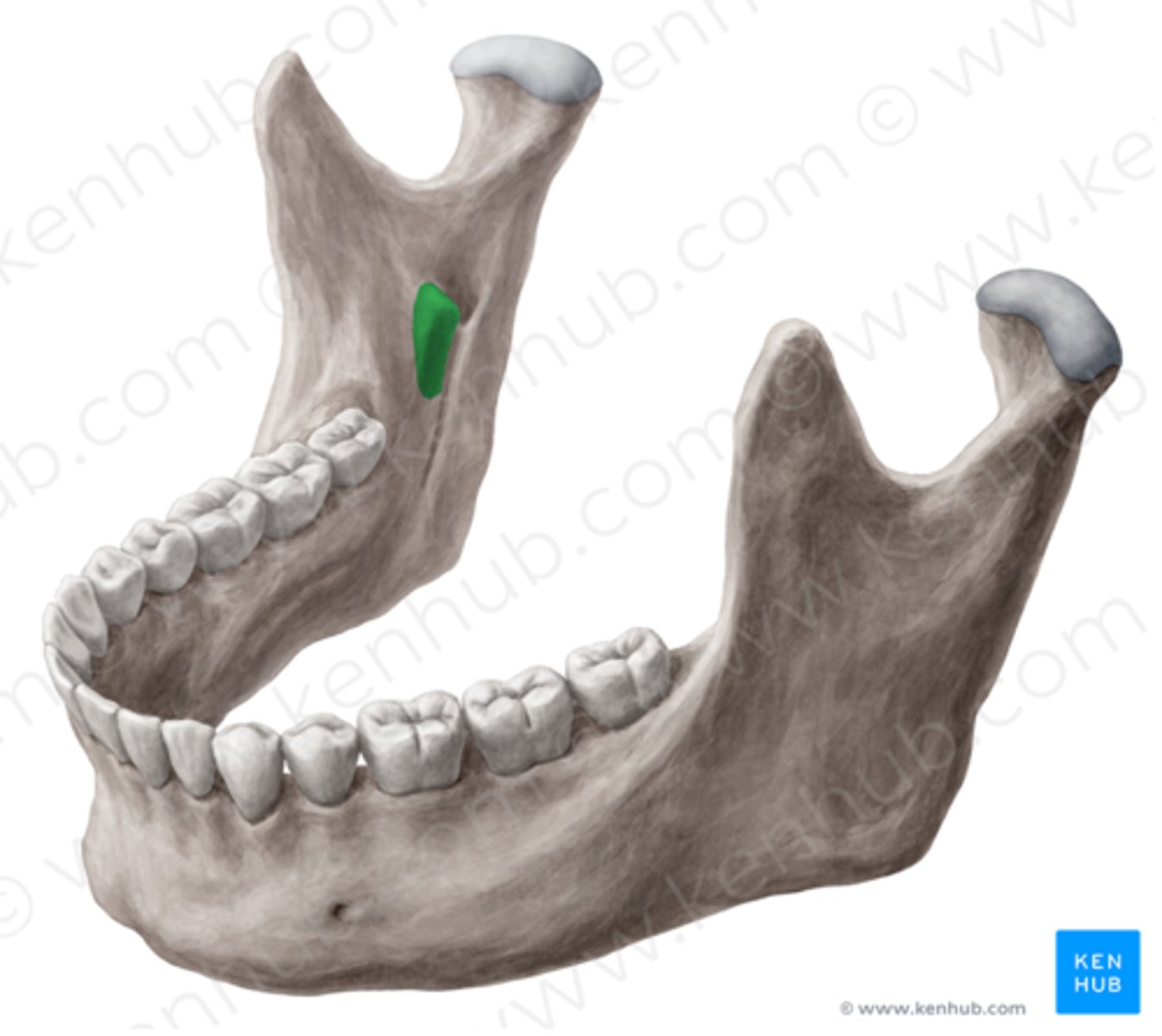
mandibular foramen
next to and guarded by the lingual
lingula
spinous process protecting the mandibular foramen and the nerve
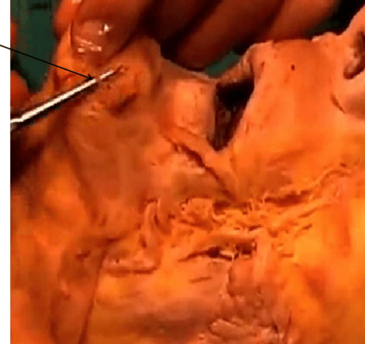
cadaver correlate
mental nerve on top of probe found after locating the mental foramen
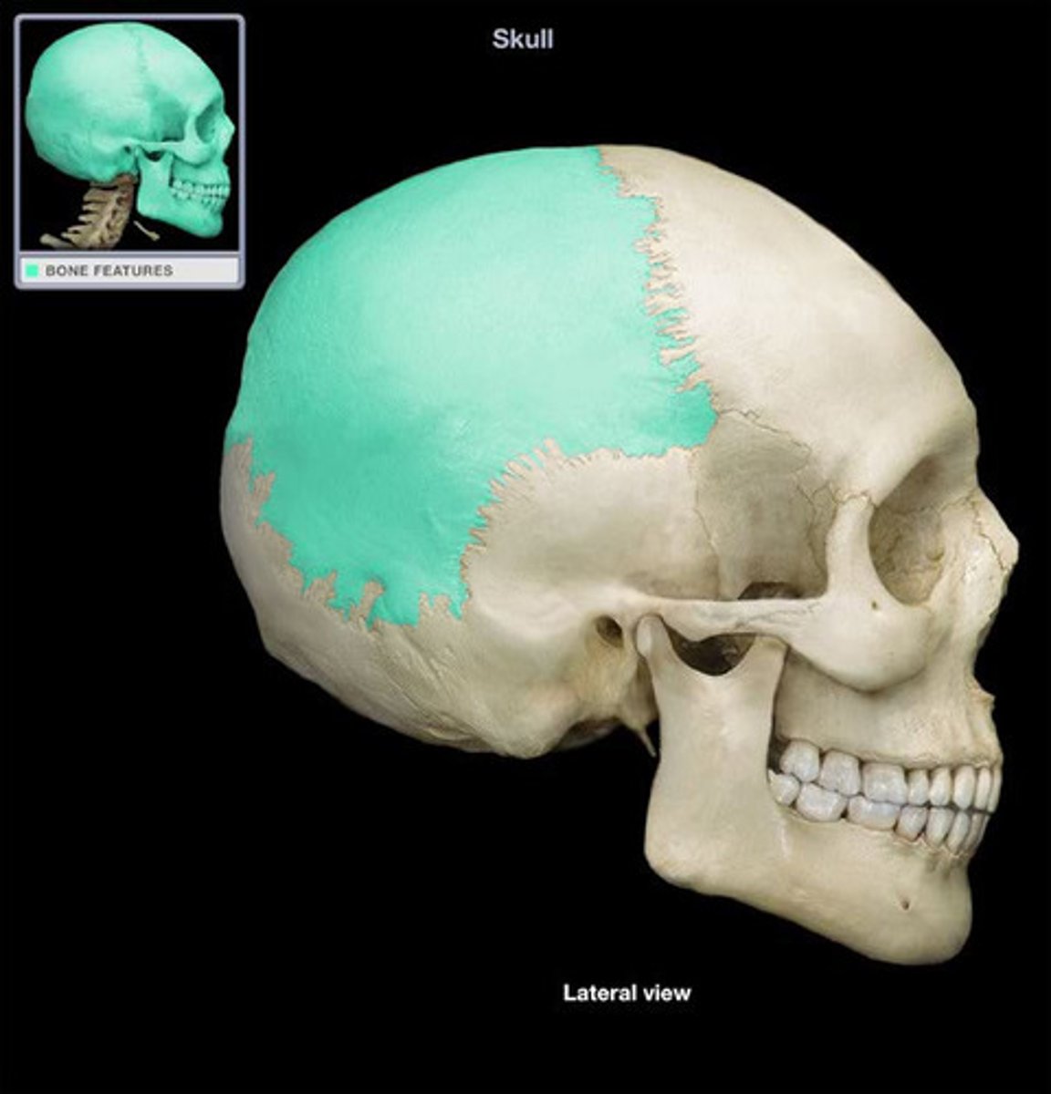
parietal bone
superior and lateral part of neurocranium
temporal bone
lies inferior to parietal bones on both sides
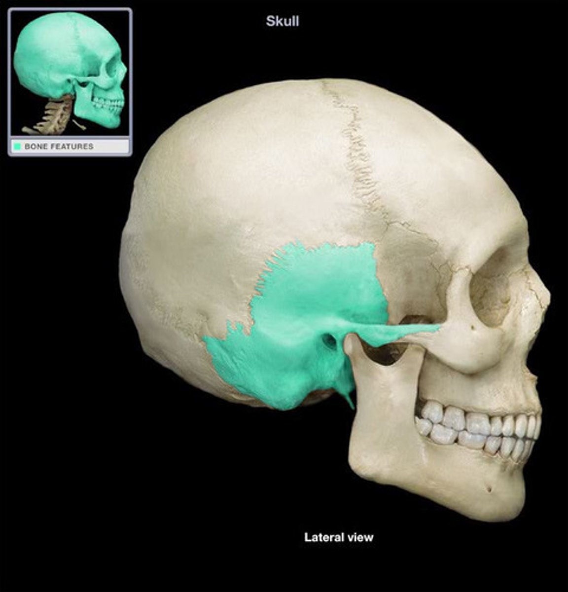
squamous part of temporal bone
anterior and upper part, contributes to temporal fossa
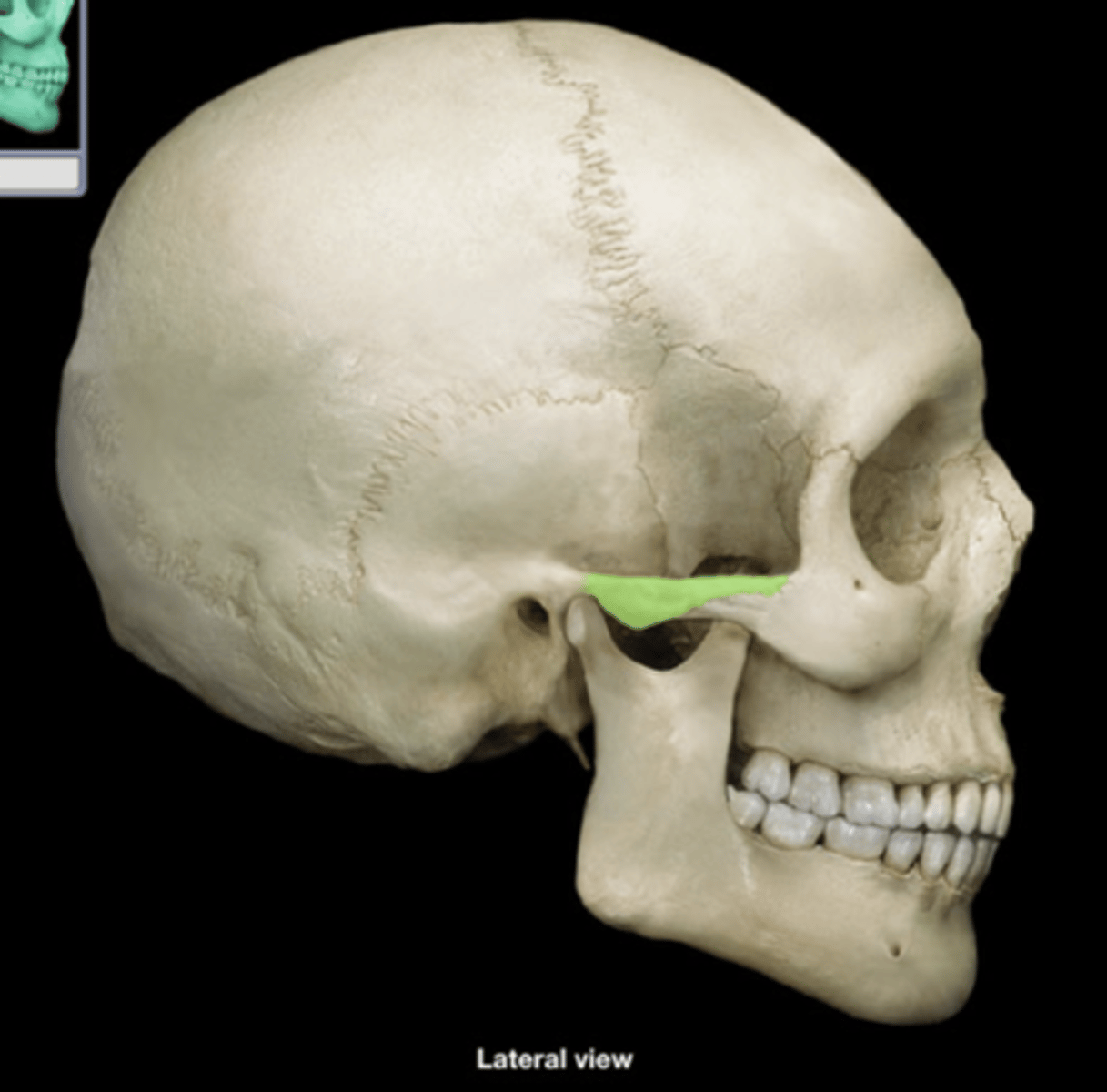
zygomatic process (lateral view)
forms part of zygomatic arch
external auditory meatus of temporal bone
external ear opening
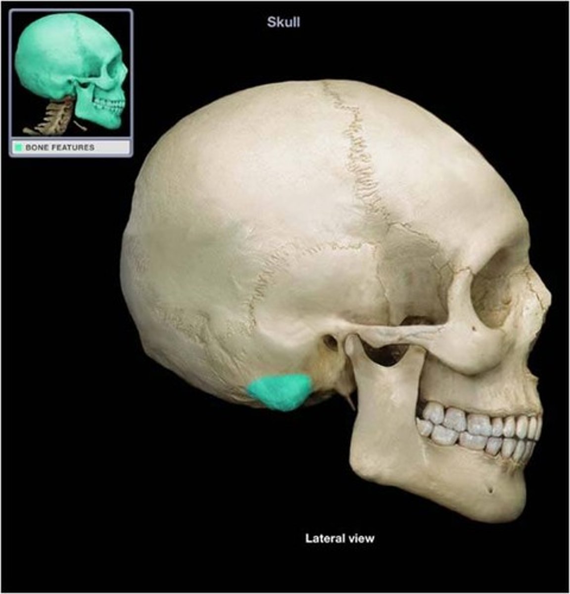
mastoid process of temporal
posteroinferior to external acoustic meatus opening, sternocleidomastoid muscle attachment
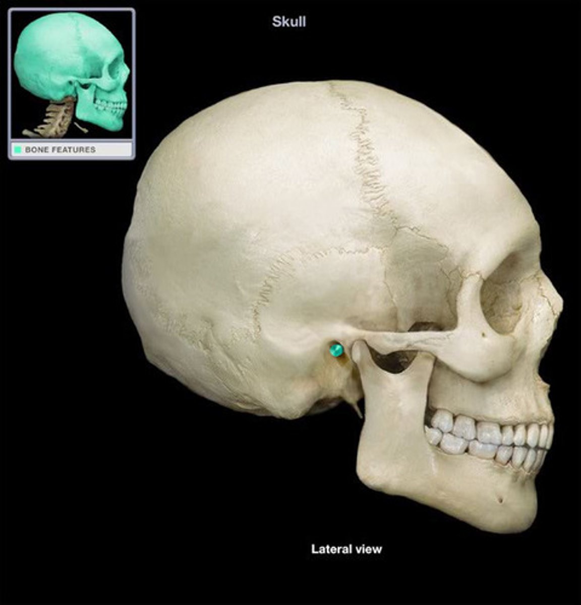
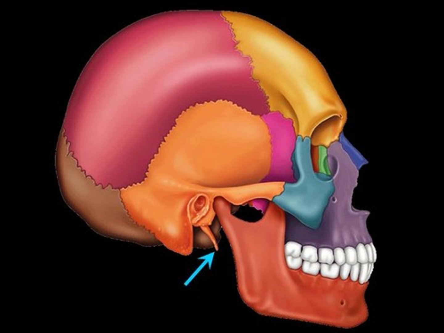
styloid process of temporal
anteromedial to mastoid process
articular tubercle of temporal bone
front boundary of mandibular fossa
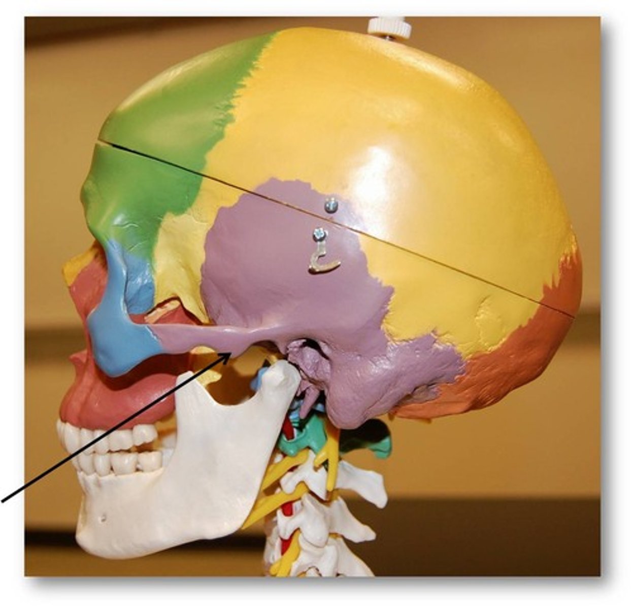
zygomatic arch
formed by zygomatic process (temporal) and temporal process (zygomatic)
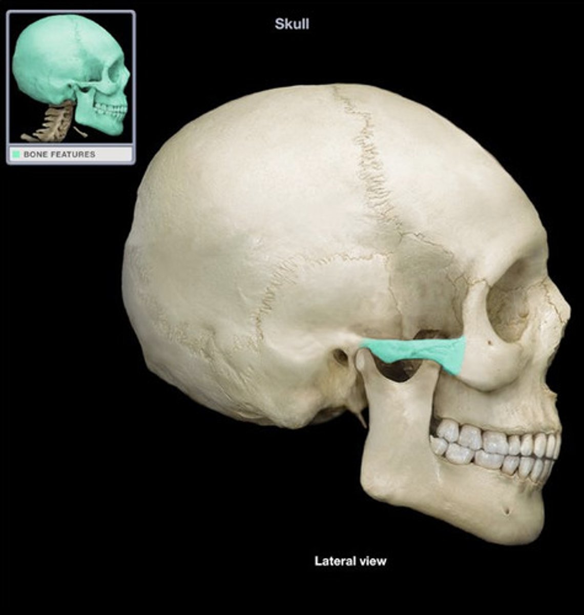
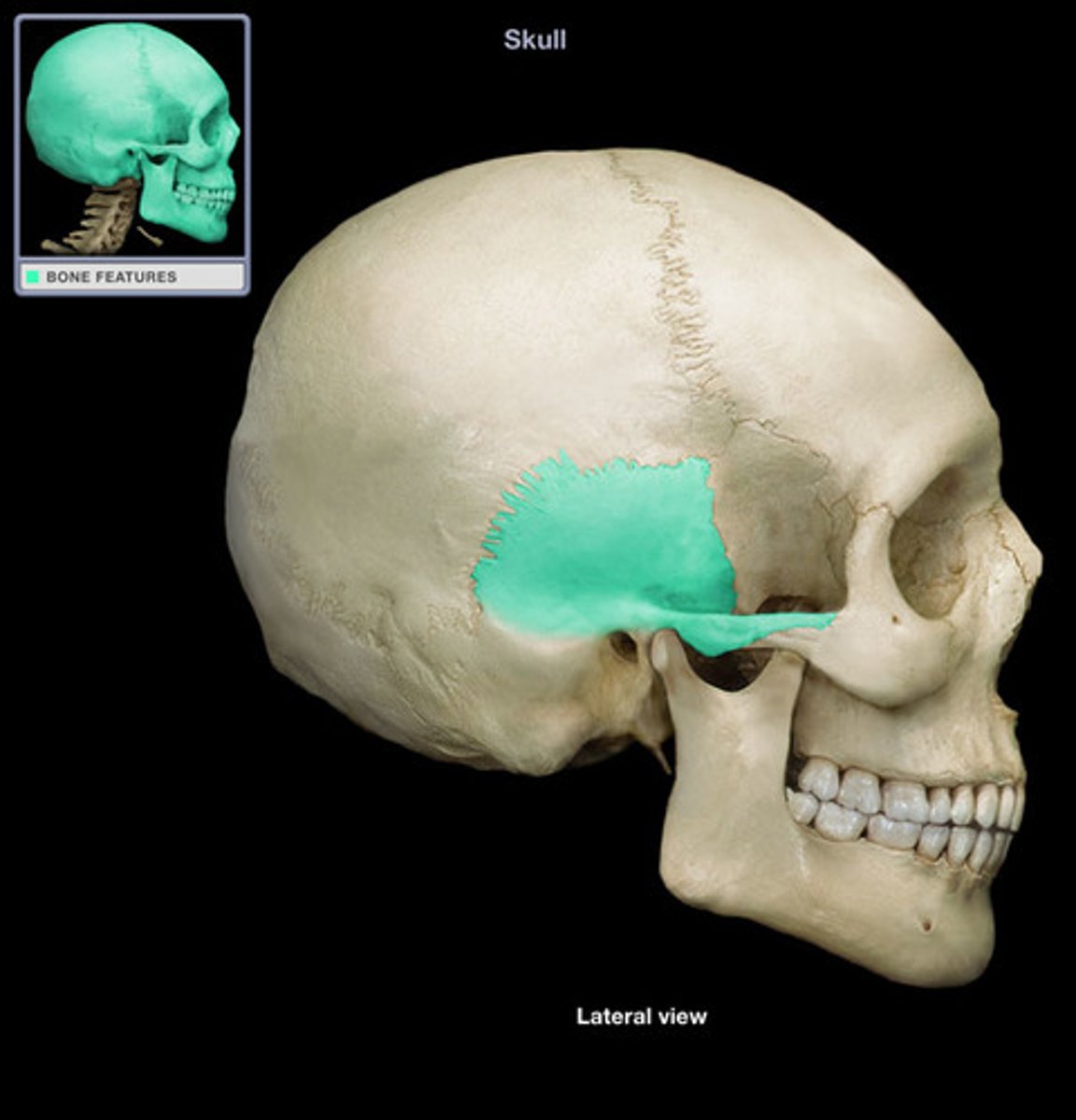
greater wing of sphenoid bone
anterior to temporal bone and inferior to frontal
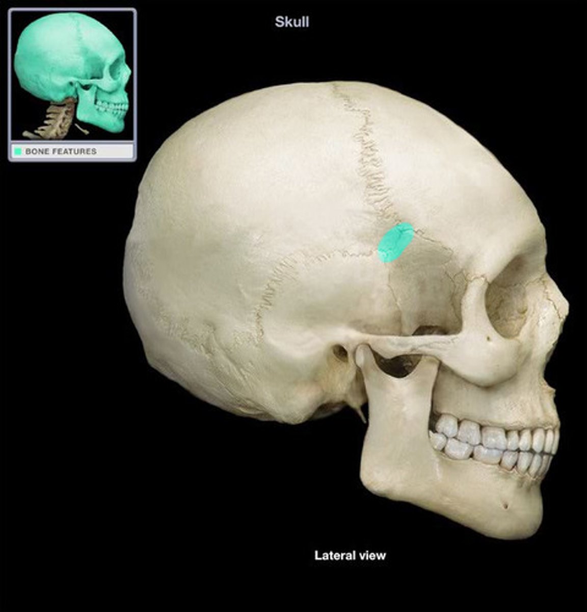
pterion
weak area of bone junction
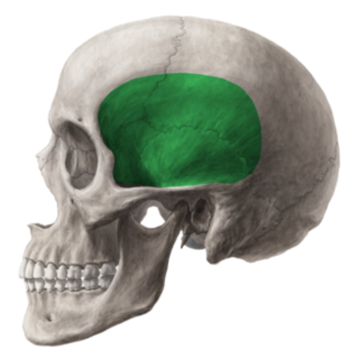
temporal fossa
muscle filled space - specifically temporals muscle
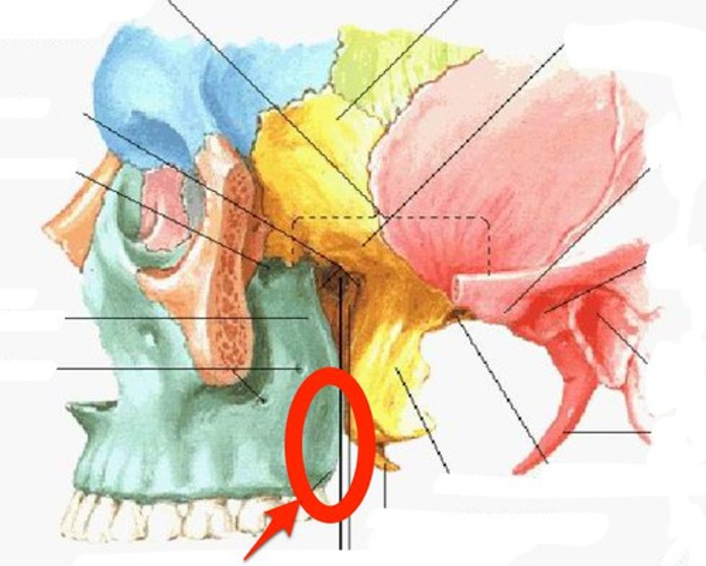
infratemporal fossa
medial to ramus of the mandible and the zygomatic arch

what are two parts of the sphenoid bone?
infratemporal crest, lateral pterygoid plate
pterygomaxillary fissure
connects infratemporal fossa and pterygopalatine fossa
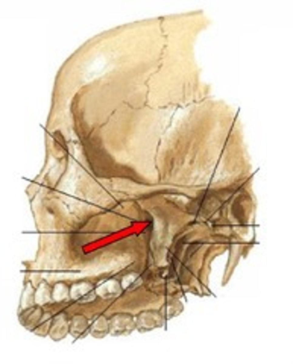
pterygopalatine fossa
pyramidal space, medial to pterygomaxillary fissure
sphenopalatine foramen
opening on medial wall to pterygopalatine fossa leading to nasal cavity
inferior orbital fissure
communicates the infratemporal fossa with the orbit
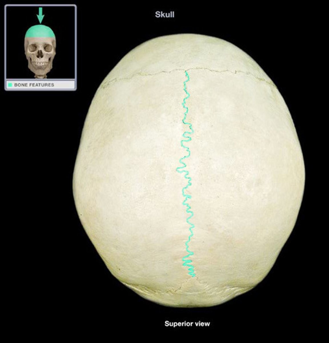
what are the bones and joints (sutures) of the superior view of the cranium?
coronal suture, sagittal suture, lambdoid suture
coronal suture
separates frontal and parietal bones
sagittal suture
separates parietal bones
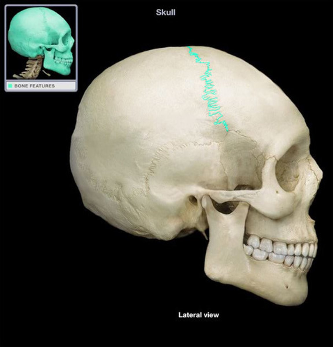
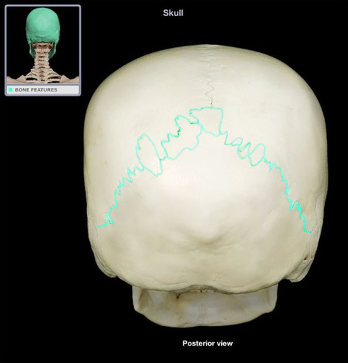
lambdoid suture
separates parietal and temporal bones from occipital bone
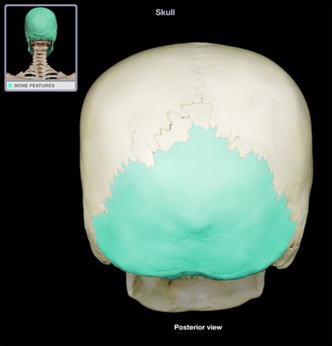
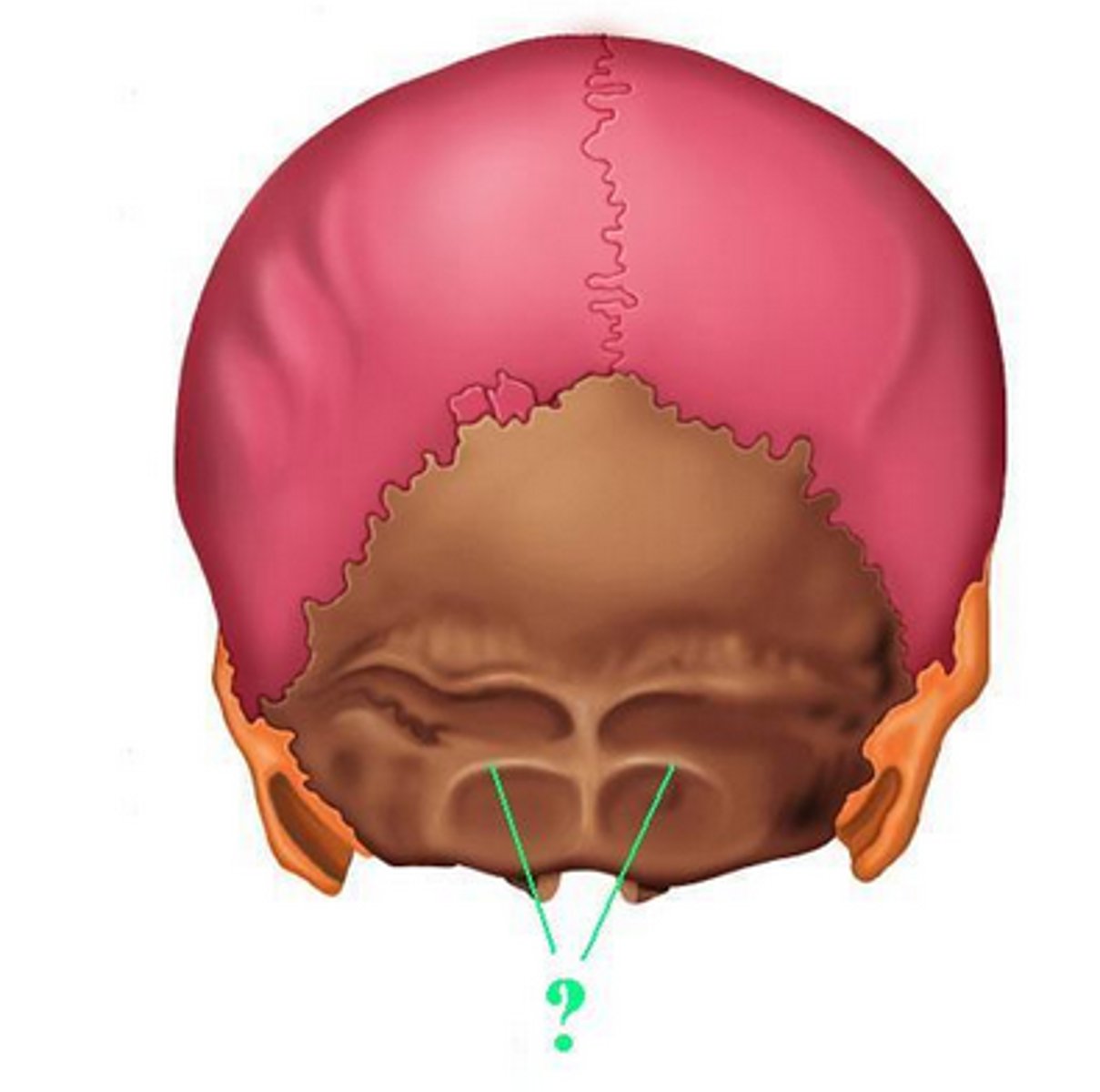
occipital bone
back of head
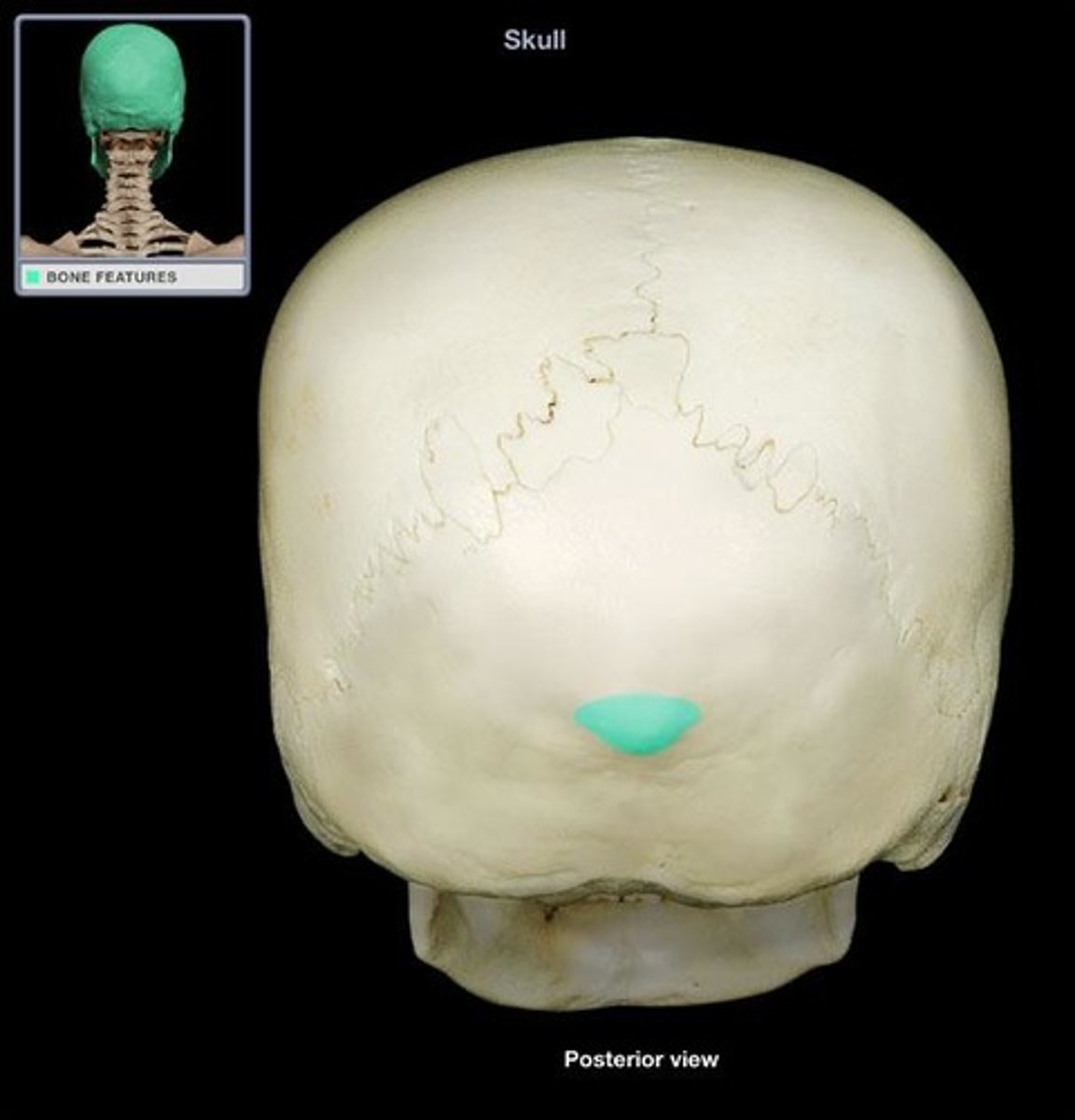
external occipital protuberance
muscle and ligament attachment
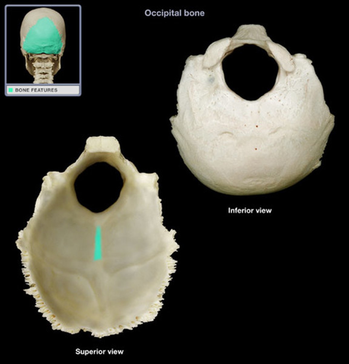
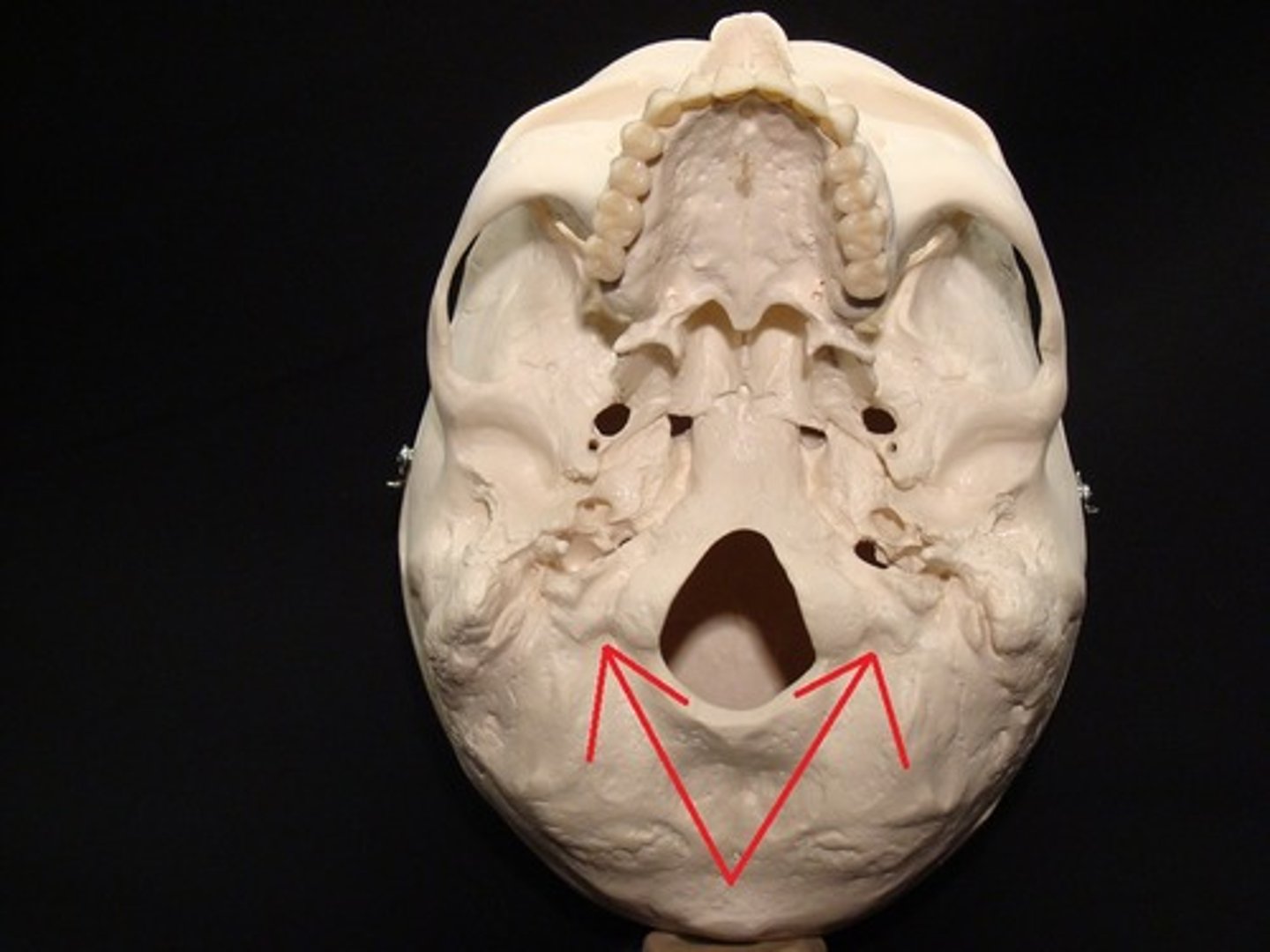
external occipital crest
extends medially from external occipital protuberance to foramen magnum
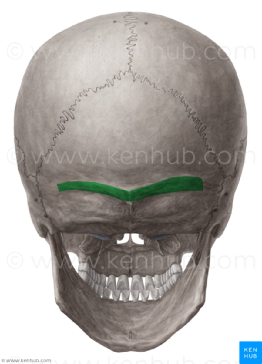
superior nuchal line
extends laterally from external occipital protuberance, marks the superior limit off neck posteriorly
inferior nuchal line
anterior to superior nuchal line and posterior to posterior rim of foramen magnum
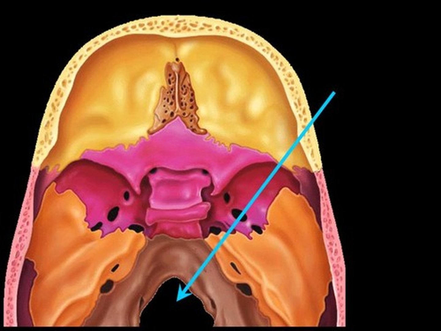
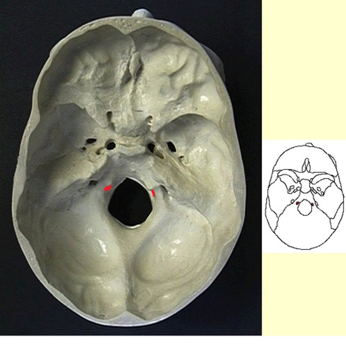
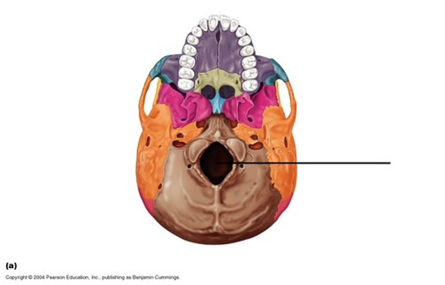
foramen magnum
transition point between brain and spinal cord
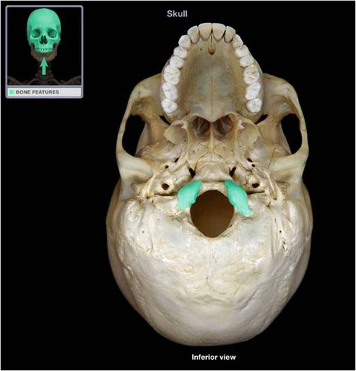
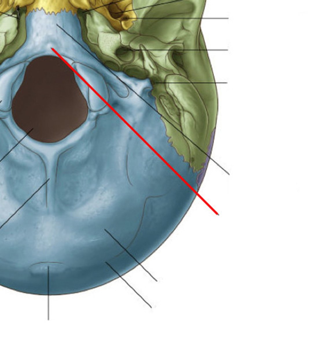
occipital condyles
protuberances on each side of foramen magnum, articulates with cranium with vertebral column
condylar fossa
depression posterior to foramen magnum and contains condylar canal
hypoglossal canal
anterior to occipital condyles
basilar part of occipital bone
extends forward from the foramen magnum
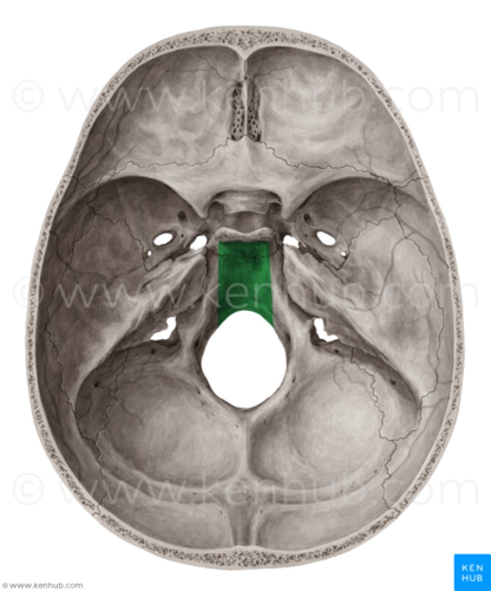
pharyngeal tubercle
eminence on basilar part, attachment for pharyngeal
formen lacerum
exist only in the diseased, dry cranium, between occipital, temporal, sphenoid bone
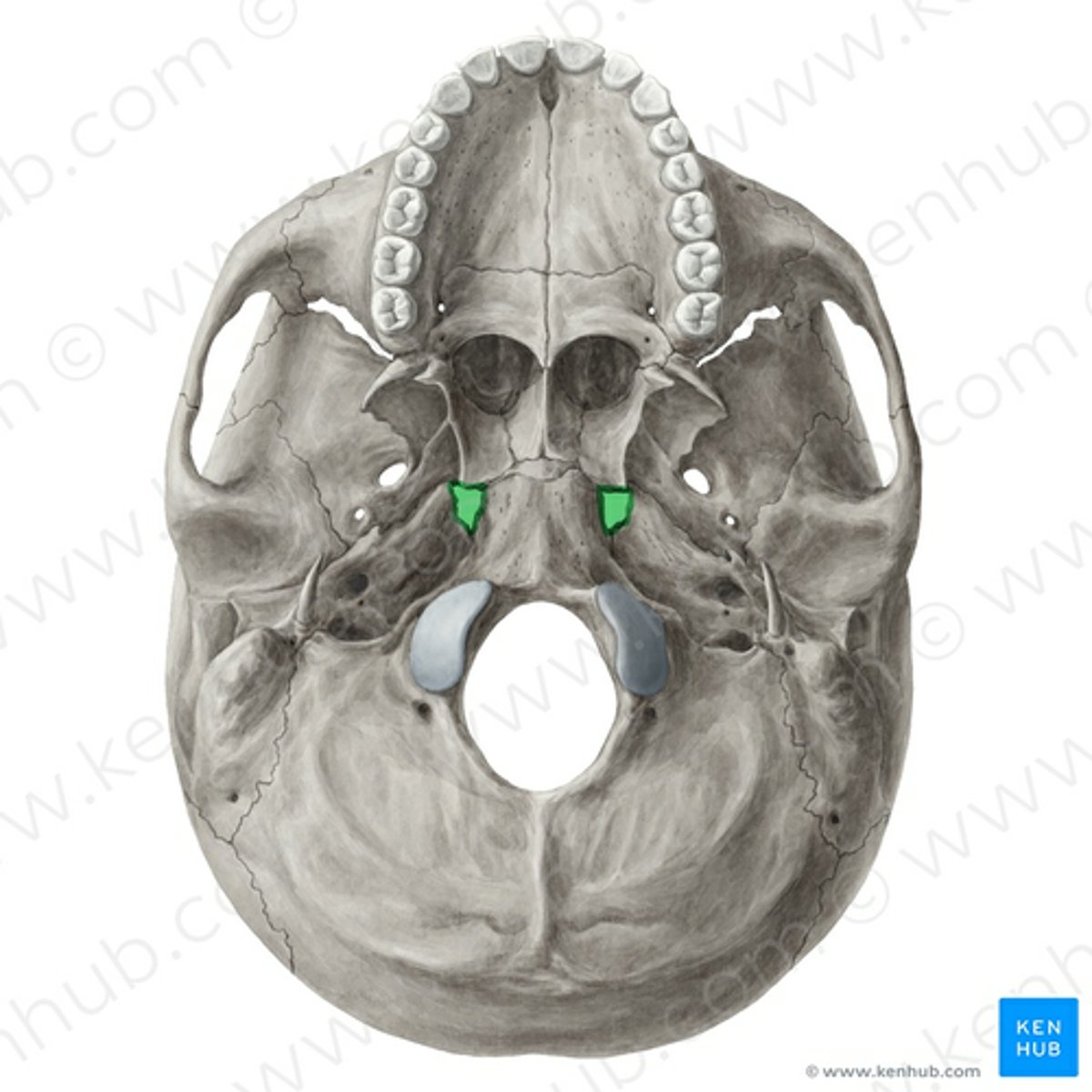
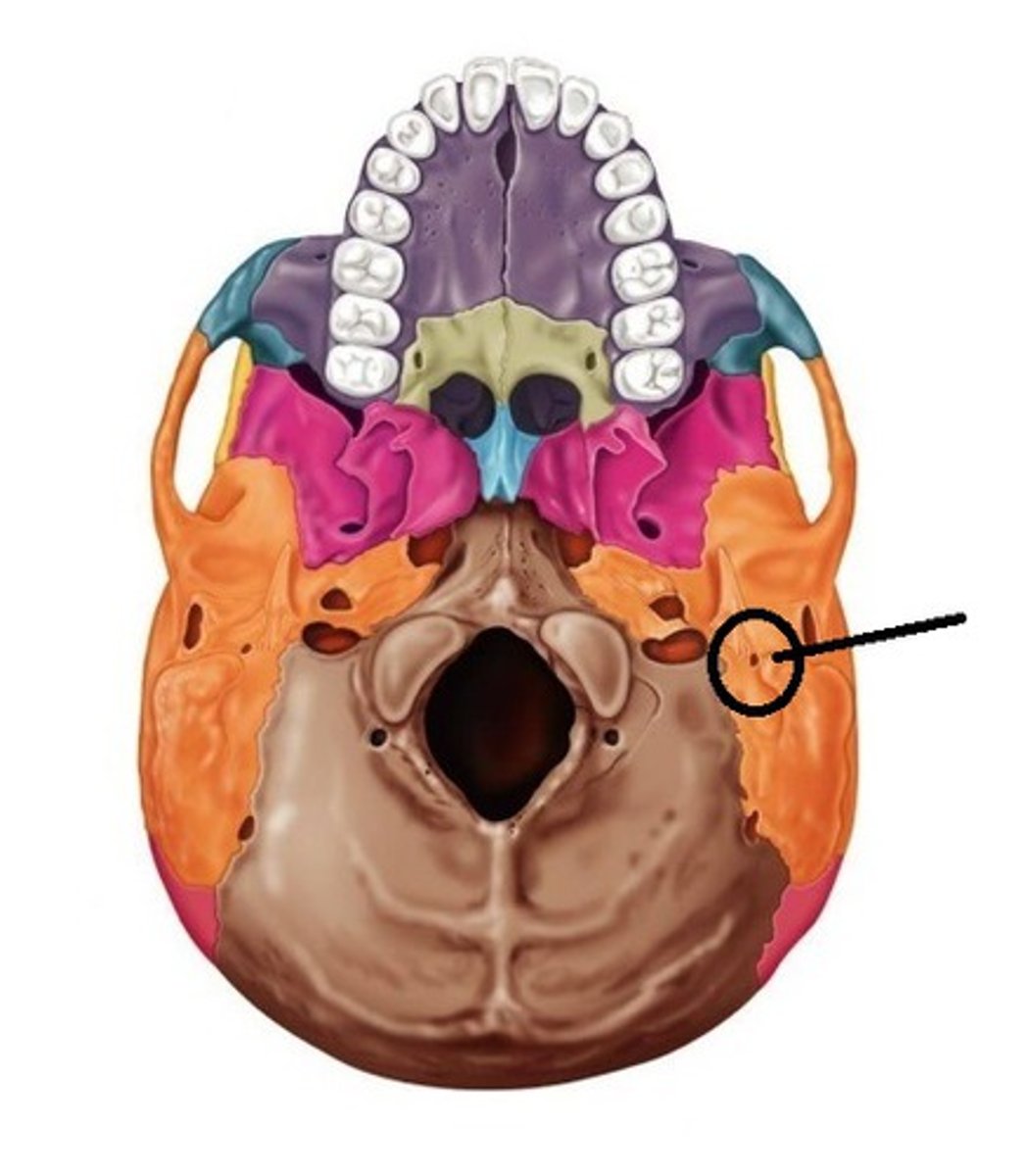
external surface - temporal bone (orange)
anterolateral to occipital and posterolateral to sphenoid (orange)
stylomastoid foramen
posterolateral to styloid process and anteromedially to mastoid process