CHAPTER 16: DENTAL TRAUMA
 Call Kai
 Learn
Learn Practice Test
 Spaced Repetition
 Match
Match Flashcards
 Knowt Play
1/49

Earn XP
Description and Tags
ppt / sas
50 Terms
maxillary incisors
most frequently injured teeth in the primary dentition
2-4 years → peak age for injuries to the primary teeth
crown fractures → are the most common injuries to the primary teeth
battle sign
bruising of the mastoid region; is associated with base-skull fracture
anatomic risk
vector: maxillary incisors are the most frequently injured teeth.
vulnerability: protruding incisors (class II malocclusion) increase trauma risk by 2-3x
primary vectors
indirect trauma → blows to the chin
automobile accidents → major cause of dental injuries
pacifier use → deflects force, causing displacement rather than fracture
primary incisor
lower crown-to-root ratio (less leverage resistance against impact forces)
spongy bone & highly resilient (absorbing impact by allowing the tooth to move rather than crack)
primary teeth luxate (displace) far more often than they fracture when compared to permanent teeth.
permanent incisor
dense alveolar bone
higher crown-to-root ratio firmly anchor the toothh (making coronal fracture the path of least resistance during impact)
routine pulp vitality testing
is NOT reliable in the primary dentition due to patient age and reporting capability. Instead, rely on percussion:
2 reasons to percuss:
sensitivity in response to percussion gives information about the extent of damage to the apical tissues
the sound in response to percussion is also an important indicator of the likelihood of ankylosis
radiographic baseline
3 weeks: periapical radiolucencies (necrosis) become detectable
6-7 weeks: inflammatory root resorption ankylosis or becomes visible.
6 months: wait if asymptomatic
1 month: postoperative
all traumatized teeth should be radiographed to assess:
stage of root development
injuries to root and developing structures
ellis & davey fracture classification I
presents smooth sharp edges
simple fracture involving enamel only
tx: restore primarily for esthetics or smooth to prevent soft tissue irritation.
ellis & davey fracture classification II
extensive fracture involving considerable dentin, but the pulp is intact
tx: requires covering exposed dentin to prevent pulpal irritation
ellis & davey fracture classification III
extensive fracture exposing the dental pulp
appears as a red dot or bleeding point
tx: requires immediate endodontic intervention or extraction.
ellis & davey fracture classification IV
necrotic tooth
ellis & davey fracture classification V
complete loss of the clinical crown or avulsion
ellis & davey fracture classification VI
root fracture
ellis & davey fracture classification VII
displaced tooth
ellis & davey fracture classification VIII
crown fracture
ellis & davey fracture classification IX
pediatric traumatic injuries
cervical or partial pulpotomy
management for class III crown fracture with vital pulp + thin root dentin (open apex) to preserve vitality
complete pulpectomy (fill with resorbable paste)
management for class III crown fracture with necrotic or neglected pulp
immediate extraction
management for class III crown fracture with massive resorption or follicle at risk to protect permanent bud
remove loose fragment & perform pulpotomy on main stable fragment
management for crown-root fracture (split tooth) with shallow splitting (supracrestal)
extract both fragments; unrestorable
management for crown-root fracture (split tooth) with deep splitting (creates periodontal pocket)
2 ways to determine fracture depth in crown-root fracture / split tooth
shallow splitting → supracrestal
deep splitting → creates periodontal ocket
apical fragment rule
never attempt to surgically dig out or retrieve a retained apical root fragment
surgical intervention risks catastrophic damage to the succedaneous permanent tooth bud
the fragment must be left alone to undergo physiologic resorption
concussion
clinical presentation:
NOT mobile. NOT displaced
tender to biting and percussion
PDL status:
PDL absorbs impact and is inflamed (leaves the tooth tender to biting pressure and percussion)
radiographic findings:
normal
no changes
management:
no tx
monitor pulpal condition
soft diet meticulous hygiene
subluxation
clinical presentation:
LOOSENED, but NOT displaced
bleeding from gingival crevice
PDL status:
PDL damaged but attachment remains largely intact.
radiographic findings:
widened pdl space
management:
monitor, soft diet
chlorhexidine application
intrusion
clinical presentation:
tooth is DRIVEN INTO ITS SOCKET
compressed pdl and crushed alveolar socket
management:
if the root of the primary tooth is pushed against the crown of the permanent incisor, it may severely damage the developing tooth bud → immediate removal of the primary tooth to relieve the pressure and minimize damage
intruded incisors that do not pose a risk the permanent teeth can be left to spontaneously reerupt
extrusion
clinical presentation:
ELONGATED from socket
increased mobility, central dislocation
pdl status:
pdl is severely torn
high risk of ischemia
radiographic findings:
increased periapical radiolucency / apical gap
management:
reposition and splint
extract based on severity and apex closure
lateral luxation
clinical presentation:
displaced labially, lingually, or laterally
pdl is torn, alveolar bone is contused or fractured
management:
mild: allow spontaneous repositioning
severe: reposition and splint (7-14 days), initiate endodontic therapy, or extract if labial bone is heavily fractured.
lingual luxation
clinical presentation:
crown moves palatally (often interfering with occlusion)
apex is pushed labially → labial bone plate is fractured
management:
reposition immediately before coagulum forms
utilize intentional open bite (composite buttons on molars) to clear occlusion
extract if severely displaced
labial root displacement
80% of cases are safe
clinical signs:
palatal crown inclination
hard swelling palpated in the labial vestibule
radiograph:
tooth appears foreshortened and highly opaque
management:
safe to preserve
allow spontaneous re-eruption (typically begins within 2 to 6 months)
monitor for rotation
palatal root displacement
dangerous
clinical signs:
root is pressed directly against the developing permanent tooth bud
radiograph:
tooth appears elongated
management:
immediate extraction
extract immediately to relieve pressure and minimize irreversible damage to the permanent successor
avulsion
clinical signs:
the tooth is COMPLETELY DISPLACED from the alveolus
pdl is severed
fractures of alveolar bone may occur
general rule:
avulsed primary incisors are NEVER replanted.
the biomechanical risk:
complete displacement from the alveolus severs the pdl
reinsertion forces the contaminated root or coagulum directly into the delicate follicle of the developing permanent successor, irreversibly irreversibly damaging it in up to 75% of cases.
next steps:
account for the avulsed tooth immediately to rule out aspiration.
if a well-meaning parent has already replanted the tooth at home, advise them of the extreme risk
if retained, it requires splinting and immediate root canal treatment with resorbable paste to prevent rapid inflammatory resorption.
pathologic sequelae of trauma to the teeth
reversible pulpitis
coronal discoloration
infection of the periodontal ligament
pink / red discoloration
observed shortly after the injury may represent intrapulpal hemorrhage
early presentation:
intrapulpal hemorrhage
rupture of blood vessels causes extravasation of RBCs into dentinal tubules.
late presentation:
internal resorption
the coronal dentin is destroyed from within
apical root can be left to resorb spontaneously if the crown is lost.
yellow discoloration
can be seen when the dentin is thick and the pulp chamber narrower than usual → condition termed as pulp canal obliteration
pathology:
pulp canal obliteration (PCO)
accelerated deposition of thick dentin narrows the pulp chamber.
pathology:
pathologic, but requires NO treatment.
monitor radiographically.
dark (gray / black)
RBC hemolysis, often indicates pulp necrosis.
when the pulp becomes necrotic or when pulpal hemorrhage occurs, red blood cells lyse and release hemoglobin—hemoglobin and its derivatives such as hematin molecules that contain iron ions, invade the dentin tubules and stain the tooth dark.
management:
this color alone does NOT mandate extraction
over 70% of asymptomatic dark primary incisors exhibit no radiographic pathology
extract or treat endodontically ONLY if swelling, fistula, mobility, periapical radiolucency are present
internal resorption
can lead to root perforation
odontoclasts attack dentin from within due to chronic inflammation

external (atypical) resoprtion
apical half
coronal half remains unaffected.
eventually:
apical half is completely resorbed.
root becomes short with a rounded apex.
clinically resembles early natural exfoliation.
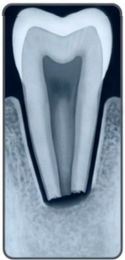
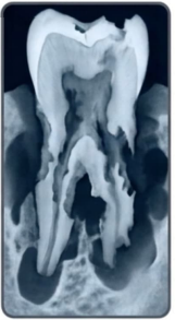
rapidly progressing (inflammatory)
extremely rapid destruction
related to necrotic pulp and inflamed pdl
requires immediate endodontic filling with resorbable paste
replacement external root resorption
aka: ankylosis
results from irreversible PDL injury.
alveolar bone directly contacts and fuses with the root.
root is gradually resorbed and replaced by bone.
more common in intruded primary teeth.
clinical result:
tooth eventually becomes infraoccluded (below the normal occlusal level).
management:
extract if it causes:
delayed eruption of the permanent tooth, or
ectopic eruption of the permanent tooth.

complications for the permanent dentition
enamel defects
follicle deflection
arrested development
space loss & ectopic eruption
arrested development
loss of pulp vitality at an early age halts dentin apposition, apposition, leaving the primary tooth with thin, fragile root walls.
follicle deflection
an infected, expanded primary follicle can physically push and deflect the permanent central incisor away from its normal eruption path.
enamel defects
permanent incisors succeeding infected primary teeth often present with enamel hypoplasia caused by the inflammatory cascade or over-instrumentation during primary endodontics
at-home care protocols
diet modification:
maintain a strictly soft diet for 7-14 days to stabilize luxated or repositioned teeth.
topical disinfection:
apply 0.2% chiorhexidine gluconate to the injured gingiva twice daily.
application technique:
use a cotton-tipped applicator to swab the crevices
do not use as a rinse to prevent the toddler from swallowing the solution while effectively disinfecting the torn pdl
monitoring & clinical alerts
vigilance for infection:
return to the clinic immediately if facial swelling, red mucosal tracking, or a "pimple" (fistula) appears on the gums.
watch closely for suddenly increased tooth mobility or expressions of chewing sensitivity.
reassurance on esthetics:
warn parents that the tooth may transition to a dark gray or black color over the coming weeks.
reassure them that color change without associated pain or swelling often requires monitoring only, not immediate surgical extraction.
practice question in sas
