Unit 9: Visual System
 Call Kai
 Learn
Learn Practice Test
 Spaced Repetition
 Match
Match Flashcards
 Knowt Play
1/17

There's no tags or description
Looks like no tags are added yet.
Name | Mastery | Learn | Test | Matching | Spaced | Call with Kai | Chat |
|---|
No analytics yet
Send a link to your students to track their progress
18 Terms
LO: Identify and describe the cells of the retina, and discuss the pattern of their interconnections
Photoreceptors – rods and cones; transduce light into electrical signals.
Bipolar cells – connect photoreceptors to ganglion cells; can be “ON” or “OFF” types depending on their light response.
Ganglion cells – final output neurons of the retina; their axons form the optic nerve.
with the exception of amacrine cells, ganglion cells are the only retinal cells that fire APs and provide the only exiting axons from the retina to the brain via optic nerve
Horizontal cells – connect laterally between photoreceptors and bipolar cells; mediate lateral inhibition for contrast.
Amacrine cells – connect between bipolar and ganglion cells; modulate motion and temporal aspects of vision.
Pathway: Photoreceptors → Bipolar cells → Ganglion cells → Brain
Lateral modulation by horizontal (outer retina) and amacrine (inner retina) cells refines the signal
Anatomy of the retina
3 important points → (1) only light sensitive cells in the retina are rod and cone receptors (2) ganglion cells are the only source of output from the retina (3) with exception of amacrine cells, ganglion cells are the only retinal neurons that fire APs. All other cells depolarize or hyperpolarize with a rate of NT release that is proportional to the membrane potential
The path that visual information takes is from the…
Photoreceptor →(horizontal cells) biopolar cell → (amacrine cells) ganglion cell
Ganglion cells are the sole output of the retina (in a few slides we will discuss where the information goes next!)
Retinal processing also influenced by lateral connections
Horizontal cells
Receive input from photoreceptors and project to other photoreceptors and bipolar cells
Amacrine cells
Receive input from bipolar cells and project to ganglion cells, bipolar cells, and other amacrine cells
Seemingly inside-out layers
Light passes through ganglion cells and bipolar cells before reaching photoreceptors.
Many nocturnal animals have reflective layer beneath photoreceptors.
LO: Compare the similarities and differences between cones and rods
BOTH:
Outer segment
Inner segment
Synaptic terminal
Cell body
Mesopic conditions (Intermediate light levels)
Rods
Long, cylindrical Outer Segment with many discs
Higher photopigment concentration
1000 times more sensitive to light
92 million of these in each human retina
Contribute to vision in scotopic (dark) conditions
Bulk of contribution in nighttime lighting
Absent in the fovea
express just the photopigment rhodopsin and help us see in dark conditions. They are NOT found in the fovea and are more numerou son the periphery of the retina
Rhodopsin – pigment
Cones
Shorter, tapering outer segment with fewer disks
Lower photopigment concentration
5 million of these in each human retina
Bulk of contribution in photopic (light) conditions
Bulk of contribution in daytime lighting
Concentrated in the fovea
Perception of Color (because they can express on of three different kinds of opsons that are sensitive to short, medium, or long wavelength of light)
Contain one of three opsins (short, medium, and long wavelength activated roughly equivalent to blue green red)
Photoreceptors are the light detectors in the retina.
They use photopigments in the disk membranes to detect light (absorb light) and are able to convert the electromagnetic radiation into neural signals (i.e. changes in photoreceptor membrane potential)
Rods detect light using the photopigment rhodopsin
Cones detect light using three different photopigments called opsins (the three different varieties detect short, medium, or long wavelengths of light). This allows us to see color!
As cells depolarize and hyperpolarize, the rate of NT release is proportional to the membrane potential
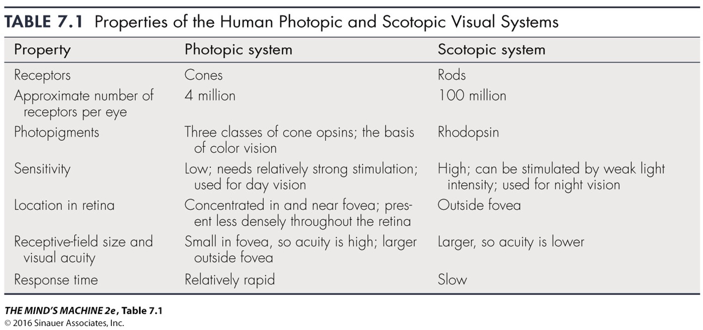
Duplex Retina
Duplex Retina: two complementary systems in one eye (some animals only have rods or cones)
The structural differences between rods and cones correlates with their functional differences
Rods, for example, have a lot of photopigment stacked in the outer segment (rhodopsin) which makes them very sensitive even in low light.
Cones detect light using three photopigments called opsins (short medium or long wavelengths of light) allow us to see color
Think about how at night time our ability to distinguish colors diminishes. This is because in these scotopic conditions our vision relies primarily on rods which are the more sensitive photoreceptor & able to detect low light and our cones which enable our color vision are not sensitive enough
Conversely in the daytime, cones do most of the work
LO: Explain why our vision is much better at the fovea than elsewhere
Fovea is the retinal region specialized for high-acuity vision.
Cones are densely packed, no rods present.
Light directly hits photoreceptors (inner retinal layers displaced sideways).
Low convergence: each cone connects to a single bipolar and ganglion cell → high spatial resolution.
No blood vessels in the foveal pit → minimal light scattering.
Only cones are present, rods are absent as well as the other cellular layers are pushed out of the way
In photopic conditions our visual acuity is much higher in our central retina because of the sole presence of cones there
In scotopic conditions, our central vision is blind because there are no rods here!
textbook
Rods and cones differ in number and distribution across the retina, affecting vision.
Photopic (daylight) vision relies on cones concentrated in the fovea, giving high spatial sensitivity in central vision.
Visual acuity is highest when light falls directly on the cone-rich fovea.
The fovea is a pit in the retina where inner cell layers are pushed aside so light directly hits photoreceptors, reducing light scattering and image blur.
Peripheral vision has lower acuity because it contains fewer cones and more rods.
Color discrimination is also poorer in the periphery due to fewer cones.
Example: you need larger letters or objects to see clearly or distinguish colors using peripheral vision.
At dim (scotopic) light levels, vision relies only on rods, not cones.
Peripheral retina is more sensitive to low light because it has more rods and greater rod convergence onto bipolar and ganglion cells.
Central (foveal) vision is essentially blind in the dark because there are no rods in the fovea.
Rods are specialized for low-light detection, unlike cones, which function in bright light.
Example: A dim star can be seen better with peripheral vision than when looked at directly, showing the higher rod sensitivity outside the fovea.
LO: Summarize and illustrate the steps in phototransduction
In darkness:
Photoreceptor membrane potential ≈ –30 mV.
cGMP keeps Na⁺ channels open → continuous glutamate release.
When light hits photopigment (rhodopsin):
11-cis-retinal → all-trans-retinal (isomerization).
Activates transducin (G-protein).
Transducin activates phosphodiesterase (PDE).
PDE breaks down cGMP → GMP.
↓ cGMP closes Na⁺ channels → hyperpolarization.
Reduced glutamate release signals “light detected.”
Recovery: enzymes restore cGMP and retinal to dark state.
Transduce light energy into changes in membrane potential
Most of what we know about phototransduction in rods applies to cones as well.
In the darkness there is a steady flow of sodium into the photoreceptor dark current. This maintains that high (resting membrane potential) RMP at -30mV
Transduction- light is HYPERPOLARIZING!
Light bleaches rhodopsin (changes conformation of retinal from inactive to active) → G-protein becomes active → PDE is activated which reduces cGMP levels → reduced cGMP levels closes Na channels (cGMP helped keep Na channels open)
This causes a receptor potential in the photoreceptor (NOT an action potential)
Ganglion cells (with exception of amacrine cells are the only ones that fire APs and their axons exit from the brain via optic nerve!)
In bright light, cGMP levels in rods fall to the point where the response to light becomes saturated; increasing the light level causes no additional hyperpolarization. Thus, vision during the day depends entirely on the cones, whose photopigments require more energy to become bleached.
The process of phototransduction in cones is virtually the same as in rods; the only major difference is in the type of opsins in the membranous disks of the cone outer segments.
In bright light, cGMP levels in rods drop so low that rods become saturated and cannot respond to additional light.
Daytime vision depends entirely on cones, which require more energy for photopigment bleaching.
Phototransduction in cones works the same way as in rods, except for the type of opsins present.
Cones contain three types of opsins, each with different spectral sensitivities:
Short-wavelength (“blue”) cones: peak at ~430 nm
Medium-wavelength (“green”) cones: peak at ~530 nm
Long-wavelength (“red”) cones: peak at ~560 nm
Each cone type responds to a broad range of wavelengths, and there is overlap among their sensitivities.
The terms blue, green, and red can be misleading because color perception depends on the combined activation of multiple cone types.
Using short, medium, and long wavelength terminology is more accurate and less confusing.
What is a receptive field?
Area of retina or visual space that when light is applied the firing rate of the neuron changes
LO: Plan how you would find the receptive field of a ganglion cell
Present small spots of light or dark on a screen while recording from a single ganglion cell.
Move the light to find the region where it changes the firing rate.
Identify:
Excitatory (ON) areas → increase firing with light.
Inhibitory (OFF) areas → decrease firing with light.
The ON and OFF regions together define the cell’s receptive field.
Textbook
A receptive field is the area of the retina where light changes a neuron's firing rate.
Light outside this area has no effect on the neuron’s activity.
In the visual system, receptive fields can be described as areas on the retina or areas in visual space, since the two correspond.
The term “receptive field” applies broadly to other sensory systems (e.g., in touch, it refers to areas of skin that activate a neuron when stimulated).
LO: Describe the differences between receptive field center and receptive field surround
Center – direct input from a small group of photoreceptors via bipolar cells.
Surround – indirect input through horizontal cells providing lateral inhibition.
Center-surround organization:
ON-center/OFF-surround: light in center increases firing, light in surround inhibits.
OFF-center/ON-surround: opposite pattern.
This arrangement enhances contrast and edge detection.
Textbook
Bipolar cells have receptive fields shaped like concentric circles and are classified as ON or OFF types.
Classification depends on how they respond to glutamate released by photoreceptors.
Their receptive fields are shaped by direct input from photoreceptors and indirect input through horizontal cells.
In the direct pathway (no horizontal cells):
Light hyperpolarizes some bipolar cells → these are OFF bipolar cells (light turns them off).
Light depolarizes other bipolar cells → these are ON bipolar cells (light turns them on).
The cone-to-bipolar synapse inverts the signal: cones hyperpolarize in light, but ON bipolar cells depolarize in response.
Retinal ganglion cells have center-surround receptive fields, like bipolar cells.
ON-center and OFF-center ganglion cells get input from the matching type of bipolar cell.
Unlike bipolar cells, ganglion cells fire action potentials continuously, even in darkness.
ON-center cells:
Increase firing when light hits the center of the receptive field.
Decrease firing when light hits the surround.
OFF-center cells:
Decrease firing when light hits the center.
Increase firing when a dark spot covers the center.
Center and surround responses oppose each other, canceling out when both are stimulated equally.
Ganglion cells are mainly sensitive to differences in illumination (contrast), not uniform brightness.
(A) Bipolar cells receive direct synaptic input from a cluster of photoreceptors constituting the receptive field center. In addition they receive indirect input from surrounding photoreceptors via horizontal cells.
(B) On-center bipolar cell is depolarized by light in the receptive field center via the direct pathway
(C) Light in the receptive field surround hyperpolarizes the On center bipolar cell via the indirect pathway. Because the intervening horizontal cell, the effect of light on the surround photoreceptors is always opposite the effect of light on the center photoreceptors
This same organization applies to ganglion cells
How can different bipolar cells give opposite responses to direct cone input?
The answer is that there are two kinds of receptors that receive glutamate released by the photoreceptors.
OFF bipolar cells have ionotropic glutamate receptors, and these glutamate-gated channels mediate a classical depolarizing excitatory postsynaptic potential from the influx of Na+. Hyperpolarization of the cone causes less neurotransmitter to be released, resulting in a more hyperpolarized bipolar cell.
On the other hand, ON bipolar cells have G-protein-coupled (metabotropic) receptors and respond to glutamate by hyperpolarizing. Each bipolar cell receives direct synaptic input from a cluster of photoreceptors. The number of photoreceptors in this cluster ranges from one at the center of the fovea to thousands in the peripheral retina.
LO: Predict the site of a lesion in the retinofugal pathway based upon the visual field deficit
Optic nerve lesion (one side): blindness in that eye.
Optic chiasm lesion: loss of peripheral (temporal) fields in both eyes → bitemporal hemianopsia.
Optic tract lesion: loss of contralateral visual field in both eyes → homonymous hemianopsia.
LGN or visual cortex lesion: similar contralateral field loss, sometimes with macular sparing (due to dual blood supply).
Textbook
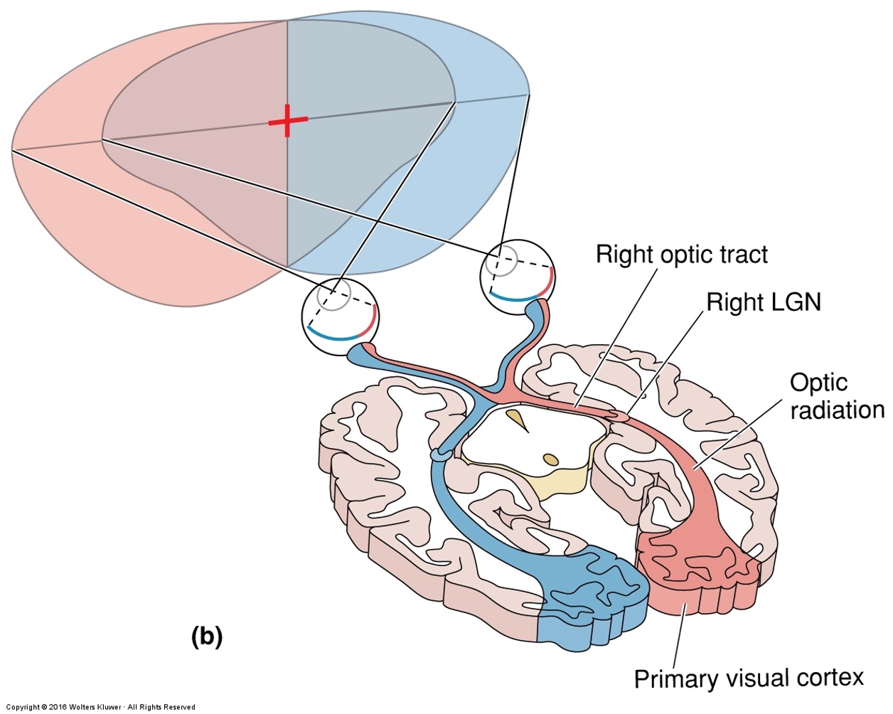
Objects in the left visual field are seen by the nasal retina of the left eye and the temporal retina of the right eye.
Nasal retinal fibers cross at the optic chiasm, sending all left visual field information to the right hemisphere.
Conversely, the right visual field is processed by the left hemisphere.
Rule of thumb: left field → right brain; right field → left brain.
This crossing (decussation) also occurs in motor pathways, where each brain side controls the opposite body side.
Remember…
left visual field is imaged on the nasal retina of the left eye and the temporal retina of the right eye (RED in figure)
right visual field is image on the nasal retina of the right eye and the temporal retina of the left eye (BLUE in figure)
A)Transection of the left optic nerve (#1)
Left Optic Nerve → all vision contributed by the left eye, but remember most of the left visual field is also viewed by right eye, so you only lose the monocular vision portion of the left hemifield (far left peripheral vision)
B)Transection of the left optic tract (#3)
Left Optic Tract → all of right visual hemifield
C)Transection of the optic chiasm (#2)
Optic Chiasm → only crossing fibers (from nasal retinas) are loss→ peripheral vision
ex: Left nasal retina views the left peripheral vision (part A of the figure)
LO: List the structures of the retinofugal pathway including non-thalamic targets and their purpose
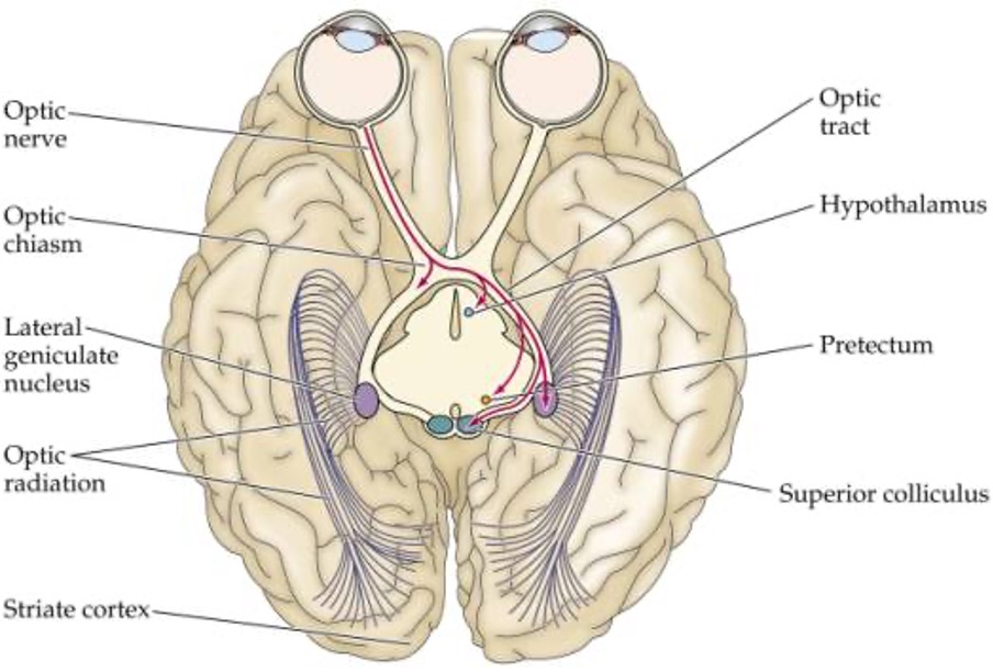
The structures in the retinofugal pathway, in order they receive visual information, are…
(1) Optic nerve
(2) Optic Chiasm
(3) Optic Tract
Now if let’s trace the pathway all the way through the cells in the pathway. Remember, the path that visual information takes is from the…
Photoreceptor →(horizontal cells) biopolar cell → (amacrine cells) ganglion cell which are the sole output of the retina à optic nerve à optic chiasm à LGN à V1 (also known as striate cortex or primary visual cortex)
Simplified..
Photoreceptor à bipolar cell à ganglion cell à optic nerve à optic chiasm à optic tract à LGN à V1
Non-thalamic targets
hypothalamus (Suprachiasmatic nucleus) - entrain our circadian rhythm to the earth’s light/dark cycle
Pretectum- control the size of the pupil & certain eye movements
Superior colliculus - orients the eyes in response to new stimuli (move fovea to objects of interest)
LO: Describe the organization of retinal inputs to the LGN
Six layers: inputs are eye-specific (3 from each eye).
Magnocellular (M) layers: motion, luminance info from M-type ganglion cells.
Parvocellular (P) layers: color, fine detail from P-type ganglion cells.
Koniocellular layers (between them): color contrast, blue cone input.
LGN acts as a relay and gate, modulated by cortical feedback.
Lateral Geniculate Nucleus (LGN of thalamus) gateway to visual cortex “like a little knee”
Input from the eyes is segregated! And input from different cells is segregated
6 distinct cell layers; major target of optic tracts
1 contralateral eye input
2 ipsilateral
3 ipsilateral
4 contralateral
5 ipsilateral
6 contralateral
Most ventral layer = 1
**Remember…
each eye views parts of both your left & right visual fields
information from the LEFT visual field is viewed COMPLETELY by the RIGHT LGN
this means the right LGN needs information from BOTH eyes!! And that input is kept separate
The right and left lateral geniculate nuclei, located in the dorsal thalamus, are the major targets of the two optic tracts. Viewed in cross section, each LGN appears to be arranged in six distinct layers of cells
LO: Compare and contrast P-type and M-type ganglion cells
The only output from the retina comes from ganglion cells. There are three types of ganglion cells M-type, P-type and non-M/non-P type. M-type and P-type ganglion cells process different kinds of information, therefore they….
M and P ganglion cells carry different kinds of information to the LGN from the retina
LGN organized by GC input- Different types of ganglion cells appear to play different roles in visual perception!
M (magno)-type Ganglion cells
5% of ganglion cell type population
Larger receptive fields
Larger cells
Conduct AP more rapidly
MORE sensitive to low-contrast stimuli
Respond to stimulation of their receptive fields with a transient BURST of APs
Not sensitive to differences in wavelength of light, still detects green, red, etc. (respond to color just responses are just not color specific!). Still on/off or off/on cells
P (parvo) -type Ganglion cells COLOR
90% of ganglion cell type population
Sustained discharge as long as stimulus is on
Some are Sensitive to differences in wavelength of light (color detection!) - color opponent cells
Red versus green
Blue versus yellow
Red -560 nm (long); Green - 530nm (intermediate); Blue -430nm (short)
R+G- → optimally excited by red in the receptive field center and inhibited by green in the surround
nonM-nonP ganglion cell
Remaining 5%
Some are Sensitive to differences in wavelength of light (color detection!) - color opponent cells
Textbook
M and P ganglion cells send different visual information to the LGN.
M cells:
Large receptive fields
Fast conduction
Sensitive to low contrast
Respond with brief bursts (transient response)
No color opponency — respond to light changes, not color
P cells:
Small receptive fields
Slower conduction
Respond with sustained firing (continuous response)
Many show color opponency:
Red–green (R+G− or G+R−)
Blue–yellow (B+Y− or Y+B−)
Color-opponent cells: center and surround respond to opposite colors, canceling each other with white light.
Overall, ganglion cells convey three main comparisons:
Light vs. dark (M cells)
Red vs. green (P cells)
Blue vs. yellow (nonM–nonP cells)
LO: Explain retinotopy
Spatial organization where neighboring retinal cells project to neighboring neurons in the LGN and visual cortex.
Preserves the spatial map of the visual field.
Foveal region occupies disproportionately large cortical area → cortical magnification.
Retinotopy - organization of input
Neighboring cells in the retina feed information to neighboring places in their target structures (LGN & striate cortex)
2D surface of retina is mapped onto the 2D surface of subsequent structures
3 key points:
Distorted because visual space is NOT uniformly sampled by cells in retina (many more cells with receptive fields in or near the fovea so the striate cortex receives input from more of the neurons) so the central field of vision seen by the fovea is overrepresented!
that a discrete point of light can activate many cells in the retina, and often many more cells in the target structure, due to the overlap of receptive fields
(single point of light in retina = broad distribution of peak in cortex)
There are NO pictures in the primary visual cortex. Don’t be misled by the term map
Perception is based on brain’s interpretation of distributed patterns of activity NOT snapshots of the world
Textbook
Retinotopy means that neighboring retinal cells send information to neighboring locations in the LGN and primary visual cortex (V1) — creating a spatial map of the retina.
1⃣ Mapping distortion: The fovea has many more ganglion cells, so the central visual field is magnified in V1.
2⃣ Overlap: A single point of light activates many neurons because receptive fields overlap, forming a broad pattern of activity in the cortex.
3⃣ No literal image: The “map” in V1 is not a picture — perception comes from the brain interpreting neural activity patterns, not viewing images directly.
LO: Explain the characteristics of receptive fields in the striate cortex (V1)
Simple cells: respond to bars or edges of specific orientation and location.
Complex cells: respond to oriented bars anywhere within their field, sensitive to motion.
Binocular cells: receive input from both eyes → depth perception (stereopsis).
Orientation columns: organized vertical columns where all cells respond to the same orientation.
Textbook
Layer IVC (input layer):
Receives input from magnocellular (IVCα) and parvocellular (IVCβ) LGN neurons.
Small, monocular, center-surround receptive fields.
IVCα: not color-sensitive.
IVCβ: shows color opponency (e.g., red–green).
Outside Layer IVC:
Neurons gain new properties — binocularity, orientation selectivity, and motion sensitivity.
Inputs from both eyes mix, forming binocular neurons that respond to either eye.
Ocular dominance columns organize V1 so neurons are more driven by one eye, yet remain binocular.
Binocular receptive fields align in both eyes → preserve retinotopy and enable depth perception (stereopsis).
Orientation selectivity:
Many V1 neurons respond best to a bar of light at a specific angle; perpendicular bars give weak responses.
Found mainly outside layer IVC.
Orientation columns: cells stacked vertically (across layers II–VI) share the same preferred orientation.
Moving tangentially across cortex → orientation gradually shifts, forming a mosaic pattern.
Specialization: analyzes object shape.
Direction selectivity:
Subset of orientation-selective neurons responding to motion in one direction only.
Simple cells:
Have distinct ON and OFF regions; orientation-selective.
Complex cells:
No distinct ON/OFF zones; respond to light anywhere in the field.
Likely formed from multiple simple cells.
Summary:
Layer IVC handles basic monocular input, while upper layers integrate signals into binocular, orientation-, and motion-sensitive responses, forming the basis for shape, depth, and motion perception in vision.
LO: Describe the hierarchy of receptive fields in the visual system
Retina: spots of light/dark (center-surround).
LGN: similar center-surround fields.
V1 simple cells: combine LGN inputs to detect oriented edges.
V1 complex cells: detect motion and direction.
Higher areas (V2, V4, IT): respond to complex shapes, faces, and objects.
Each level integrates signals from lower levels → increasing complexity.
LO: Describe the functional differences between the dorsal stream and the ventral stream
Stream | Pathway | Function |
|---|---|---|
Dorsal (“where/how”) | V1 → V2 → MT (middle temporal) → Parietal lobe | Spatial vision, motion detection, guiding actions |
Ventral (“what”) | V1 → V2 → V4 → Inferior temporal cortex | Object recognition, color, form, faces |
Damage to dorsal: motion blindness (akinetopsia).
Damage to ventral: visual agnosia or prosopagnosia (face blindness).
🧭 Dorsal Stream – “Where/How”
Function: Motion analysis & visual control of action.
Pathway: V1 → V2 → V3 → MT → MST → parietal lobe.
Neurons: Like magnocellular (motion-sensitive, not color).
Receptive Fields: Motion-selective (linear, radial, circular).
Key Areas:
MT: Direction-selective; responds to object motion. Electrical stimulation alters perceived motion (Newsome).
MST: Roles in navigation, eye movement, motion perception.
Damage: Loss of motion perception (akinetopsia).
Role: Spatial awareness & movement guidance.
🎨 Ventral Stream – “What”
Function: Object recognition, shape & color perception.
Pathway: V1 → V2 → V3 → V4 → IT → temporal lobe.
Neurons: Like parvocellular (detail, color).
Receptive Fields: Complex; respond to color, shape, patterns.
Key Areas:
V4: Shape & color processing; damage → achromatopsia.
IT: Complex form & visual memory; includes Fusiform Face Area (FFA) for face recognition.
Damage → prosopagnosia (can’t recognize faces).
Role: Identifying and remembering what we see.
🧭 Dorsal = motion & location | 🎨 Ventral = form & color
LO: Describe the inputs and outputs of the striate cortex
The LGN has a single major target à primary visual cortex
Primary visual cortex is also known as…
Broadmanns area 17
V1
Striate cortex (dense stripe of myelinated axons running parallel to the surface)
Segregation of neurons into layers suggests that there is a division of labor in the cortex, similar to what we saw in the LGN
LO: Describe the function of area IT
Area IT (Inferior Temporal Cortex) – Summary
Location: At the far end of the ventral (“what”) stream, receiving major input from V4.
Function: Involved in visual perception and visual memory; connects to temporal lobe areas for learning and memory.
Receptive Fields: Complex; respond to a wide range of colors and abstract shapes.
Face Processing:
Some IT neurons (found by Charles Gross) respond strongly to faces or face-like patterns.
Human fMRI studies (Nancy Kanwisher) identified a Fusiform Face Area (FFA) on the fusiform gyrus — highly responsive to faces.
Disorders:
Prosopagnosia – face blindness caused by damage to the FFA or nearby extrastriate areas, even when general vision remains normal.
Key Idea: Area IT combines sensory input with stored memory to recognize and identify complex visual objects, especially faces.