SBVAC Week 2 Anatomy and Physiology Terms (Revised)
 Call Kai
 Learn
Learn Practice Test
 Spaced Repetition
 Match
Match Flashcards
 Knowt Play
1/90

There's no tags or description
Looks like no tags are added yet.
Name | Mastery | Learn | Test | Matching | Spaced | Call with Kai | Chat |
|---|
No analytics yet
Send a link to your students to track their progress
91 Terms
Anatomy
Study of the body and its parts and their relationships to one another.
Physiology
The way a living organism or body part functions
Homeostasis
Maintaining stable equilibrium
What is the normal anatomical position?
Standing, facing forward, arms at sides and palms facing up

All Anatomical Planes
Transverse, Coronal/Frontal, Sagittal, Mid-axillary, Mid-clavicular
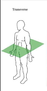
Transverse Plane
Divides the body from top and bottom (head-waist and legs)
Coronal/Frontal
Divides the body from front and back

Sagittal
Divides the body into left and right

Mid-axillary
Divides the body in half by your armpit area (middle)
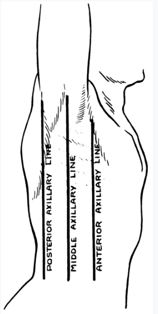
Mid Clavicular
Line down the middle of your clavicular bones

Supine
Position lying on spine, face-up

Prone
Lying flat on chest, face down

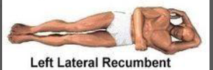
Left/Right Lateral Recumbent
Lying on the side

Recovery Position
Left lateral recumbent, laying on left side
Trendelenburg (“shock position”)
Supine with legs raised 6”-12” above head, not used anymore
Fowler’s
Sitting upright in a 90 degree angle

Semi-Fowler’s
Sitting 45 degrees

Tripoding
Bent over with hands on knees, like after running

Superior
Closer to head
Inferior
Closer to feet
Anterior/ventral
Towards the front
Posterior/dorsal
Towards the back
Medial
Closer to the midline

Lateral
Further from the midline

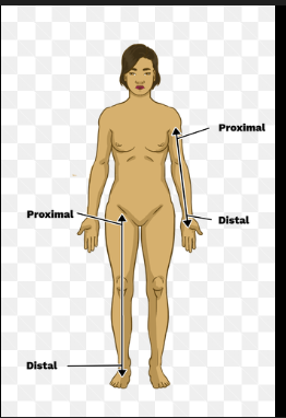
Distal
Farther from the core (original point of limb)
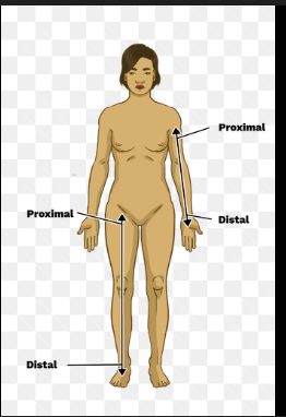
Proximal
Closer to the core (original point of limb)
Superficial
Towards the surface

Deep
Farther from surface

Flexion
Decreasing angle at a joint (Think of contracting)

Extension
Increasing angle at a joint (Think of resting)

Adduction
Moving towards midline/body

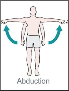
Abduction
Moving away from the midline/body
Body Cavities and Separation
Thoracic and Abdominal/Peritoneal, separated by the diaphragm
Retroperitoneal and Pelvic

What organs are in the Thoracic Cavity?
Lungs, trachea, heart, great vessels
Describe the four quadrants of the GI System
Umbilicus divides the cavity into 4 quadrants:
Right Upper: Liver, gallbladder, stomach, pancreas
Left Upper: Spleen, most of stomach, pancreas
Right Lower: Appendix
Left Lower: Small/Large Intestine (Also in all other quadrants)

What organs are in the Retroperitoneal Cavity?
Kidneys, descending aorta (heart), inferior vena cava
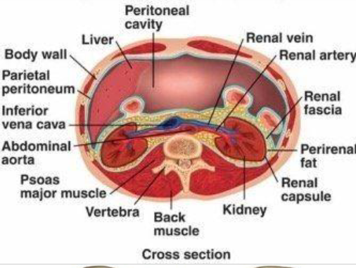
What organs are in the Pelvic?
Reproductive Organs and Bladder

Functions of the Skeletal System
Support and Structure, Protection, Movement, Production of Red Blood Cells (RBC), Mineral Storage
What does the Skeletal System consist of? What connects bone-to-bone and muscle-to-bone? How many bones does an adult have?
Ligaments: Connect bone-to-bone
Tendons: Connect muscles to bone
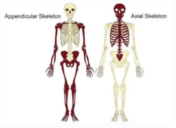
Two Divisions:
Axial Skeleton: Bones of head and trunk
Appendicular Skeleton: Bones of appendages
There are 206 bones in the body made of cartilage!
List out all parts of the skull?
Cranial Bones:
Frontal
Parietal
Occipital
Temporal
(FPOT)
Maxilla:
Upper Jaw Bone
Mandible:
Lower Jaw Bone
Zygomatic:
Cheekbone
Foramen Magnum:
Opening at skull base where brain connects to spinal cord

Describe all vertebrae that the spinal column consists of? What specific vertebrae are important?
33 vertebrae that protect the spinal cord
C 1-7 (Cervical)
T 1-12 (Thoracic)
L 1-5 (Lumbar)
Sacrum (5 fused vertebrae)
Coccyx (4 fused vertebrae)
C1 and C2 are the Atlas and Axis. They support and allow skull movement.
C 3,4, and 5 protect the phrenic nerve and “keep the diaphragm alive”

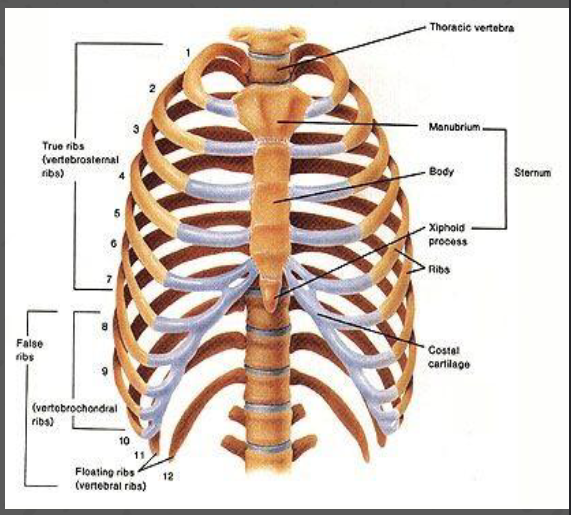
What does the Rib Cage Consist of?
Sternum
Manubrium, body, xiphoid process
Ribcage: 12 pairs of ribs
True Ribs 1-7: connect directly to sternum vial costal cartilage
False Ribs 8-10: connect indirectly to sternum through 7th rib by cartilage
Floating Ribs 11-12: do not connect to sternum, only to spine
What bones are part of your upper extremities (shoulders, arms, hand)
Shoulder:
Clavicle, Scapula (shoulder blade), Humerus
Arm:
Humerus, Radius (Lateral), Ulna (Medial)
Hand:
Carpals, Metacarpals, Phalanges

What bones are in the Pelvis?
Pelvic Bones (Fused):
Illium
Ischium
Pubis

What bones are in the Lower Extremity (legs and feet)?
Leg Bones:
Femur
Patella
Tibia (anterior) / Fibula (lateral)
Foot:
Tarsals
Metatarsals
Phalanges

What’s the pathway of air flow? (Upper and Lower)
Upper Airway:
Oro/nasopharynx
Pharynx
Epiglottis
Larynx
Lower Airway:
Trachea
Bronchi
Bronchioles
Alveoli
Capillaries

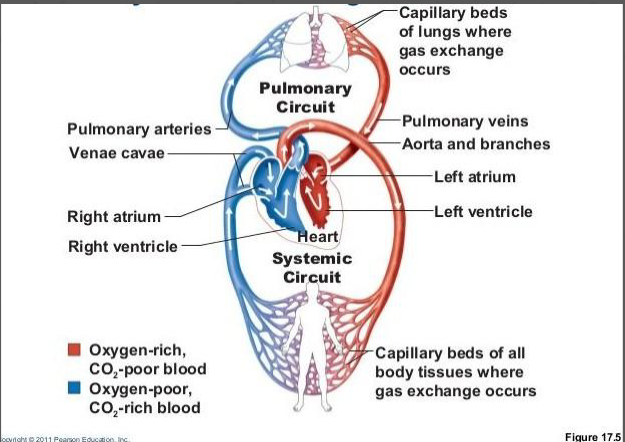
Pathway of Blood through the Heart
Superior/Inferior Vena Cava
Right Atrium
Tricuspid Valve
Right Ventricle
Pulmonary Valve
Pulmonary Artery (Deoxygenated)
Lungs
Pulmonary Vein (Oxygenated)
Left Atrium
Mitral/Bicuspid Valve
Left Ventricle
Aortic Valve
Aorta (to systemic circulation)

Pathway of Blood through the Body (Systemic)
Heart (aorta)
Arteries
Arterioles
Capillaries
Venules
Veins
Heart (vena cavae)
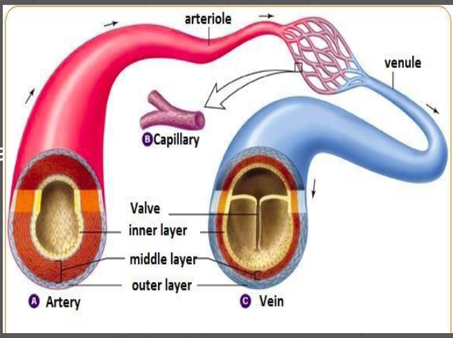
What are the three main cardiovascular vessels and what do they do?
Arteries:
Carry blood AWAY from the heart’s thick, muscular walls, operate at a higher pressure
Veins:
Carry blood TOWARDS the heart, less muscular walls, operate at a lower pressure, contain valves to prevent backflow
Capillaries:
Delivers blood to/from tissues/cells, thin walls, 1 cell thick
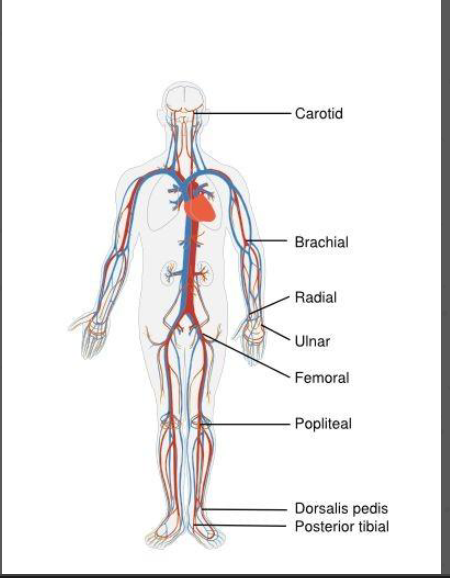
What are the six main types of pulses?
Carotid
Radial
Brachial
Femoral
Posterior Tibialis (inside ankle)
Dorsalis Pedis (top of foot)
Blood Pressure
Systolic: The pressure against the walls of the arteries as the left ventricle CONTRACTS
Diastolic: The pressure against the walls of the arteries as the left ventricle RELAXES
Pathway of food through GI tract
Mouth
Pharynx
Epiglottis
Esophagus
Stomach
Small Intestine
Large Intestine
Rectum
Anus
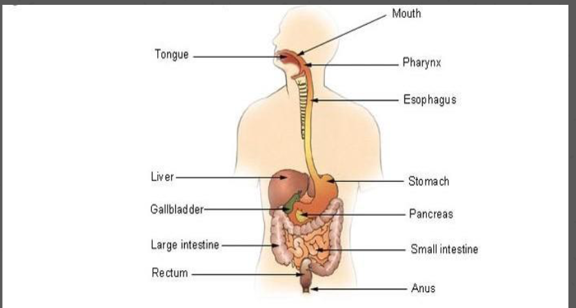
What are the three accessory organs in the digestive system?
Liver
Produces bile(digest fats), lipoproteins for cholesterol, metabolizes food, detoxifies blood, stores vitamins/nutrients
Gallbladder
Stores bile, secretes it into the small intestine
Pancreas
Produces enzymes and secretes hormones

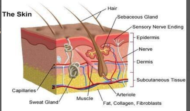
What are the layers of the integumentary system (skin)?
Consists of skin and its appendages (ex. hair and nails)
Layers of the Skin:
Epidermis: Outer Layer, functions as a barrier
Dermis: Middle Layer, contains blood vessels, nerve endings, hair follicles, sweat and sebaceous glands
Subcutaneous: Deepest Layer, contains blood vessels and fat for insulation
How is the nervous system broken down? What is in each part?
Central Nervous System:
Brain and spinal cord
Peripheral Nervous System:
Sensory (afferent) nerves
Carry info from the body towards the brain
Motor (efferent) nerves
Carry info away from the brain to the body

Define hormones and glands
Hormone: Chemical messengers that act on particular organs to stimulate a response
Gland: A group of cells that produce and secrete hormones

What organs are in your urinary system? What is the order in which urine is excreted from, and what does each organ do?
Kidneys:
In the retroperitoneum; filter blood, remove waste, maintain electrolyte and fluid balance, generate urine
Ureters:
Carry urine from the kidney to the bladder
Bladder:
Can hold up to ~700 mL of urine
Urethra
Carries urine from bladder to outside body
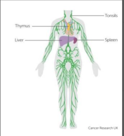
What is the Lymphatic System and what does it consist of?
A network of lymphatic vessels that filters blood and produces antibodies
Spleen, thymus, and bone marrow produce lymphocytes (white blood cells)
Lymph: Fluid containing lymphocytes and waste products
Lymph nodes: small tissue masses along lymphatic vessels that house lymphocytes, may swell during infection
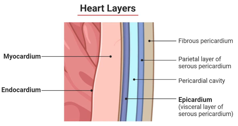
What are the layers of the heart?
Please Excuse My E- 😳
Pericardium:
Sac-like layer surrounding the heart to reduce friction with each heartbeat
Epicardium:
Outermost layer of the heart
Thin layer of connective tissue and fat protecting the heart
Myocardium:
Thick middle layer of the heart
Pumping muscle that causes heartbeats
Endocardium:
Innermost layer of the heart
Thin Lining covering the inner chambers of the heart and valves
Describe the electrical pathways through the Heart and the important nodes.
Sinoatrial(SA) Node
Heart’s natural pacemaker
Sends electrical signals to start heart contractions
Atrioventricular(AV) Node
Delays the SA electrical signal (ensures atrial contraction and blood pumping)
Located by the tricuspid valve
Bundle of HIS (atrioventricular bundle)
Carries the electrical signal to the Purkinje fibers
Branch of fibers extending from the AV node running down the length of the septum
Bifurcating/dividing into the left and right bundle branches which run across respective ventricles
Purkinje Fibers
Quickly sends electrical signals to the heart’s left and right ventricles, causing contraction

What are Arrhythmias?
Any abnormal electrical activity in the heart. Can range from harmless quirks (Sinus Arrhythmia) to deadly ones. It’s often considered a heart disease
Difference between heart attack and cardiac arrest
Heart Attack: Due to Myocardial Infarction (MI)
Occurs when blood flow to the heart muscle is blocked, causing tissue damage and death
Cardiac Arrest: Sudden and unexpected loss of heart function
Can be caused by an MI
What are Electrocardiograms (ECG/EKG)? Describe the components of the PQRST wave. Can we interpret ECG results as BLS providers?
Records the electrical activity (as a waveform) of the heart using electrodes placed on skin
Waveform of heartbeat is:
P-wave: atrial contraction/depolarization
QRS complex: ventricular contraction/depolarization
T-wave: ventricular reset/repolarization
As BLS providers we CANNOT interpret these results, must transmit to a physician or ALS member to interpret
12-lead placement (not too necessary to know, but should take time to memorize)
Place Limb Lead/3-lead on arms and leg:
Right Arm, Right Arm, Left Leg, Left Leg
Leg refers to the abdomen
V1 at the 4th intercoastal space (ICS) on the right sternum
V2 at the 4th ICS on the left sternum
V4 at the 5th ICS on the mid-clavicular line
V3 midway between V2 and V4
V5 at the 5th ICS on the anterior axillary line
V6 at the 5th ICS on the mid-axillary line
Can be flipped as a “right sided” 12-lead to visualize right side of heart
ICS are spaces below the numbered ribs, 1 being the top rib.

12-lead vs 3-lead
12-lead: Uses 10 electrodes to generate 12 views of the heart and provides a comprehensive diagnostic picture for ischemia, infarction, and arrhythmia b/c it has more electrodes for imaging
3-lead: Uses 3 electrodes for continuous monitoring, used to track heart rate and rhythm, useful for real-time detection of arrhythmia
Paramedics often run 12-lead AFTER 3-lead flags an arrhythmia as the full view guides diagnosis and indicates what advanced interventions can be used.
Sinus Arrhythmia
Harmless electrical disturbance in the heart where it speeds up during inspiration but slows down during expiration, felt by palpation

What does a normal heart rhythm look like and where does it originate?
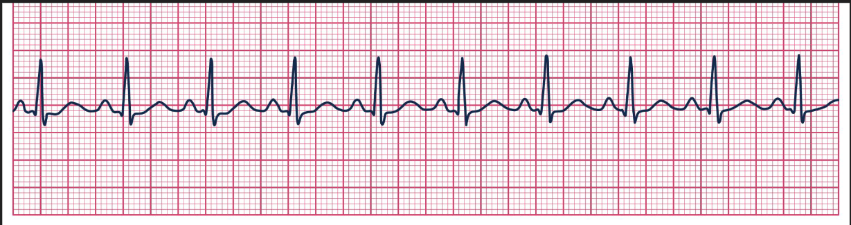
Equal spacing between beats and decent height, originates from SA
Ventricular Tachycardia (V-Tach) (Waveform and cause)
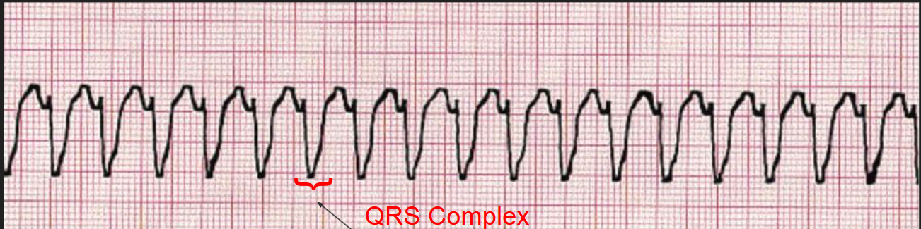
Ventricles contract quicker than atrial blood can fill them, causing no pulse.
EKG shows a fast regular rhythm, but with wide QRS complexes, and tachyarrhythmia originates in the ventricles. Without intervention, this will lead to death.
Purkinje cells themselves can become the source of an abnormal electrical impulse: Catecholaminergic polymorphic ventricular tachycardia (CPVT)
Shockable rhythm!
Ventricular Fibrillation (V-Fib) (Waveform and cause)
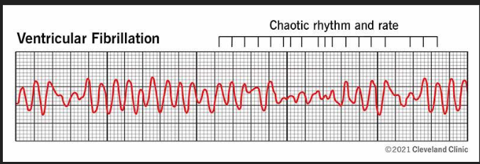
Fibrillation refers to an irregular and rapid beating of the hearts chambers, which fails to pump blood effectively.
Instead of a coordinated depolarization, the ventricular muscle fibers “quiver” or fibrillate independently
Shockable rhythm! (Disorganized rhythm, Chaotic Pattern)
Pulseless Electrical Activity (PEA) (Waveform and cause)
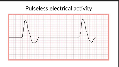
There is no pulse despite electrical activity in the heart, most likely due to muscle death or weakness. Commonly caused by hypovolemia, cardiac tamponade, and hypoxia.
NOT shockable, electrical activity is present, AED doesn’t help
*Note that a PEA may have different rhythms than the picture
Asystole
There is no electrical activity in the heart, rendering defibrillation ineffective. Continue compressions and take to higher care.
NOT shockable, won’t be of any use

Describe the AHA Out of Hospital Cardiac Chain of Survival
Activation of Emergency Response
High-Quality CPR
Defibrillation
Advanced Resuscitation
Post-Cardiac Arrest Care
Recovery
Age Parameters
Neonate: < 28 days
Infant: <1 year old
Child: Between 1 year old and puberty
Adult: Past Puberty
How to do Compressions and Ventilations
Compressions:
Push Hard (1/3 patient’s chest)
Push Fast (100-120 BPM), using feedback device or metronome
Allow full recoil on chest
Ventilations:
Open airway with head-tilt-chin-lift and insert OPA
Give 2 quick ventilations
Good ventilations produce chest rise and are delivered over 1 second
Rotate compressors every 5 CPR cycles or 2 minutes
What’s the Compression to Ventilation Ratio for adults and pediatrics with 1 and 2 rescuers?
Adults: 30:2
Infants and Children: 30:2 with 1 rescuer, 15:2 with 2 rescuers
What should you do with a patient with an advanced airway?
Ventilations are ASYNCHRONOUS, breaths can be administered during compressions
iGEL is an advanced airway device that improves CCF
Why does Defibrillation work? (For V-Fib and V-Tach)
Shocking with an AED sends a massive depolarizing current, forcing all myocardial cells into a refractory period at once.
This hopefully allows the SA node to restart an organized rhythm, kind of like putting them in time out.
What are some pad placement considerations?
Medical patch: Removed with gloved hand and wipe away any residual moisture
Hairy Chest: Shave it or “wax” it with additional pads
Pacemaker: Place pads below it
Adult Pads for Pediatrics: Apply Anterior and Posterior
Wet or in water: Pull out of water and dry chest
Witnessed vs Unwitnessed Cardiac Arrest (Diff. between adult and child)
Witnessed
In an Adult:
Activate emergency response and get AED, initiate 5 CPR cycles
In Infants/Children:
Activate emergency response and get AED, initiate 5 CPR cycles
Unwitnessed
In an Adult:
Activate emergency response and get AED, initiate 5 CPR cycles
In Infants/Children:
Perform 5 CPR cycles, then activate emergency response
Difference in infant/children is b/c cardiac arrest in such patients is more likely due to a respiratory issue, their brain has been deprived of oxygen longer.
What are Advanced Directives?
MOLST and DNR are honored by SBVAC, including:
Signed eMOLST or MOLST form
Signed NYS-approved document, bracelet, or necklace
Properly documented nursing home or nonhospital DNR
We CANNOT perform:
Compressions
Ventillation
Defibrillation
Intubation or resuscitative medical administration
What does ROSC stand for? What should you do if it occurs?
ROSC: return of spontaneous circulation
If it occurs:
Deliver supplemental O2, continue rescue breathing if the patient regains pulse without adequate breathing
Take vitals and BGL
Acquire a 12-lead EKG, send to ALS for transmission
Perform a full assessment
5 Reasons to STOP or Not Start CPR
Patient is obviously dead
ROSC occurs
Scene is unsafe
Valid DNR or MOLST provided
After 20 minutes, consider calling medical control for CPR termination
Obvious Signs of Death (TRI EMS 3D) MEMORIZE this
Rigor Mortis
Stiffening of Body caused by chemical changes after death
Dependent Lividity:
Skin discoloration due to blood pooling due to lower body parts from gravity
Submersion of over an hour
Drowning and submersion underwater for >1 hour
Decomposition:
Natural process of organic matter breaking down after death
Mass Crush injury to vital Area
Catastrophic trauma of head or torso with extreme force, causing immediate and often fatal destruction of vital organs and major structures
Transection:
Completely cut through
Decapitation:
Full or partial separation of head from body
Evisceration of brain matter
Brain tissue (white or gray matter) outside skull
Incineration
Bodily destruction through burning
How do Traumatic Arrests occur and what to do? What is “commotio cordis”?
Possible mechanisms due to falls, MVAs, or blunt trauma
Commotio Cordis: Blunt trauma to chest on uprise of T-wave, causing instant v-fib
When performing CPR, try to maintain spinal stability, use Jaw Thurst manuever, and take SMR precuations, like a backboard

What are considerations for hypothermic patients?
Check pulse and breathing for 60 seconds
Vasoconstriction makes pulse harder to feel
Dramatically slowed heart rate and respiration may delay accurate reading
Shock them with 1 time MAX with AED
Heart’s electrical velocity affected by the cold, repeated shocks may damage it further
Dependent Lividity, Rigor Mortis, and Fixed Pupils are NOT obvious signs of death
Cold temperatures affect normal bodily systems to appear as such
Injuries incompatible with life and chest wall rigidity preventing effective chest compressions ARE signs of death
Describe High-quality Chest Compressions
100-120 Compressions/Minute
1/3 of chest depth
Full recoil of chest between compressions
Limit hands off time to less than 10 seconds
What is CCF? What’s the optimal CCF and how can it be failed to accomplish?
Proportion of CPR where high-quality chest compressions are actually being delivered
Time compressions are performed
Also synonymous with minimized hands off time
AHA recommends an optimal CCF of greater than 80%
Threats to this include:
Long pulse checks
Prolonged intubation attempts
Poorly organized provider/team care
Higher CCF: higher ROSC and survival chances
Reversible Causes of Cardiac Arrest
H’s and T’s
H:
Hypoxia
Hypovolemia (severe blood plasma loss of dehydration)
Hydrogen ion (acidosis)
Hyper/Hypoglycemia
Hypo/Hyperkalemia
T:
Tension pneumothorax
Tompanade (cardiac)
Toxins
Thrombosis:
Pulmonary embolism
Myocardial infarction
Rescue Breathing rates for Adults and Children
Give rescue breathing if patient breathing isn’t enough for them
Adults: 1 breath every 5-6 seconds
Infants and Children: 1 breath every 2-3 seconds
These rates are the same with anyone with advanced airway from ALS
Describe Dual Sequential Defibrillation (not too important, just take a look)
Delivery of two defibrillation shocks simultaneously from two different defibrillators with pads placed in different positions:
Anterior Lateral Anterior Posterior
Two shocks may depolarize more myocardium cells
Sequence:
Apply AED PADS anterior posterior position, don’t delay compressions
Shock 3 times using such placement, if the patient is still in a shockable rhythm after second shock, preemptively set up second pair of pads
For fourth shock change time in Anterior Lateral pad placement, vector change
Shock using both at same time
What does the decision to transport consist of? What is Plat 10 and the Golden Hour?
Decision to transport:
Load and go: Unstable patient (high priority)
Play and stay: Stable patient (low priority)
Plat 10: 10 minutes between on scene arrival and departure
Golden Hour: 1 hour between time injury and definitive hospital care
Update ALS about patient condition
Only applicable if they were called in scene size up
Briefly summarize patient and what you’ve done so far
Decide on intercept or straight to hospital