Case 8: Is That a Rock in my Shoe?
 Call Kai
 Learn
Learn Practice Test
 Spaced Repetition
 Match
Match Flashcards
 Knowt Play
1/35

There's no tags or description
Looks like no tags are added yet.
Name | Mastery | Learn | Test | Matching | Spaced | Call with Kai | Chat |
|---|
No analytics yet
Send a link to your students to track their progress
36 Terms
Soft tissue and/or fluid
What is the normal opacity of the urinary bladder?
Liver
What is A?
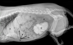
Small intestines
What is B?
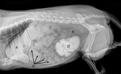
Left kidney
What is C?
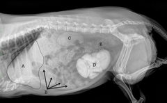
Urinary Bladder
What is D?
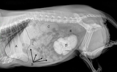
Descending colon
What is E?
Liver
What is A?
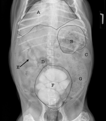
Fundus of the stomach
What is B?
Spleen
What is C?
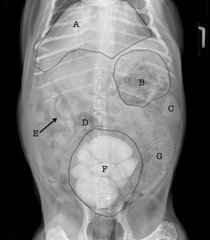
Cecum
What is D?
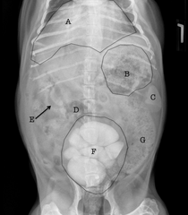
Small Intestine
What is E?
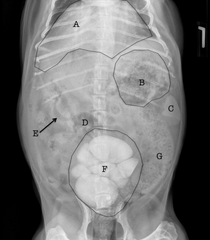
Urinary bladder
What is F?
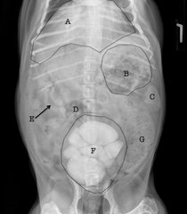
Descending colon
What is G?
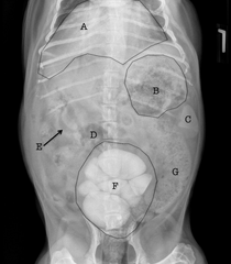
A = body
B = neck
C = apex
What are the 3 anatomic regions of the urinary bladder?
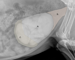
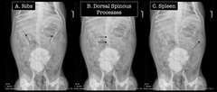
B, dorsal spinous processes
What is the best anatomic structure to use to determine whether or not the patient is rotated?
Lateral and VD abdominal projections are available for review.
What study is this: Describe the views included, the anatomy being imaged and the type of patient undergoing examination.
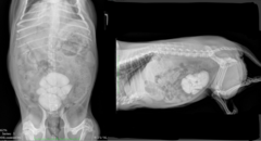
Patient positioning is adequate for interpretation.
Study technique is adequate for interpretation.
Describe patient positioning and technique
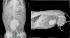
Abdominal serosal detail is within normal limits. The stomach contains amorphous granular material consistent with ingesta. Foreign material cannot be entirely ruled out, but should be clinically correlated. The liver is unremarkable. The kidneys are difficult to visualize. The spleen is unremarkable. The small intestines are uniformly fluid filled, contain small amounts of gas or are empty with no evidence of an obstructive pattern. The colon contains granular material consistent with feces. Multiple, variably sized, irregularly shaped, smoothly marginated mineral opaque structures are present within the urinary bladder, completely filling the lumen of the bladder. Two large, oval calculi are predominate at the cranial and caudal aspects of the bladder. The extra-abdominal structures, including what can be seen of the caudal thorax, are unremarkable.
Describe your findings (roentgen signs)
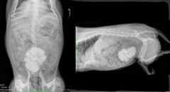
Multiple urinary calculi completely filling the urinary bladder lumen.
Describe your conclusions: Give your radiographic diagnosis and list possible differential diagnoses for all your findings. This is where you let the report reader know what you think is going on with the patient.
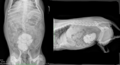
Due to the size and number of the calculi, surgical removal should be considered.
note: based on the fact these are so big and patient cannot pass them
Describe your next steps: Are there any additional imaging suggestions that would help clarify your findings? Are there critical next steps to take from a stabilization/management/treatment plan standpoint?
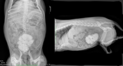
Adrenal glands and pancreas
What do you normally NOT see on radiographs?
Image on the left
Which one is the BEST positioned in terms of dorsal spinous processes?
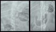
Number, size, shape, margin, opacity, position
What are the roentgen signs?
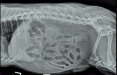
Doesn't include back end of patient = would not be able to see all stones and cannot evaluate whole penile urethra
This image is not a great image. Why?
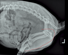
Penile urethra
Stones, if they are small enough to come out of the bladder, like to hide where (outlined in red)?
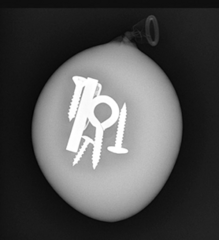
Metal = radioopaque = easy to identify
What kind of "stone" opacity is this?
Plastic = not as radioopaque but can still identify
What kind of "stone" opacity is this?
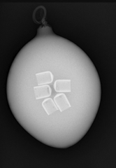
Fat opacity = minimally radioopaque but can still identify
note: was actually fish oil capsules
What kind of "stone" opacity is this?
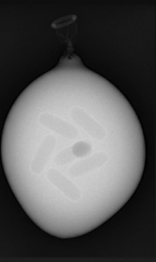
Soft tissue opacity = radiolucent
note: was actually cashews
What kind of "stone" opacity is this?
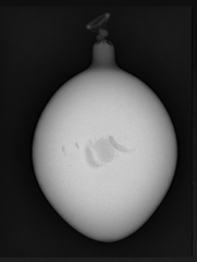
Soft tissue opacity = radiolucent
note: was actually grapes
What kind of "stone" opacity is this?
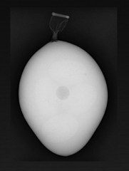
True
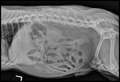
The patient in the image has radiographically evident urinary calculi.
TRUE OR FALSE
xx
For the true or false question, if the patient has radiographically evident urinary calculi = true = here
d. Utilize compression radiography to decrease superimposition so the stones can be more clearly identified.
JUSTIFICATION: Answer A is not correct because increasing kVp (increases the energy of the x-ray beam) and increasing mAs (increases the number of x-rays) results in a darker radiograph and will "burn" through the small calculi making them less conspicuous, not more conspicuous. Answer B is not correct because it requires you sending the patient home (kind of a pain) and isn't efficient. However, this is one of the reasons we like to take radiographs of fasted patients- it decreases fecal content within the descending colon, thereby minimizing superimposition of irregularly shaped mineral opaque material with the bladder that could be mistaken for urinary calculi. Answer C is not correct because horizontal radiographs are meant to identify gas within the abdomen, not necessarily radiopaque material.
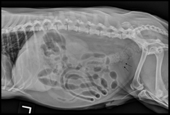
In the case presented in the Final Case Quiz, Question 1, the urinary calculi are challenging to visualize, thus, feeling confident about whether the small, round opaque structures are real is difficult. What can you do with the patient, right now, to increase your confidence level that the patient does, indeed, have urinary calculi?
a. Increase both the kVp and mAs in order to better penetrate the patient.
b. Send the patient home with instructions for the owners to fast the patient. Then have the owners bring the dog back in the morning to repeat radiographs.
c. Utilize horizontal radiography in hopes that the stones will become more obvious with the x-ray beam oriented at a different angle.
d. Utilize compression radiography to decrease superimposition so the stones can be more clearly identified.
True
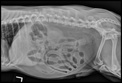
Struvite and calcium oxalate uroliths are the most common urinary calculi in the dog.
TRUE OR FALSE
False
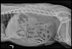
In a patient that presents for hematuria/stranguria, this is the only view we need for a complete study.
TRUE OR FALSE
Goat
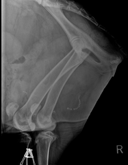
note: The correct answer is small ruminant (either a goat or sheep). The most important support for this answer is the "S" shape of the penile urethra indicating the sigmoid flexure of small ruminant species. The length of the femur as well as the flattened shape of the obturator foramen also lends support to small ruminant as the species.
This question is meant to push you outside of your comfort zone and allow you to think through how to answer the question using the Roentgen sign- SHAPE. What domestic species is this?