Distal Front Limb Radiography
 Call Kai
 Learn
Learn Practice Test
 Spaced Repetition
 Match
Match Flashcards
 Knowt Play
1/160

There's no tags or description
Looks like no tags are added yet.
Name | Mastery | Learn | Test | Matching | Spaced | Call with Kai | Chat |
|---|
No analytics yet
Send a link to your students to track their progress
161 Terms
We can control 3 parameters to alter the nature of the X Ray beam produced and so optimize our radiographic image
kVp
mA
Times
increasing kVp will —- the contrast
decrease
Higher mA increases the total number of photons and — the image overall and — contrast
DARKENS, REDUCES
what is considired the forelimb (3)
Limb distal to carpus = Dorsal/Palmar
Limb proximal to carpus = Cranial/Caudal
Trunk (scapula) = Dorsal/Ventral
what is considired the hindlimb (3)
Limb distal to tarsus = Dorsal/Plantar
Limb proximal to tarsus = Cranial/Caudal
Trunk (pelvis) = Dorsal/Ventral
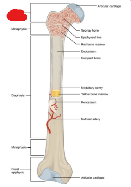
what is it
proximal epiphysis
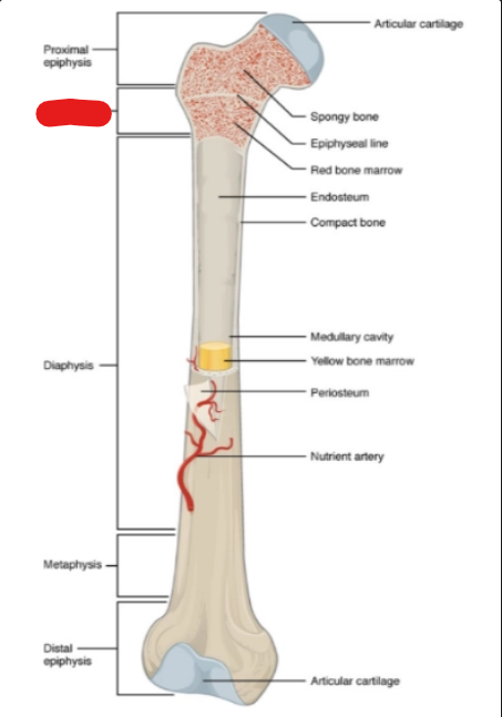
what is it
metaphysis
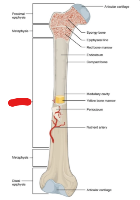
what is it
diaphysis
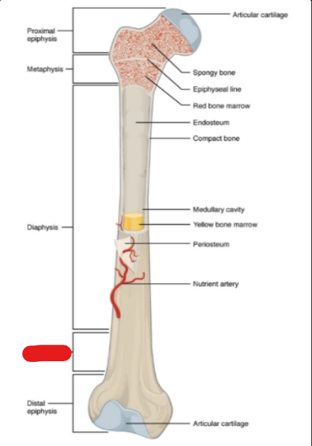
what is it
metaphysis
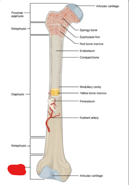
what is it
distal epiphysis
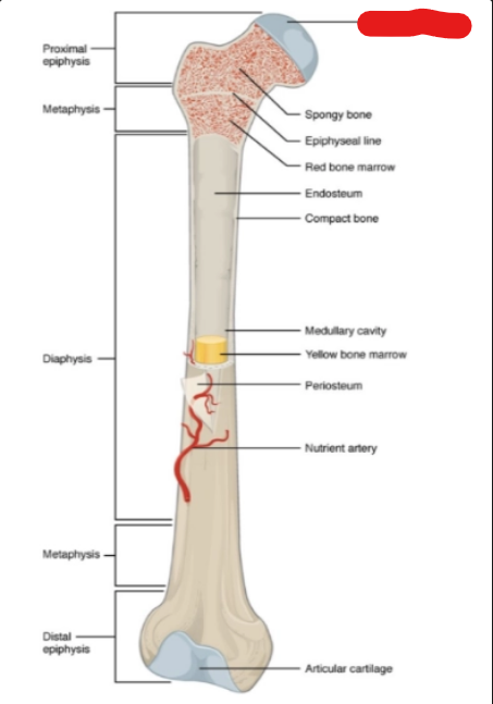
what is it
articular cartilage
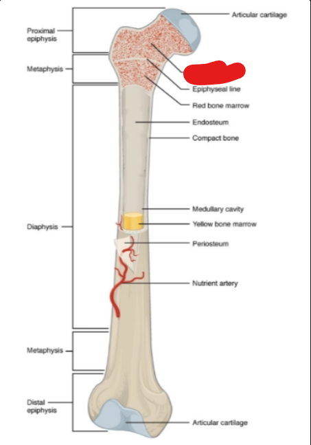
what is it
spongy bone
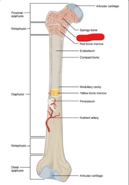
what is it
epiphyseal line
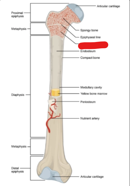
what is it
red bone marrow
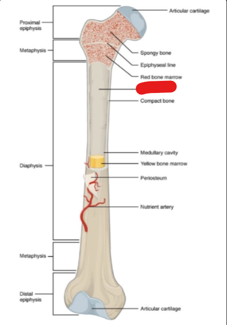
what is it
endosteum
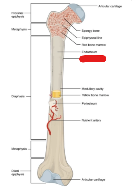
what is it
compact bone
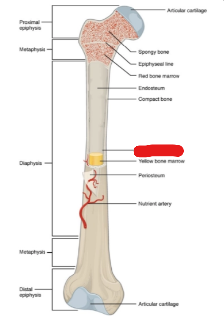
what is it
medullary cavity
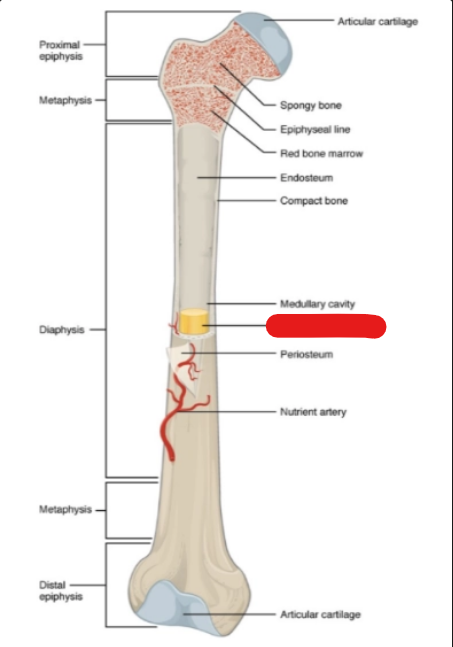
what is it
yellow bone marrow
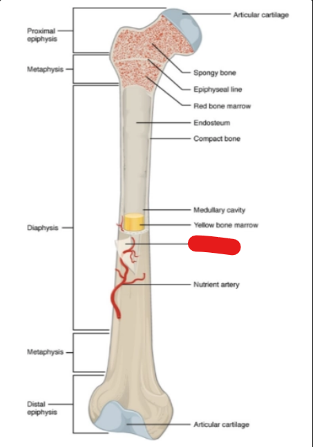
what is it
periosteum
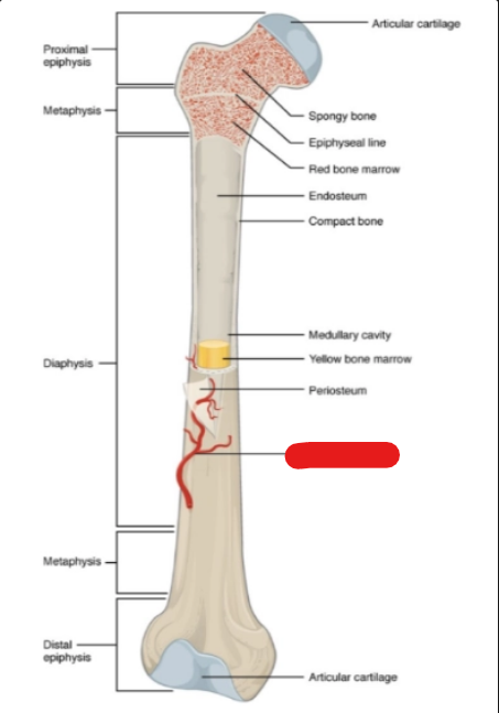
what is it
nutrient artery
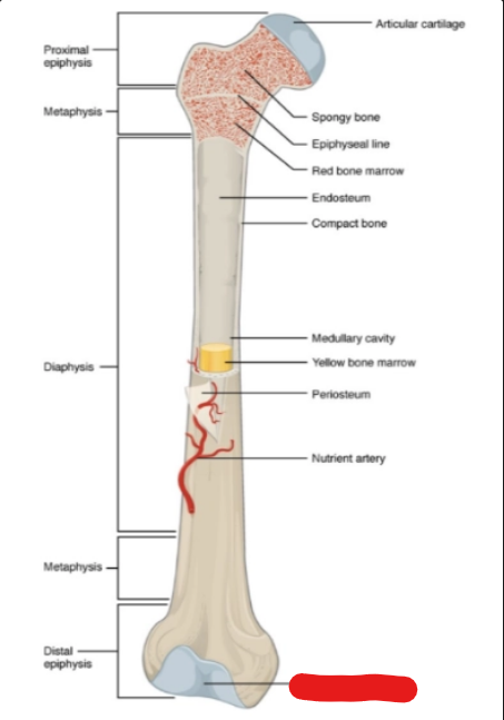
what is it
articular cartilage
It is important to collimate the field as tightly as possible to —- scatter radiation. This also results in —- contrast
reduce, higher
Use a small focal spot to maximize — —
spatial resolution.
Use —- kV and —- mAs technique to maximize radiographic contrast and not overexpose the image.
low, high
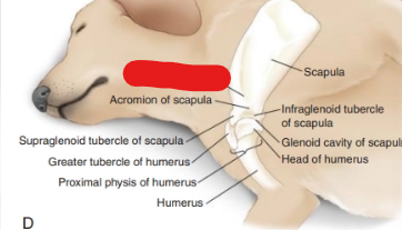
what is it
spine of scapula
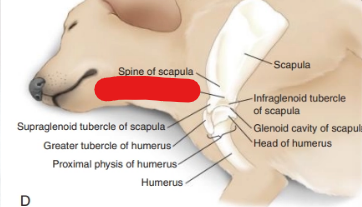
what is it
acromion of scapula
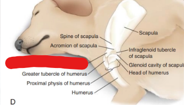
what is it
supraglenoid tubervle of scapula
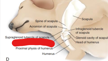
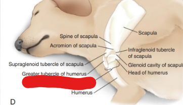
what is it
greater tubercle of humerus
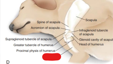
what is it
proximal physis of humerus
what is it
humerus
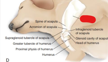
what is it
scapula
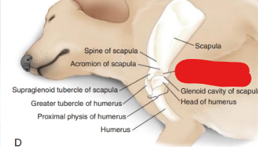
what is it
infraglenoid tubercle of scapula
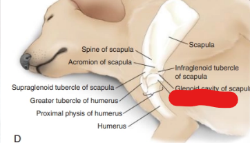
what is it
head of humerus
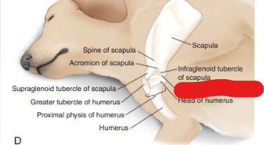
what is it
glenoid cavity of scapula
where do u measure Lateral (Mediolateral) View of the Scapula
Dorsally at the cranial border of the scapula approximately at about the site of T1 to T2 spinous process or thickest area where scapula is noted.
where is the central ray for Lateral (Mediolateral) View of the Scapula
Center of the scapula.
where are the 3 borders for Lateral (Mediolateral) View of the Scapula
Cranial: Proximal to shoulder joint.
Caudal: Past Caudal border of the scapula
Dorsally and ventrally: Include the entire scapula.
where do measure Lateral (Mediolateral) View of the Shoulder (2)
At the level of the shoulder joint.
Extend the caliper to the point of the unaffected shoulder, being careful not to include that limb.
where is the central ray for Lateral (Mediolateral) View of the Shoulder
At the scapulohumeral articulation6 (shoulder joint) by palpating the proximal head of the humerus and the glenoid fossa of the scapula.
where are the 2 borders for Lateral (Mediolateral) View of the Shoulder
Distally: Distal third of the scapula.
Proximally: Proximal third of the humerus.
where to measure Caudocranial View of the Shoulder (2)
At the level of the scapulohumeral articulation (shoulder joint).
Extend the calipers over the caudal cervical vertebral bodies, but do not include the opposite limb.
where is the central ray Caudocranial View of the Shoulder
at the center of the scapulohumeral articulation (shoulder joint).
where is the 2 border for Caudocranial View of the Shoulder
Distally: Distal third of the scapula.
Proximally: proximal third of the humerus
where to measure for Caudocranial View of the Shoulder (2)
At the level of the scapulohumeral articulation (shoulder joint).
Extend the calipers over the caudal cervical vertebral bodies, but do not include the opposite limb.
where is the central ray for Caudocranial View of the Shoulder
Palpate the proximal head of the humerus and the acromion of the scapula at the center of the scapulohumeral articulation (shoulder joint).
where are the 2 borders for Caudocranial View of the Shoulder
Distally: Distal third of the scapula.
Proximally: proximal third of the humerus
what are the routine x-rays for shoulder (2)
Lateral (Mediolateral) View of the Scapula
Caudocranial View of the Shoulder
where to measure for Lateral (Lateromedial) View of the Scapula
where is the central ray for Lateral (Lateromedial) View of the Scapula
Center of the scapula.
where are the 3 borders for Lateral (Lateromedial) View of the Scapula
Cranial: Proximal to shoulder joint.
Caudal: Past Caudal border of the scapula.
Dorsally and ventrally: Include the entire scapula.
where to measure for Skyline (Cranioproximal-Craniodistal) View of the Shoulder for the Bicipital Groove
At the head of the humerus, palpating for the bicipital groove.
where is the centre for Skyline (Cranioproximal-Craniodistal) View of the Shoulder for the Bicipital Groove
At the head of the humerus, palpating for the bicipital groove.
where is the border for Skyline (Cranioproximal-Craniodistal) View of the Shoulder for the Bicipital Groove
Proximal humerus with tight collimation.
what are the 2 ancillary views of the shoulder
Lateral (Lateromedial) View of the Scapula
Skyline (Cranioproximal-Craniodistal) View of the Shoulder for the Bicipital Groove
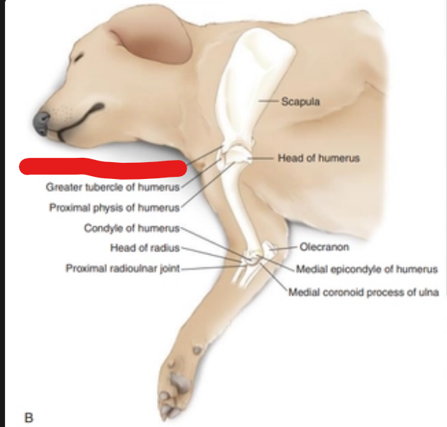
what is it
supraglenoid tubercle of scapula
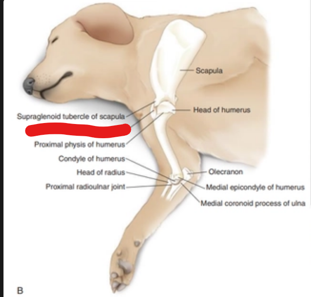
what is it
greater tubercle of humerus
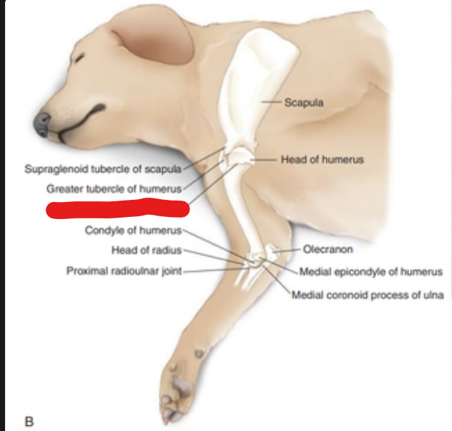
what is it
proximal physis of humerus
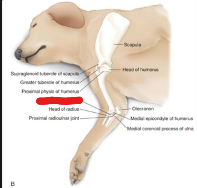
what is it
condyle of humerus
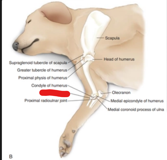
what is it
head of radius
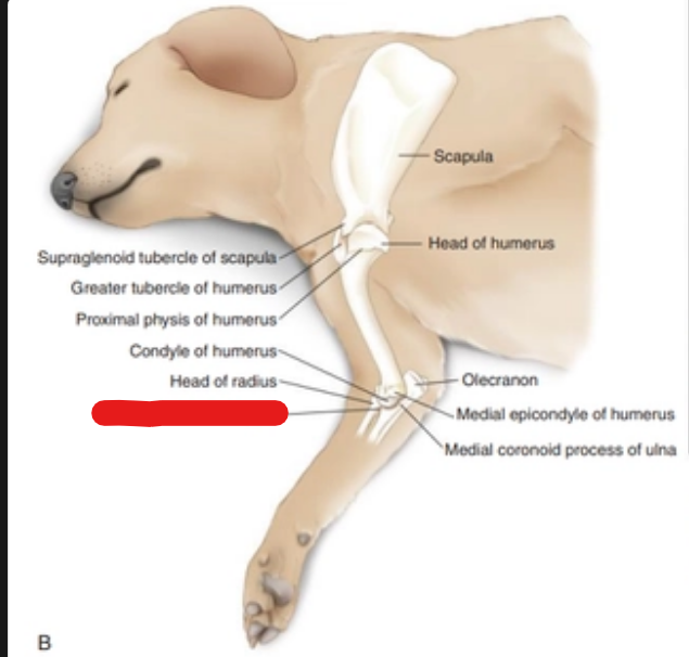
what is it
proimal radioulnar joint
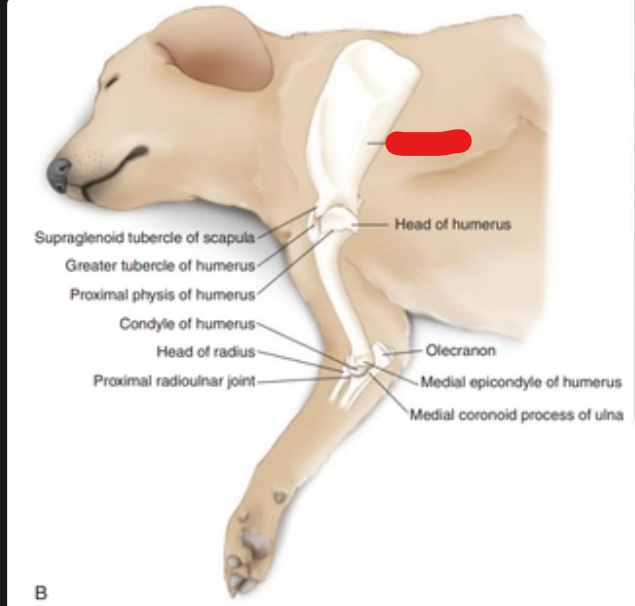
what is it
scapula
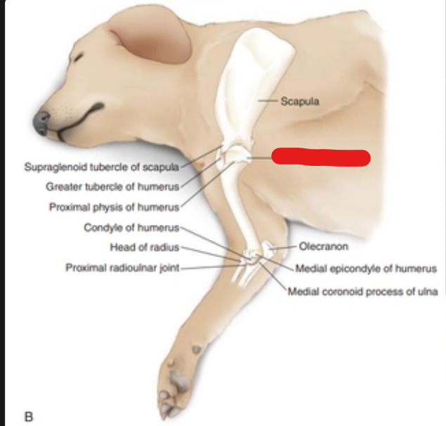
what is it
head of humerus
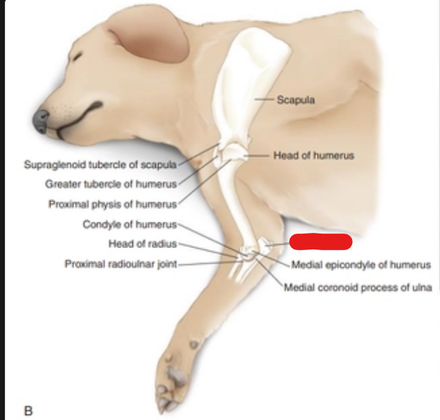
what is it
olecranon
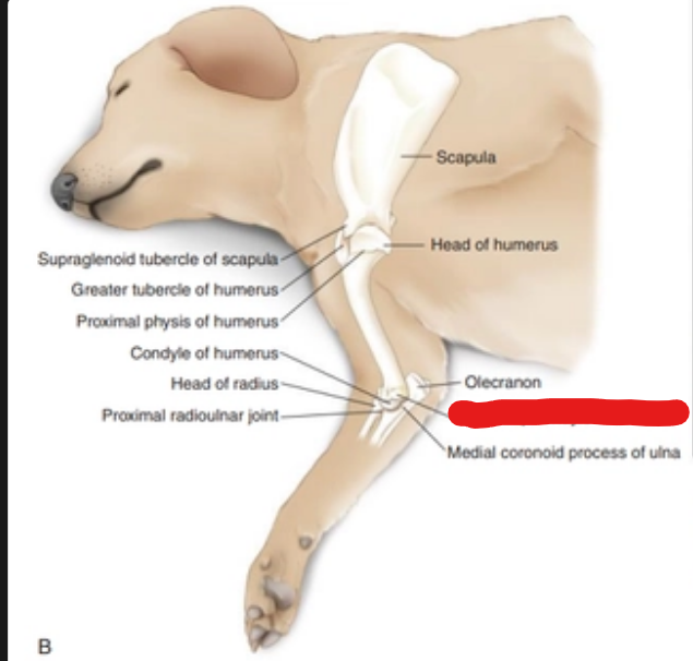
what is it
medial epicondyle of humerus
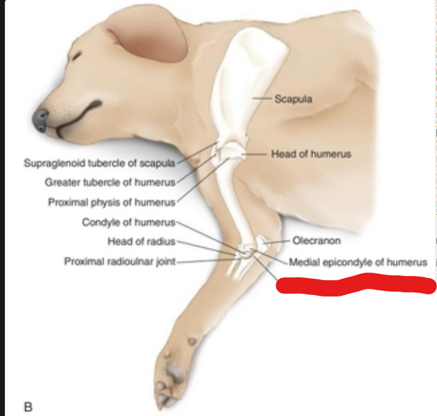
what is it
medial coronoid process of ulna
where to measure for Lateral (Mediolateral) View of the Humerus
Toward the proximal humerus at the level of the scapulohumeral articulation (shoulder joint).
where is the central ray for Lateral (Mediolateral) View of the Humerus
Midshaft of the humerus.
where are the 2 borders of the Lateral (Mediolateral) View of the Humerus
Proximal: To shoulder.
Distal: To elbow
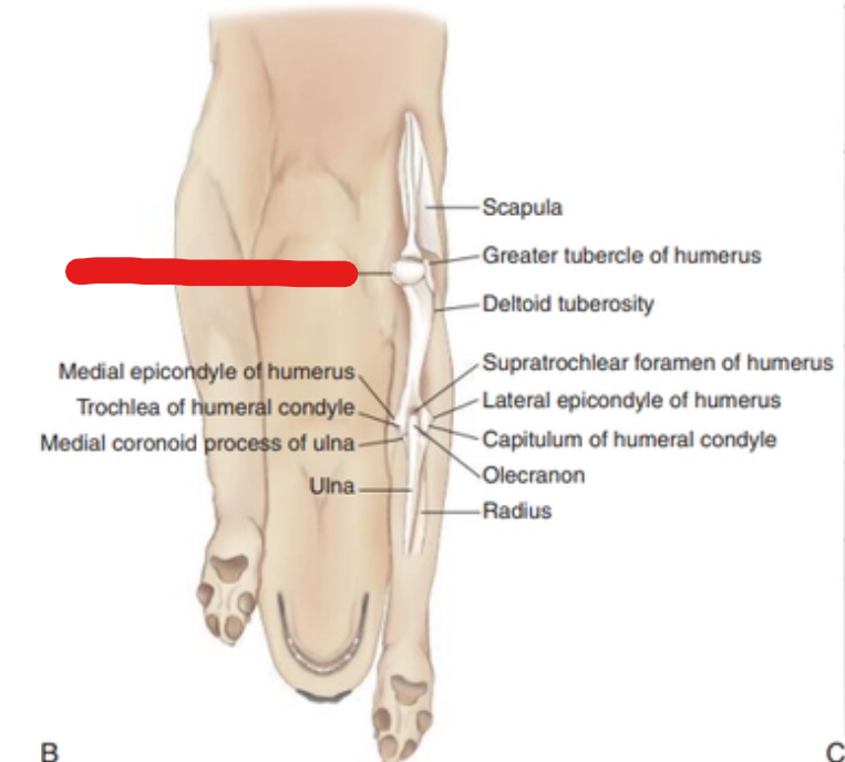
what is it
lesser tubercle of humerus
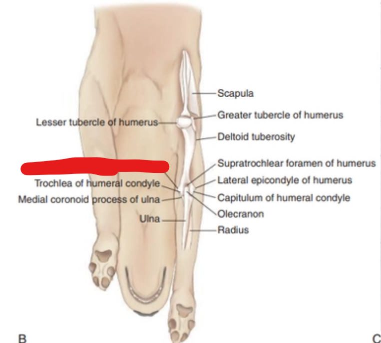
what is it
medical epicondyle of humerus
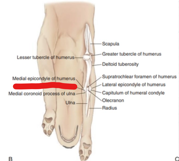
what is it
trochlea of humeral condyle
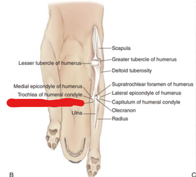
what is it
medial coronoid process of ulna
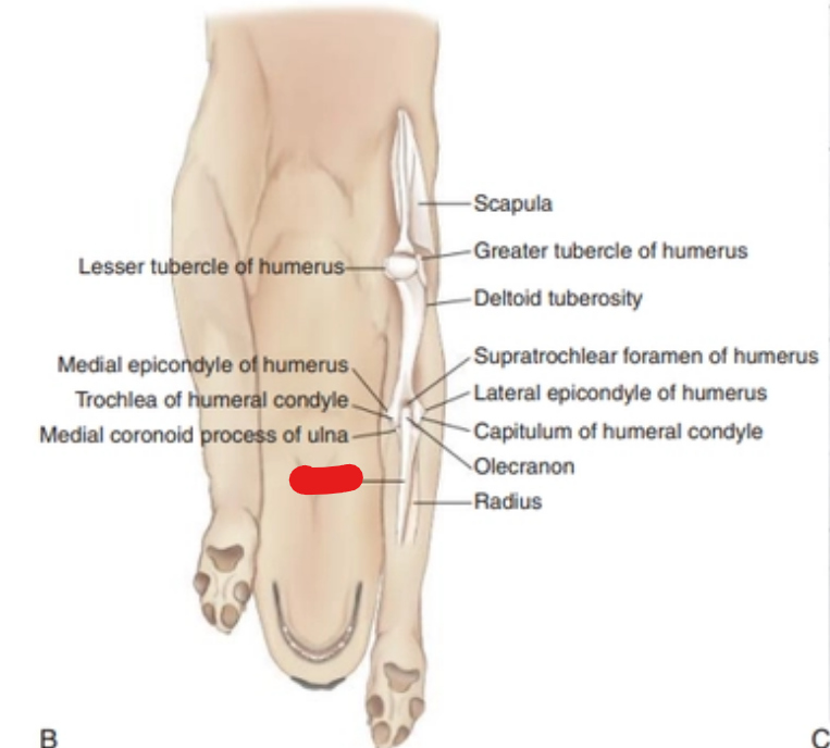
what is it
ulna
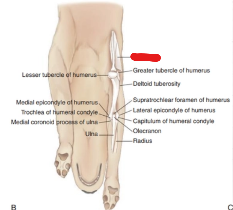
what is it
scapula
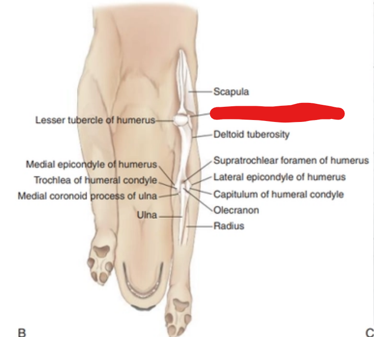
what is it
greater tubercle of humerus
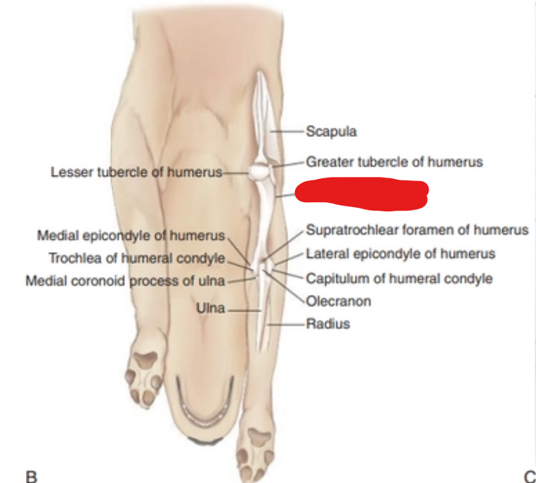
what is it
deltoid of humerus
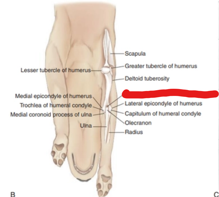
what is it
supratochlear foramen
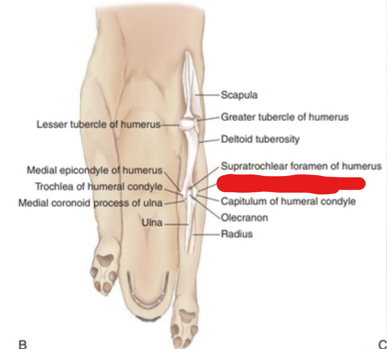
what is it
lateral epicondyle of humerus
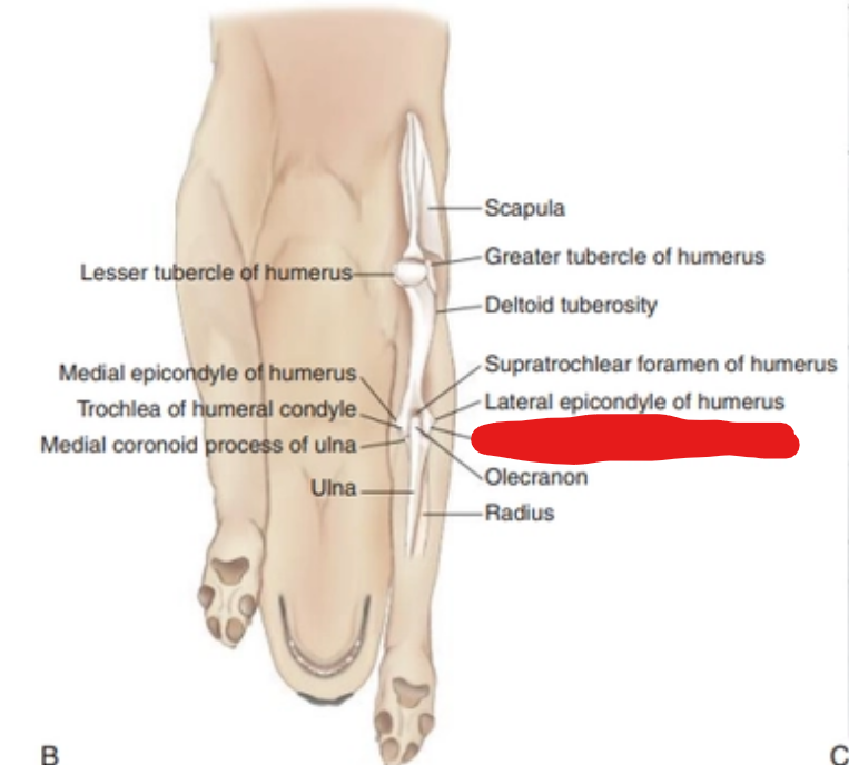
what is it
capitulum of humeral condyle
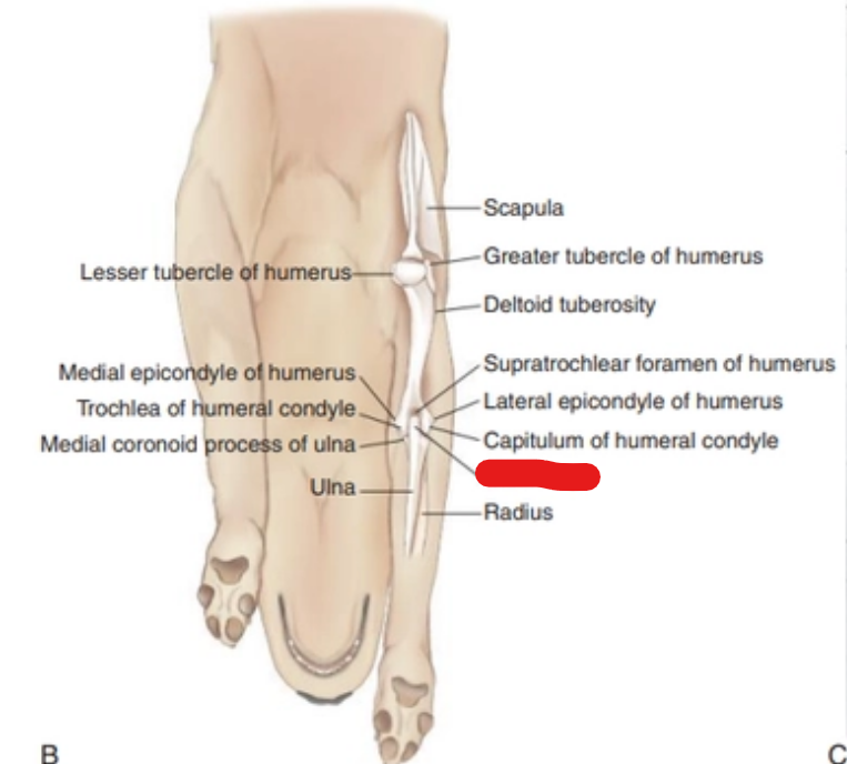
what is it
olecranon
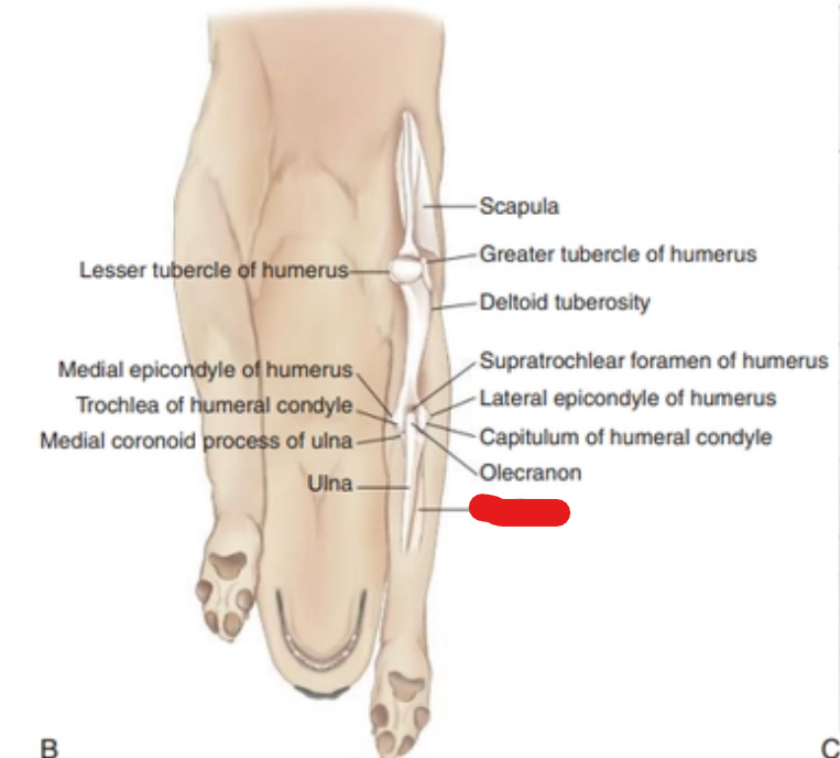
what is it
radius
what is the measure of the Caudocranial View of the Humerus
Midshaft toward the proximal aspect; across at the level of T1.
where is the central ray of the Caudocranial View of the Humerus
where are the 2 borders of the Caudocranial View of the Humerus
Proximal: Include shoulder joint.
Distal: Include elbow joint
what are the upper arm routine x-rays (2)
Lateral (Mediolateral) View of the Humerus
Caudocranial View of the Humerus
where is the measurement of the Craniocaudal View of the Humerus
Midshaft of the humerus toward the proximal aspect.
where is the central ray of the Craniocaudal View of the Humerus
Midshaft of the humerus.
where are the 2 borders of the Craniocaudal View of the Humerus
Proximal: Include shoulder joint.
Distal: Include elbow joint
what are the upper arm ancillary x-rays
Craniocaudal View of the Humerus
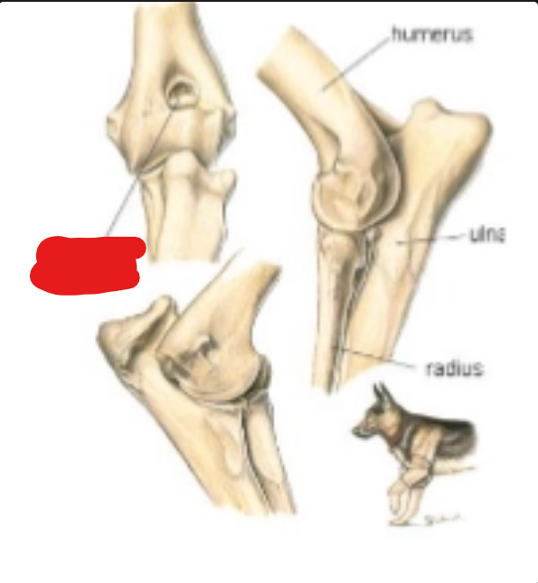
what is it
anconeal process
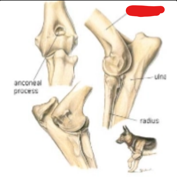
what is it
humerus
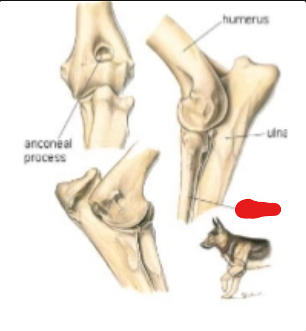
what is it
radius
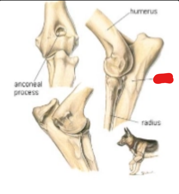
what is it
ulna
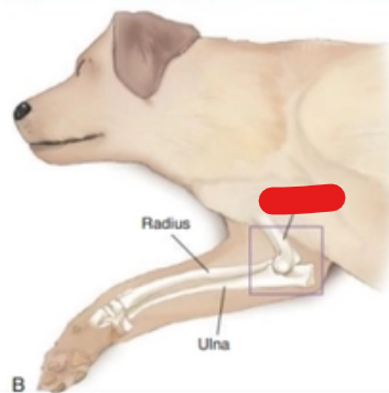
what is it
humerus
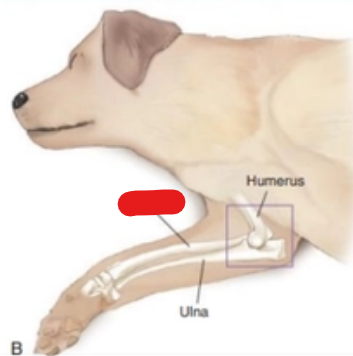
what is it
radius
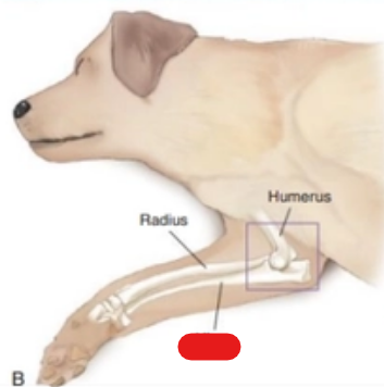
that is it
ulna
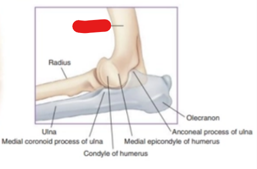
what is it
humerus
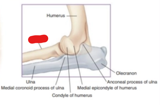
what is it
radius
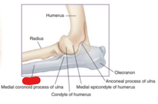
what is it
ulna
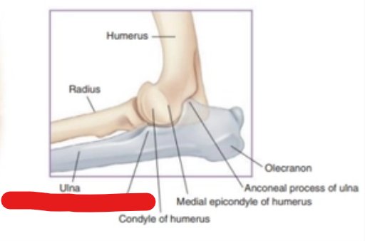
what is it
medial coronoid process of ulna