Muscle Physiology (Final)
 Call Kai
 Learn
Learn Practice Test
 Spaced Repetition
 Match
Match Flashcards
 Knowt Play
1/89

There's no tags or description
Looks like no tags are added yet.
Name | Mastery | Learn | Test | Matching | Spaced | Call with Kai | Chat |
|---|
No analytics yet
Send a link to your students to track their progress
90 Terms
Types of Muscle
- Skeletal
- Cardiac
- Smooth
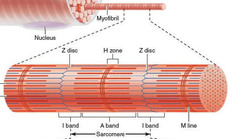
Sarcomere
- Contractile Unit of Muscle Fiber
The generation of action potentials in the sarcolemma triggers what?
- A sequence of events inside the cell that resuts in force development
Sarcolemma
- Plasma membrane of a muscle fiber (cell)
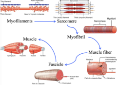
Muscle Structure: Largest → Smallest
- Muscle → Fascicle → Muscle Fiber (cell) → Myofibrils → Sarcomeres (repeating units inside myofibrils)
How do muscle cells generate force?
- Specialized motor proteins within the cell
Myofibrils consist of what?
- Sarcomeres., repeating contractual units
Components of a Sarcomere
- Z discs, A band, I band, H zone, M line
Z Disc
- Separates sarcomeres from each other
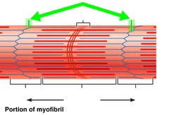
I Band
- Thin filaments only, attached to Z Disc
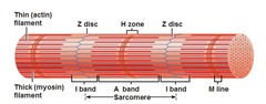
A Band
- Dark area; extends length of the thick filaments
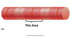
H Zone
- The region at the center of an A band of a sarcomere that is made up of myosin only. The H zone gets shorter (and may disappear) during muscle contraction.
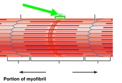
M Line
- Center of sarcomere, attaches to the thick filaments found in the H Zone
Light vs Dark Striations
- Light: I Bands → Thin
- Dark: A Bands → Thick
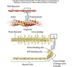
Components of Thin Filaments
- Backbone: Double helix of actin filaments
- Helical Grooves: Occupied by Tropomyosin + Troponin
Tropomyosin does what at rest?
- Obscures the binding sites between actin and myosin
What regulates skeletal muscle contraction?
- Tropomyosin + Troponin (T, C, I) Trimer
Troponin T
- Anchors trimer to tropomyosin
Troponin C
- Binds calcium
Troponin I
- Inhibits interactions between actin and myosin when intracellular calcium is low
How is skeletal muscle contraction regulated?
1. Motor neurons release Acetylcholine at a synapse
2. Action potentials sweep across the muscle fiber and into it along the transverse tubules
3. Ca2+ released from SR → myofibrils contract
Thick Filament Composition
- Head: Actin binding site + elements to bind and hydrolyze ATP for force development; cross bridges that bind to actin during muscle contraction
- Tails: Bundled together to form core of the filament
- Hinge: Connects the heads + tails
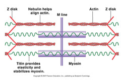
Titin
- Important for maintaining sarcomere structure, runs from Z disc → M Line
- Responsible for passive tension that is measured when a relaxed muscle becomes stretched
Elastic String of Muscle
Dystrophin
- Scaffolding protein that links thin filaments to proteins of sarcolemma
Sliding Filament Theory
- Theory that actin filaments slide towards eachother during muscle contraction, while the myosin filaments are still
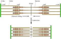
What happens if intracellular [Ca2+] is low?
- Actin binding sites are unavailable → no muscle contraction
What happens if sufficient tension is generated across muscle fibers to overcome a resistance
- Tension is transmitted via tendons to insertion points on skeleton → movement at joints
Somatic Motor Neurons: What are they? What do they release? Its result?
- Nerve cells whose cell bodies are in the brainstem and spinal cord that serve skeletal muscles
- Releases acetylcholine → Ca2+ released from SR → Voluntary control
What is total tension proportional to?
- The number of cross-bridges formed
Steps of Skeletal Muscle Contraction Phase
1. Motor neuron excites muscle fibers → Ca2+ released from SR
2. Ca2+ from SR binds to Troponin C → Troponin shifts position
3. Shifting of troponin exposes myosin binding sites on thin filaments
4. Myosin head forms tension-producing cross-bridge with the actin filament (with ADP + Pi bound to Myosin Head)
Steps of Skeletal Muscle Cross Bridge Cycle
1. Myosin is bound to ADP + Pi as well as actin filament in a cross-bridge, ready to contract
2. ADP + Pi are released, causes myosin head to pivot forward → pulling actin filaments and Z discs towards center of sarcomere (POWERSTROKE)
3. New ATP molecule binds to myosin head → head detaches from actin filament
4. ATP is hydrolyzed → ADP + Pi → Allows myosin head to reform cross-bridge with actin filament
5. Cycles of ATP hydrolysis/removal of ADP + Pi allows continuous power strokes when ATP/Ca2+ are present
Steps of Skeletal Muscle Relaxation Phase
1. Motor neuron activation ceases
2. Ca2+ dissociates from troponin C → pumped back into SR
3. Troponin-Tropomyosin complex moves back into position covering myosin binding sites → cross-bridging is unable to form and muscle tension production decreases
Motor Unit
- A motor neuron and all of the muscle fibers it innervates
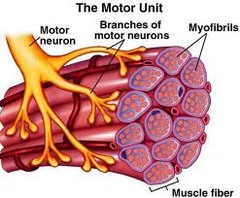
Fast vs Slow MU
- Fast: Generates tension and movement quickly
- Slow: Tension generation is slower and sustained
Muscle Fiber Types (Skeletal)
- Slow Oxidative
- Fast Oxidative
- Fast Glycolytic
Slow oxidative, Fast oxidative, Fast glycolytic: Fiber Diameter
- Slow: Smal
l- Oxidative: Intermediate
- Glycolytic: Large
Slow oxidative, Fast oxidative, Fast glycolytic: Tension Generation
- Slow: Low
- Oxidative: Intermediate
- Glycolytic: High
Slow oxidative, Fast oxidative, Fast glycolytic: Movement Velocity
- Slow: Slow
- Oxidative: Fast
- Glycolytic: Fast
Slow oxidative, Fast oxidative, Fast glycolytic: Fatiguability
- Slow: Low
- Oxidative: Intermediate
- Glycolytic: High
What do slow oxidative fibers do?
- Sustained, low intensity contractions
- Posturla muscles
- Walking and other types of "aerobic exercise"
Fast Oxidative Fibers: What do they do?
- Best for activités that requires both speed and moderate endurance
- Mid distance events, power walking, resistance training
Fast Glycolytic Fibers: What do they do?
- Powerful, maximal power, speed, and explosiveness
- Sprints
Myocardium
- Muscular, middle layer of the heart
Heart: Atria and Ventricles
- Four chambered (2 atria, 2 ventricles)
Atria and Ventricles: Steps of Contraction
1. Atria contracts first → moves blood into ventricles
2. Ventricular contractions move blood into pulmonary trunk and aorta
Intercalated Discs
- Specialized connections between myocardial cells containing gap junctions and desmosomes → excitation can spread rapidly across adjacent muscle fibers
How does cardiac muscle contract?
- Similar mechanism to skeletal
Where does the Ca2+ in cardiac muscle come from?
- Interstitium, SR
Smooth Muscle
- Involuntary muscle found inside many internal organs of the body
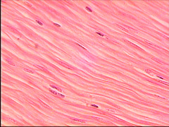
Smooth Muscle: Locations
- Blood vessels, airways
- Digestive/Urinary/Reproductive Tract
Smooth Muscle: Contraction leads to what?
- Constriction → Vasoconstriction → ↑ BP, ↓ Blood flow
Smooth Muscle: Relaxation leads to what?
- Dilation → Vasodilation → ↓ BP, ↑ Blood Flow
Morphology of Smooth Muscle Cells
- Short, spindle-shaped (fusiform) and feature a single, centrally located nucleus
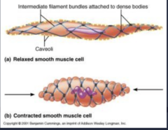
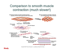
What happens to a smooth muscle cell during contraction?
- There is a network of "dense bodies:" in the cytoplasm that serve as attachment points for actin filaments, thick filaments overlapping thin in an irregular way
- Cell shortens and twists during contraction
Visceral Smooth Muscle
- Composed of sheets of several hundred spindle-shaped cells in close contact with one another in large bundles
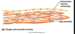
Why does each visceral (single unit) smooth muscle bundle behave as a functional syncytium?
- Gap junction connect adjacent smooth muscle cells, allows ions/electrical signals to pass directly between them → cells contract together as one unit
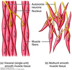
Multiunit Smooth Muscle vs Visceral
- Visceral = Team → Gap Junctions → Contract together
- Multi Unit = Solo → Little/no gap junctions → Contract Independently
When does contraction of smooth muscle begin?
- When intracellular [Ca2+] ↑↑
Source of Ca2+ in smooth muscle
- Ca2+ from Extracellular fluid
What happens when Ca2+ is introduced to smooth muscle?
1. Membrane becomes depolarized → voltage-gated Ca2+ channels open
2. Ca2+ enters from ECF → Binds Calmodulin
3. Ca2+ binds Calmodulin → Activates Myosin Light Chain Kinase → Phosphorylates myosin → muscle contraction
What can trigger depolarization of smooth muscle cells?
- A host of different hormones or neurotransmitters
What is another source of calcium for smooth muscle cells?
- Sarcoplasmic reticulum
How is smooth muscle contraction regulated?
- Different from striated, as there is no troponin in smooth muscle
- Instead, relies on phosphorylation of myosin by MLCK
Roles of Myosin Light Chain Kinase and Myosin Light Chain Phosphatase
- MLCK: Phosphorylates myosin → smooth muscle contracts
- MLCP → Dephosphorylates myosin → not able to contract
What forms when smooth muscle is stimulated and intracellular Ca2+ increases?
- Formation of Ca2+ : Calmodulin complex → Activates MLCK → Myosin phosphorylated → smooth muscle contracts
What is an isometric contraction?
- Muscle creates tension, but does not change length → no joint movement
Ex: Just holding a dumbbell
What is Isotonic Contraction?
- Muscle changes length → movement at joints
What is Concentric Contraction?
- Muscle shortens because muscle force > resistance
Ex: Lifting a dumbbell during bicep curl
What is Eccentric Contraction?
- Muscle lengthens while contracting because resistance > muscle force
Ex: Lowering the dumbbell during bicep curl
NOT RELAXATION
How are muscles generally arranged?
- As muscle pairs to balance opposing joint movements
Agonist vs Antagonist Muscles
- Agonist: Recruited to contract and cause joint movements
- Antagonist: Muscles that oppose agonists motion
Bicep Curls: Agonist vs Antagonist
- Agonist: Biceps
- Antagonists: Triceps
Tricep Extension: Agonist vs Antagonist
- Agonist: Triceps
- Antagonist: Biceps
What happens if muscle pairs contract simultaneously and equally?
- Isometric contraction → no joint movement
Muscle Stabilizer
- Helps keep your joint stable so that your agonist muscles can carry out the movement in the desired path of motion
Muscle Stabilizer: Examples
- Rotator cuff muscles
- Core abdominal muscles
- Gluteus medium
Muscle Fixator
A muscle that serves as a stabilizer of one part of the body during movement of another part.
Proprioception
- The ability to tell where one's body is in space.
Muscle Spindles: What does it detect? What does it do?
- Detects: How much and how fast a muscle is stretched
- Function: Makes the muscle contract to prevent over stretching
Stretch Sensor
Golgi Tendon Organs: What does it detect? What does it do?
- Detect: How much force/tension muscle is producing
- Function: Makes the muscle relax if the tension is too high to prevent injury.
What is a lever system?
- A lever system is a way your muscles use your bones and joints to create movement.
Like a seesaw
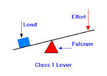
Lever Terminology: Fulcrum, Lever, Effort, Load
- Fulcrum: Joint (Pivot)
- Lever: Bone
- Effort: Muscle (pulling force)
- Load: Body part or object to be moved
First-Class Lever
- The fulcrum is positioned between the effort and resistance
- Atlanto-Occipital
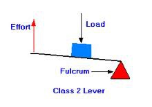
Second-Class Lever
- The load is between the fulcrum and the effort
- Standing on tiptoes
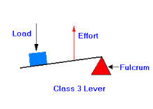
Third-Class Lever
- The fulcrum is at one end of the bar and the effort is between the fulcrum and the resistance
- Bicep curls
Muscle Force Forumla
Fm = F(resistiance) x d(resistance)/ d(muscle)
Fresistance
- Resistance/external load to be moved
dmuscle
- The point of muscle insertion and distance from the joint center
ddistance
- Length of the lever arm
Sarcopenia
- The loss of muscle mass, strength, and function that comes with aging