HMI204 - The nervous system
 Call Kai
 Learn
Learn Practice Test
 Spaced Repetition
 Match
Match Flashcards
 Knowt Play
1/69

There's no tags or description
Looks like no tags are added yet.
Name | Mastery | Learn | Test | Matching | Spaced | Call with Kai | Chat |
|---|
No analytics yet
Send a link to your students to track their progress
70 Terms
What is the three fundamental processes in the nervous system?
sensory input, integration, and motor output
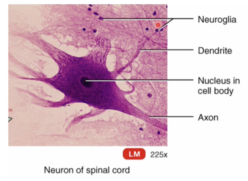
In nervous tissue, there are neurons and neuroglia (glial cells). A neuron’s function is to receive and transmit information. What is the function of neuroglia?
helps to support, nourish, and protect neurons.
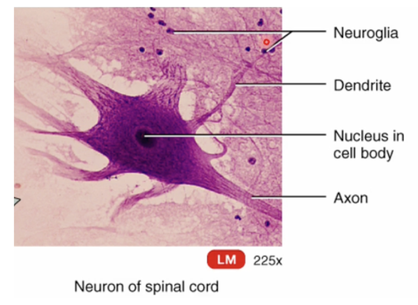
Anatomically, the nervous system is categorised as Central and Peripheral nervous systems. What nerves do they encompass and what is their function?
CNS - includes the brain and spinal cord. Their function is to process received sensory input, integrate this information and determine a motor command.
PNS - includes all nerves outside of the CNS. Their function is to transmit information to and from the CNS and the rest of the body.
Why can diseases that destroy CNS neurones produce permanent neurological deficits?
CNS neurones lack centrioles and cannot undergo mitosis, meaning they have very limited ability to replace lost cells.
Create a diagram to show how information from receptors get processed and turned into motor commands.
PNS is divided functionally into efferent and afferent. What does the afferent division do?
brings info from receptors into the CNS
What are the 3 main types of receptors?
Special Sensory receptors - smell, taste, vision, balance and hearing
Visceral sensory receptors - internal organs
Somatic sensory receptors - skeletal muscles, joints and skin
PNS is divided functionally into efferent and afferent. What does the efferent division do? This division also has more subcategories. Expand on this.
it transmits information about motor commands from the CNS to the rest of the body.
Efferent PNS can be divided into:
Somatic nervous system - voluntary/conscious control over the command. I.e. skeletal muscle
Autonomic nervous system - involuntary movements. I.e. smooth and cardiac muscles
Sympathetic division - flight or fight response. Prepares body for stress, action & danger.
Parasympathetic - ‘rest and digest’. Relaxes body, helps heal and save energy.
Following a large meal, which autonomic division predominates and why?
Parasympathetic nervous system because it promotes digestion, nutrient absorption, and energy conservation.
What tissues do parasympathetic and sympathetic nervous system innervate?
Smooth muscle, cardiac muscle, glands
Sympathetic; glands
What is the enteric nervous system?
Extensive network of neurons that line the walls of the GIT.
Controls digestion independent of CNS.
Neurulation is an the early embryonic process in vertebrates. What happens here and whys it important?
Ectoderm of the embryo folds in itself to create a neural tube, which ultimately becomes the central nervous system comprising the brain and spinal cord.
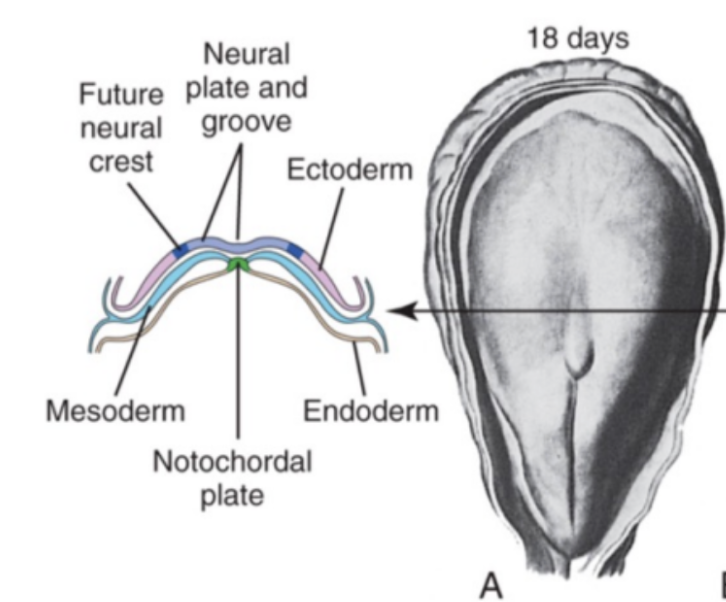
What is happening around day 18 of embryonic development?
embryo is made up of 3 layers:
Ectoderm - This is where the nervous system develops
Mesoderm
Endoderm
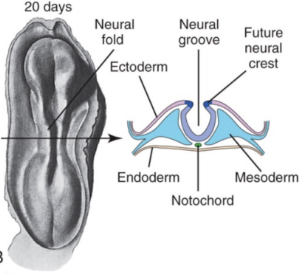
What step of neurulation is happening on day 20 of embryonic development?
Notochord induces ectoderm to fold in from the middle.
Starts to separate the ectoderm into surface ectoderm and neuroectoderm.
What step of neurulation is happening on day 21-22 of embryonic development?
edges of the neural folds meet and starts to fuse from the middle of the embryo to the superior and inferior ends (cranial and caudal ends).
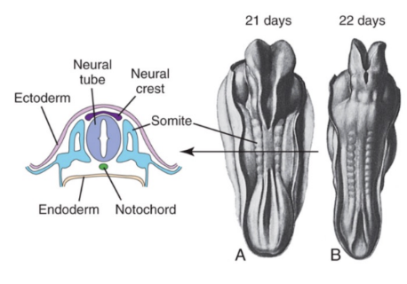
What step of neurulation is happening on day 25 of embryonic development?
Fusion of rostral/cranial neuropore
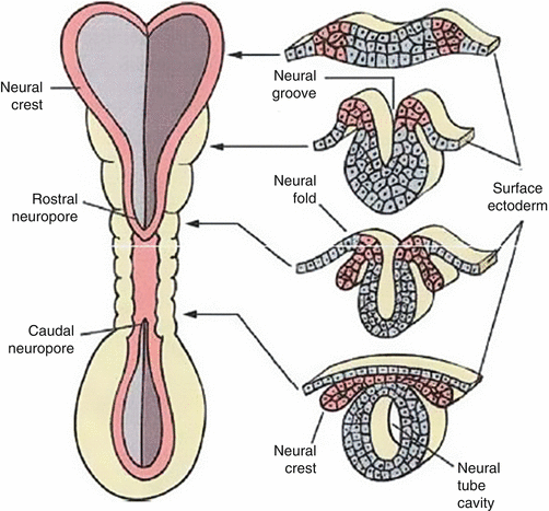
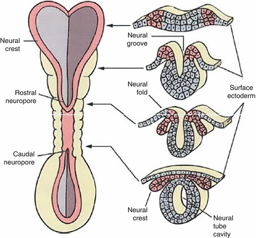
What is step of neurulation is happening on day 27-28 of embryonic development?
Fusion of caudal/inferior neuropore
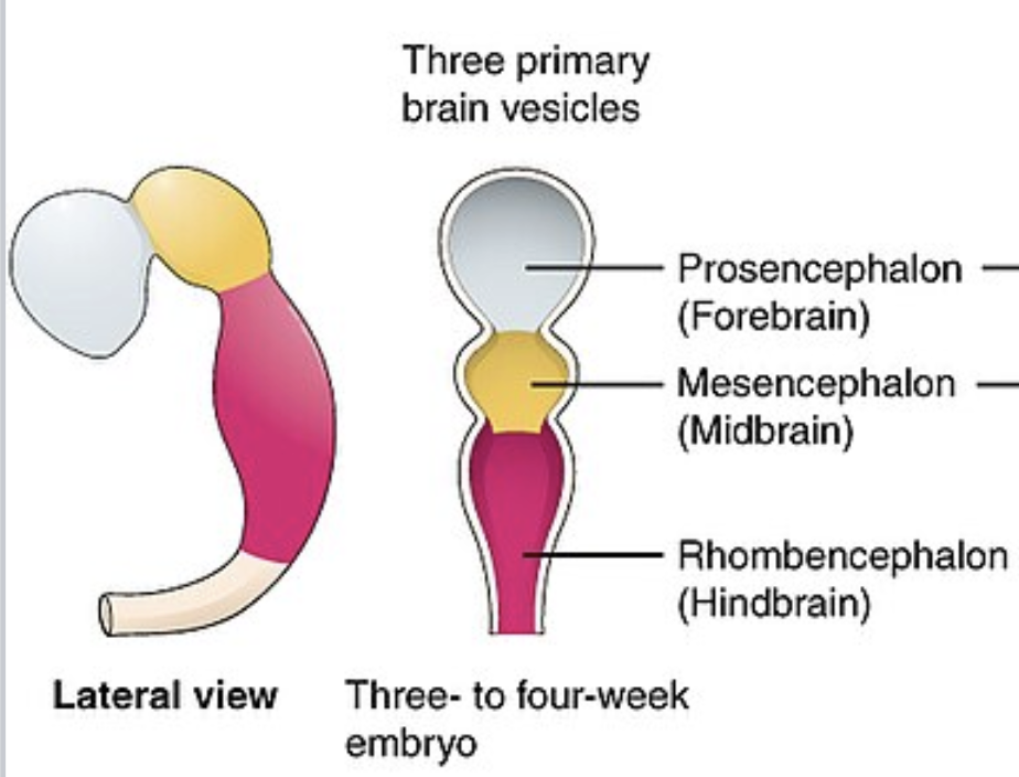
After 1 month, what stage is the embryonic brain development at?
forms the 3 primary brain vesicles:
Forebrain
Midbrain
Hindbrain
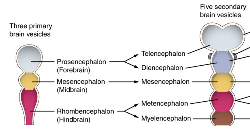
A week after forming the three primary brain vesicles, they divide and transform into secondary vesicles. List these by showing their branch from the primary vesicle.
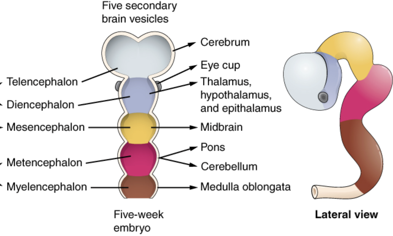
In a child, the secondary vesicles have divided further and form the different parts of the brain as we know it. List which parts branch off the secondary vesicle.
Are conditions that occur closer to the cranial end or caudal end more fatal?
cranial end due to higher risk of neurological compromise.
Describe the condition: Anencephaly (An- without, encepha- brain)
Non-closure of the cranial neuropore; non-formation of the cranial vault and brain. Non-compatible with life
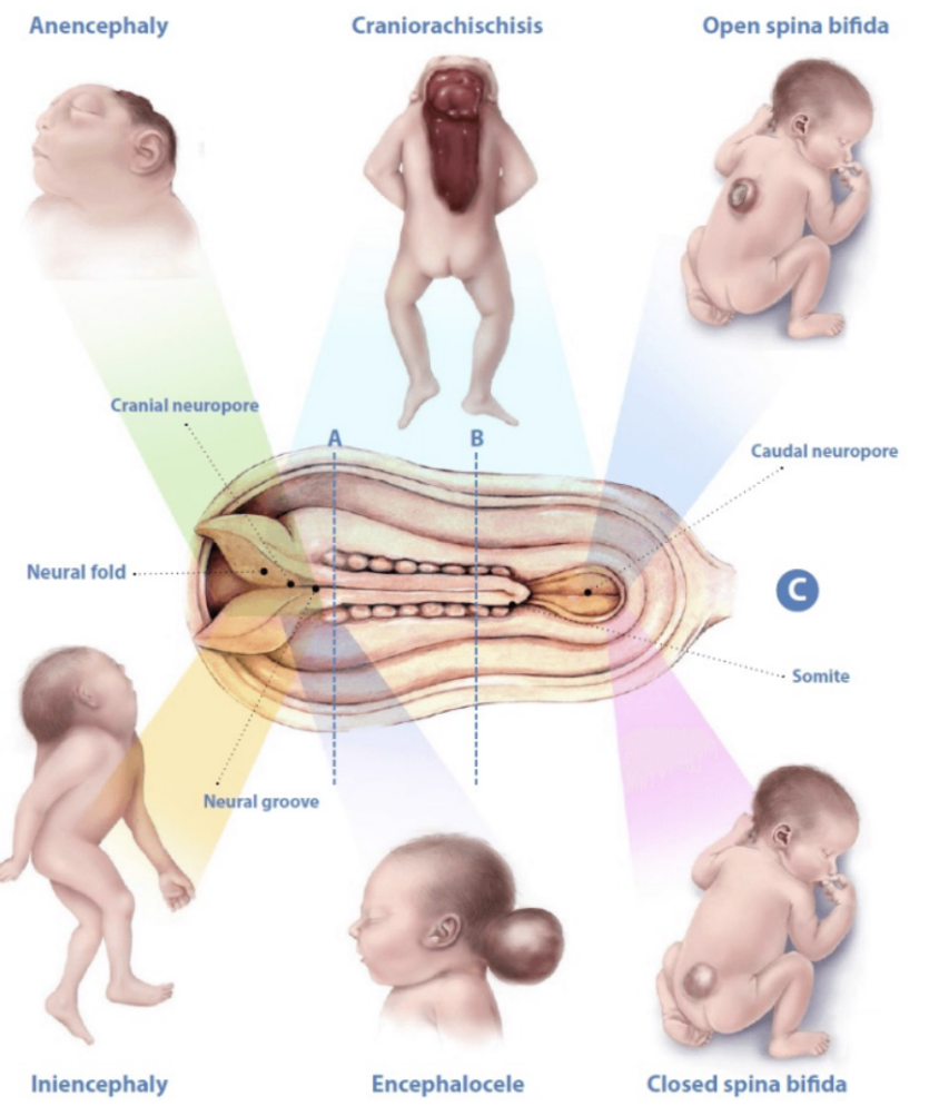
Describe the condition: Rachischisis (rachis - spine, chisis - split)
It occurs when the neural tube fails to close in the womb, leaving the spinal cord flattened and completely exposed without skin or bone protection. Major neurological compromises.
Craniorachischisis is an example.
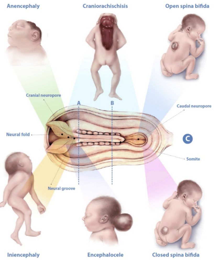
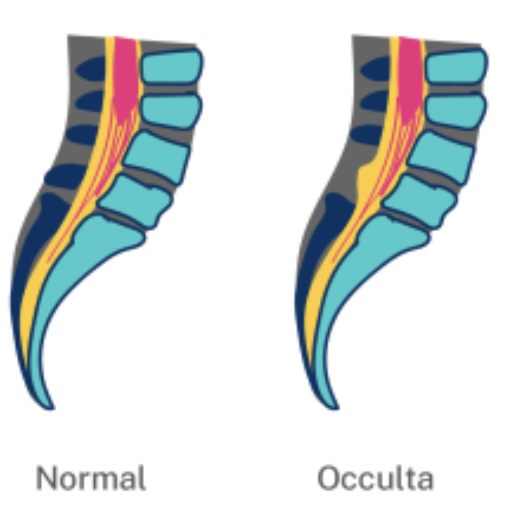
Describe the condition spina bifida occulta
Has a small gap in the vertebrae, but the spinal cord and nerves stay inside
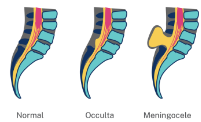
Describe the condition spina bifida meningocele
Protrusion of meninges (connective tissue surrounding the spinal cord) and CSF but not the spinal cord
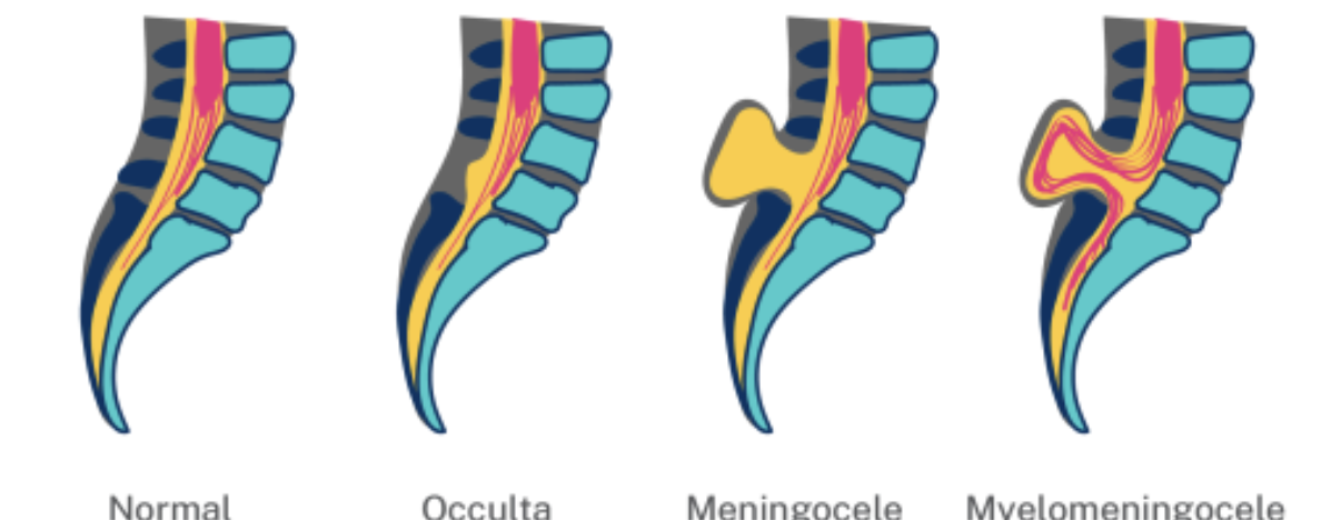
Describe the condition spina bifida myelomeningocele
Most severe. Protrsion of fluid-filled sac containing parts of the spinal cord, CSF, and meninges.
A fetus has incomplete closure of the cranial end of the neural tube at approximately day 25. Which condition would most likely develop?
Anencephaly due to failure of rostral neuropore closure.
A fetus develops a defect due to failure of caudal neuropore closure. Which anatomical structure is primarily affected?
The spinal cord region, resulting in spina bifida.
A patient asks why folic acid supplementation is recommended during pregnancy. How would you explain its relevance to the nervous system?
Folic acid supports normal neural tube development and reduces the risk of neural tube defects such as spina bifida and anencephaly.
An embryo has abnormal development of the forebrain. Which adult brain regions may be affected?
Structures derived from the forebrain, including the cerebrum, thalamus, hypothalamus, and pineal gland.
developmental abnormality affects the telencephalon. Which major adult brain structure would likely be abnormal?
The cerebrum.
A patient has abnormalities involving the pons and cerebellum. Which embryological structure was likely affected?
The metencephalon.
A disease destroys large numbers of neurones but leaves glial cells intact. Would nervous system communication remain normal? Explain.
No. Glial cells cannot replace neuronal communication because they do not generate or conduct action potentials. Loss of neurones results in impaired signalling and neurological dysfunction.
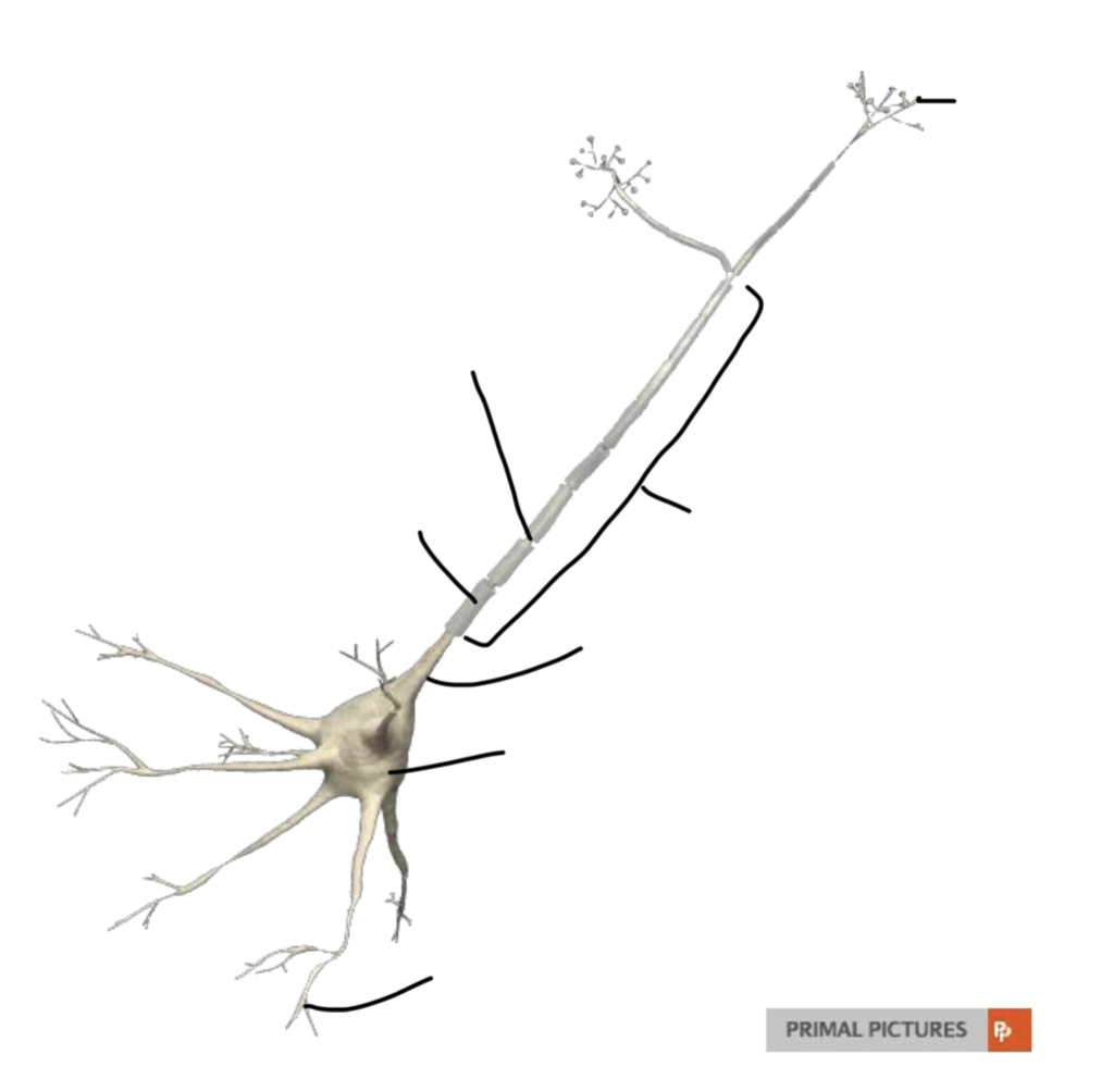
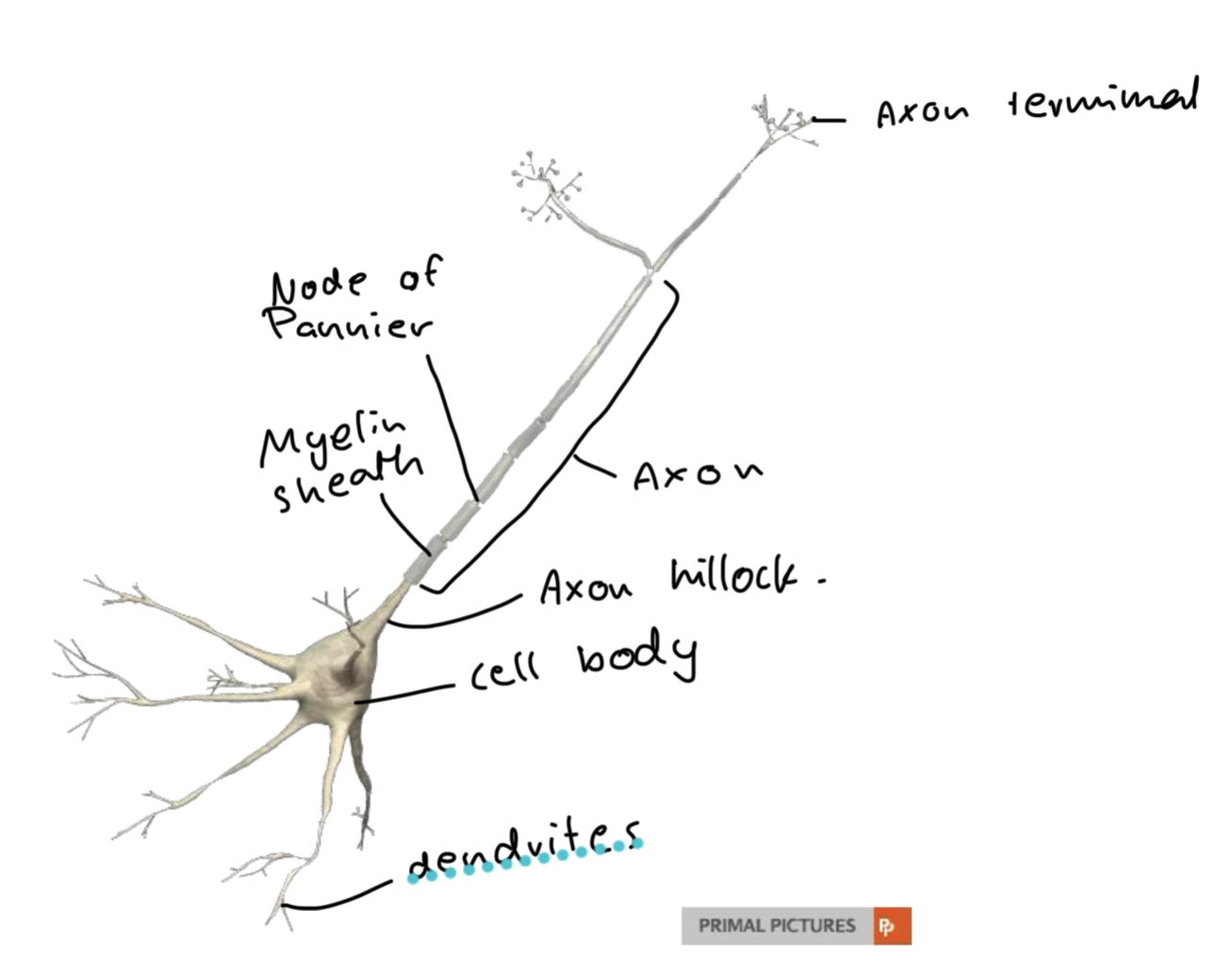
Label this neuronal structure and explain the 4 basic structure and each of their function
Dendrites - collect info from other neurons
Cell body/Soma - produces essential proteins and integrates incoming signals
Axon hillock - acts as the neural integration and trigger zone, summing incoming excitatory and inhibitory signals and initiating an all-or-none action potential if the electrical threshold is reached
Axon - transmit electrical impulses away from the neuron's cell body to other cells
Axon terminals - convert electrical signals into chemical messages and release them to communicate with other cells
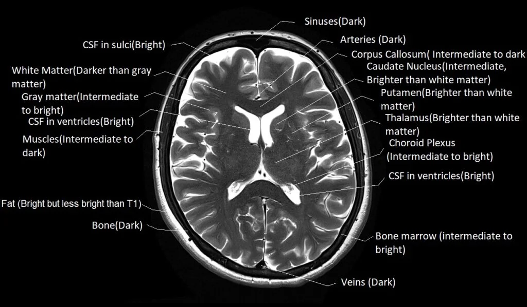
Areas that are high in cell bodies are known as… and appear … in T2 weighted image.
grey matter, hyperintense
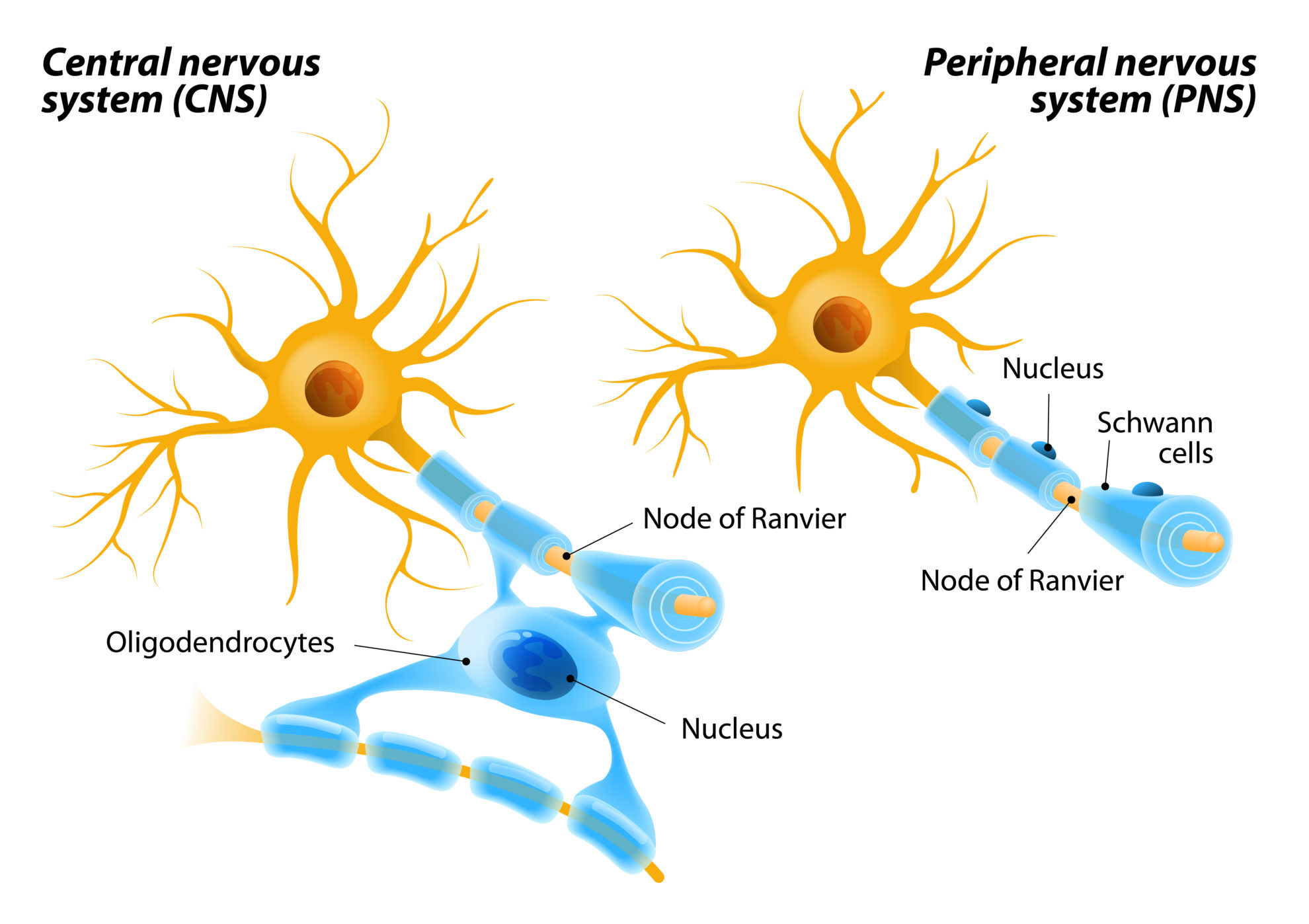
What is the purpose of myelin covering the axons?
Fatty insulation created by glial cells that speeds up action potentials (signal) from the cell body to the axon terminals through the axon.
There are two different types of glial cells. Name and categorise them and also explain how they are different.
oligodendrocytes myelinate axons in the Central Nervous System (CNS). One cell extends multiple arms to insulate up to 50 different nerve fibers simultaneously in the tightly packed brain.
Schwann cells myelinate axons in the Peripheral Nervous System (PNS). One cell dedicates its entire body to insulating just a single segment of the axon.
Areas that are high in myelin are known as… and appear … in T1 weighted image.
white matter, hyperintense
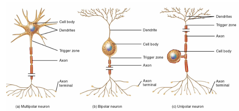
What are the 3 types of neurons and what are their functions.
Multipolar (motor)
Carry signals away from the central nervous system out to muscles and glands.
Bipolar
These are rare and found in specialized sensory areas like the retina of the eye.
Unipolar (sensory)
Carry somatic (sensory) and visceral signals from the body's sensory receptors to the CNS.
Sensory receptors are classified as:
Interoreceptors - detect information from the internal environment
Exteroreceptors - detect information from the external environment
Proprioceptors - responsible for monitoring the position and movement of skeletal muscles and joints
Glial cells have the ability to regenerate unlike neurons, what kind of risk does this create?
most primary brain tumours originate from neural glial cells
The outside of the neuron is more positive than inside, why?
higher concentration of positive sodium ions outside the cell
positive potassium ions leaking out of the cell.
Negatively charged proteins inside the cell
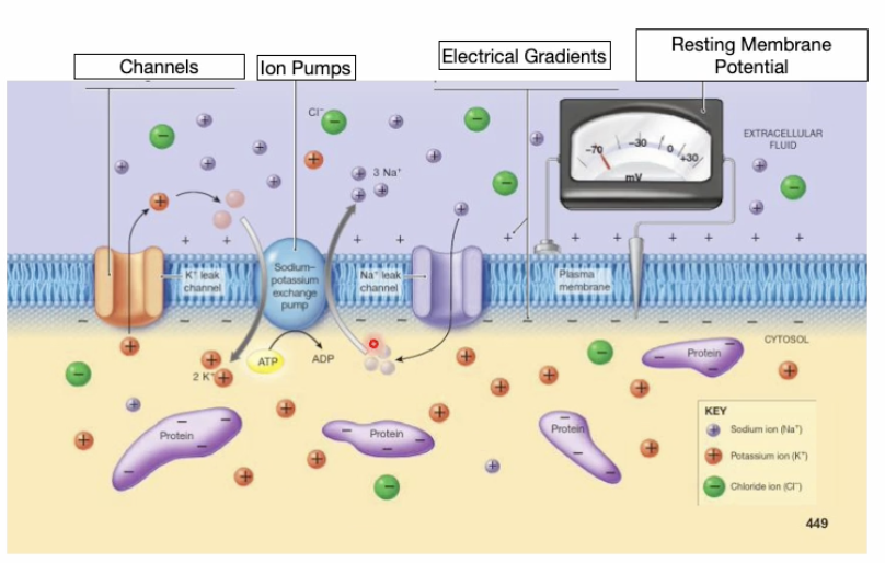
What mV is the resting membrane potential?
-70mV
Ions are going to be pressurised to move based on two gradients; electrical and chemical. What are they in relation to the neuron.
Chemical: high concentration of sodium outside the cell → want to move into the cell
Electrical: higher negative charge on the inside → positively charged ions want to move in
Sodium ion really wants to move inside the cell due to the electrochemical gradient, but can only do so via channels. What are the 4 channels?
Ligand-gated
Always open/leak
Mechanically-gated
Voltage-gated
There is one type of channels on the cell membrane when a neurotransmitter binds to it, what is it called? Where is it most abundant on a neuron and why?
Ligand-gated channels.
Most abundant in dendrites and cell bodies because it is where neurotransmitters are caught.
Then the channel opens to let ions pass through → creates a localised change in voltage → graded potential.
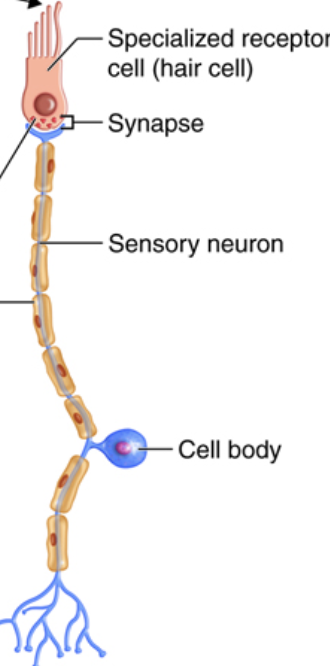
Mechanoreceptors are cells that detect physical changes and mechanical forces like touch, pressure, vibration, stretch, and sound. A certain type of channel is abundant in mechanoreceptors
Mechanically gated channels
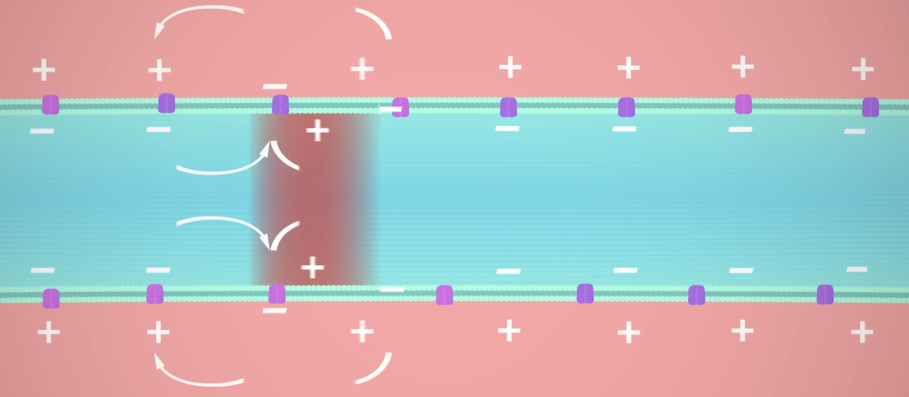
A change in the membrane potential from -70mV to -55mV causes what channels to open? Where are these channels most abundant in the neuron and why?
Sodium voltage-gated channels.
They are most abundant in the axon because this helps to propagate the local electrical signal down the axon.
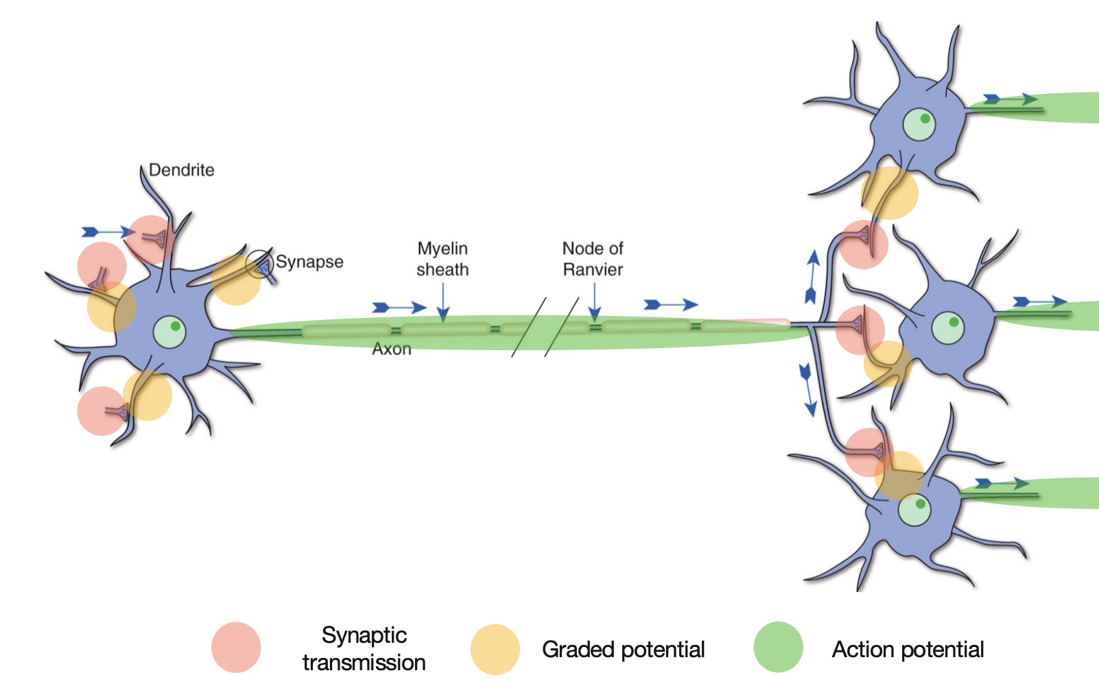
How are different channel types distributed across parts of a neuron?
Dendrites/cell body (plasma membrane): chemically/ligand-gated channels
Axon: many voltage-gated channels
Axon terminals: many voltage-gated calcium channels
What signaling events occur in dendrites/cell body, axon, and axon terminals?
Dendrites/cell body: graded potentials, usually driven by ligand-gated channels
Axon: action potentials, especially involving voltage-gated sodium channels
Axon terminals: synaptic transmission, requiring voltage-gated calcium channels for neurotransmitter release
Sodium-potassium exchange pump helps to maintain the sodium and postassium ion gradient via active transport. How many potassium and sodiums are exchanged? and what does active transport mean for the blood supply in these areas?
2 potassium in, 3 sodium out
Active transport → requires energy → requires lots of blood supply.
A stroke is when blood flow to a part of the brain stops. Since there is a lack of blood supply, what would that mean for the neurons?
Decreased oxygen delivery → decreased ATP production → less ATP for exchange pumps → accumulation of sodium inside the cell → disrupted signaling → cascade of intracellular events → cell death.
Outline the process of how action potential forms
local stimulus is needed i.e. axon terminal releasing neurotransmitter to the dendrites
sodium-gated channels open, sodium will rush in. That causes a local change in the membrane potential called a graded potential.
If the graded potential is strong enough via temporal or spatial summation → produce an action potential
Temporal summation