Basic principles of radiographic practice
 Call Kai
 Learn
Learn Practice Test
 Spaced Repetition
 Match
Match Flashcards
 Knowt Play
1/17

There's no tags or description
Looks like no tags are added yet.
Name | Mastery | Learn | Test | Matching | Spaced | Call with Kai | Chat |
|---|
No analytics yet
Send a link to your students to track their progress
18 Terms
Radiation
Energy that travels in high energy electromagnetic waves
ALARP
As Low As Reasonably Practicable
Three functions of ALARP
Identifying the risk vs benefit to the patient
Getting it right the first time
Optimising radiation dose
Production of x-ray imaging
Requires the right amount of x-ray beams with the right amount of energy
Kilovoltage peak
Amount of energy of the x-ray beam
Contrast
Affected by kVp because energy of the x-ray beams must be high enough
Milliampereseconds
Amount of x-ray photons in the x-ray beams
Density
Affected by mAs because the greater the amount of x-ray photons, the greater the density of the x-ray subject
Image noise
Scattered x-ray photons increasing the graininess of the image
Attenuation
When energy is lost due to obstacles
Source Image Distance (SID)
Instance from the x-ray tube to the imaging detector
110cm for most imaging
180cm for chest x-rays
Source Object Distance
The larger it is and closer it is to the detector, the less magnified the image
Grids and Bucky mechanism
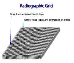
Grids used between detector and patient to absorb scattered radiation and reduce image noise
Absorbs more primary radiation so requires a greater dose
Automatic Exposure Chambers
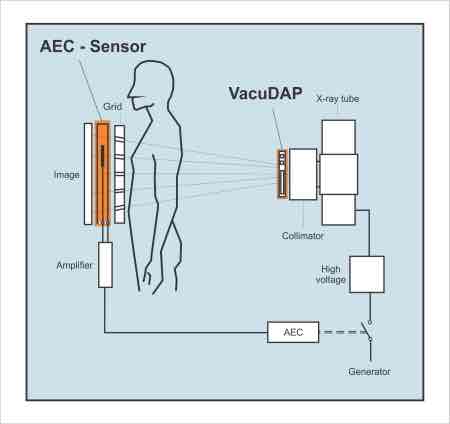
Used when radiation dose is uncertain
Used when a large area with different densities is being image
Radiation cuts out once detector has received a given density
Located between patient and detector
Light Beam Diaphragm
Cross light produced where the centering point is aimed
Collimation
Size of the field imaged
Three reasons for limiting collimation
Reduces radiation dose to patient
Improves image quality
Reduces scatter
Anatomical side markers
Mark which side of the body has been scanned
Allows for radiographer identification