Biology 1106 Module 3
1/246
There's no tags or description
Looks like no tags are added yet.
Name | Mastery | Learn | Test | Matching | Spaced |
|---|
No study sessions yet.
247 Terms
Hydrostatic skeleton
Animal has a soft body with fluid-filled cavities surrounded by muscles. Example: Earthworms
Exoskeletons
Consists of rigid outer covering that protects internal organs and provides an attachment site for muscles. Completely encases the muscles and internal organs.
Endoskeletons
Muscles attach to the internal bones or cartilage; makes framework
Bones
Specialized connective tissue, composed of organic and inorganic components. Primary skeletal component in most vertebrate.
Cartilage
Specialized connective tissue that is designed to withstand compression and tension. Found in joints. Cells are surrounded by a matrix. Flexible.
Fibroblast
Produce collagen
Chondroblast
Form chondrocytes (cartilage). They secrete extracellular matrices and become stuck inside lacunae, creating the chondrocytes (cartilage cells).
Osteoblast
Form bones (osteocyte formation). They secrete an enzyme which causes calcium phosphate to form the crystaline hydroxypaptide which accounts for hardness of bones.
Mesenchyme
Cells that make up bone and cartilage and are derived from embryonic connective tissue
Osteoclasts
Remove bone
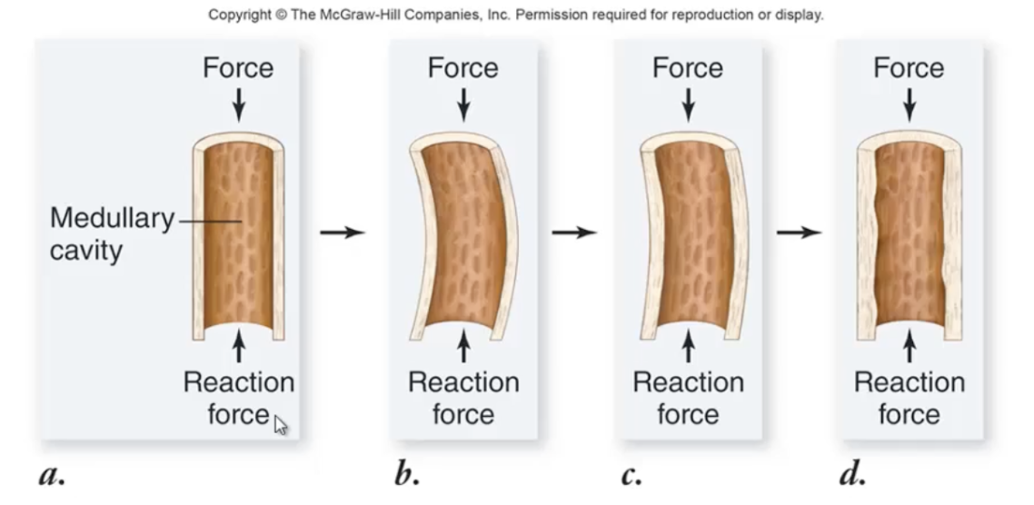
Remodeling and repair
Living bones are capable of responding to demands.
Epiphysis
The widened ends of the bone
Growth plates
It is where lengthening occurs. Made of cartilage. When you are growing, they form new cartilage and thicken. As it thickens, it lengthens. It pushes the epiphysis away from bone shaft and at the same time, the cartilage closest to the shaft calcifies.
Bone remodeling
response to stress. Osteoblasts are depositing new bone (producing more matrix) while osteoclasts are reabsorbing established bones. This new bone will restrict bending.
Ball and socket joints
Can achieve movement in all directions
Joint
provide motion powered by muscles. The orgin remains stable while the insertion is attached to the portion of the bone that moves.
Flexion
Antagonist movement produced by contraction of the hamstring muscles, which causes lower leg to move backwards
Extension
Contraction of quadriceps, pulling leg forward
Skeletal muscle
A type of muscle that has a bundle of muscle fibers/cells. The muscle cells are multinucleated. Each individual muscle fiber contains a bundle of 4 - 20 myofibrils.
Myofibrils
Composed of thick and thin myofilaments. Account for contraction of skeletal muscles.
Myosin
Thick myofilaments
Actin
Thin myofilaments
Sarcomeres
Smallest subunit of muscle contraction, which is measured from Z-line to Z-line. They shorten during muscle contraction
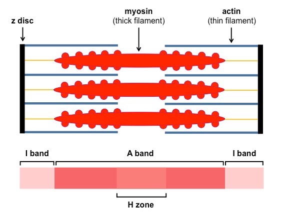
A Band
Composed collectively of both actin and myosin
H Band
Composed of myosin (thick filaments)
I Band
Composed of actin (thin filaments)
“M line”
Dotted line in middle of sarcomere where myosin filaments meet
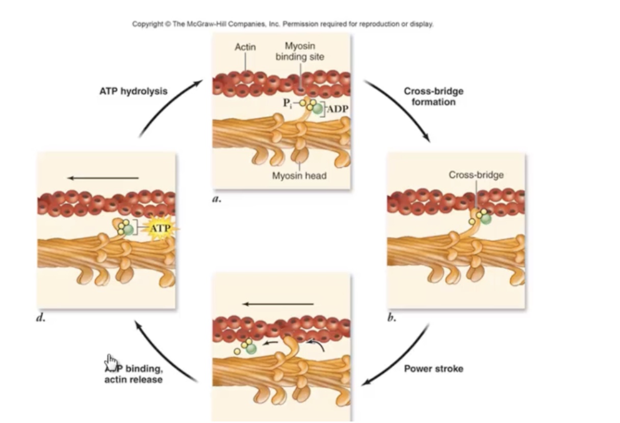
Cross bridge
Myosin heads temporarily attach to actin filaments, pulling the actin filaments which results in muscle contraction
Cross Bridge overview
First, the hydrolysis of ATP to ADP and Pi causes a conformational change that moves myosin head into energized or ready state.
Next, the energized head forms a cross bridge with the actin filaments.
Then, during the powerstroke, ADP and Pi are released and the myosin head shifts into a low energy postion that pulls the attached myosin filament in one direction
Finally, following the power stroke, ATP can bind to the myosin head which weakens the link between myosin and actin and the cross-bridge breaks. ATP hydrolysis returns myosin to the energized conformation and cycle begins again. The cycle continues as long the muscle is stimulated to contract. It takes energy to break the cross bridge between actin and myosin.
Ligament
Flexible and tough fibrous tissue. Connects bone to bone or catilage to cartilage or holds together a joint
Tendon
Flexible and tough fibrous connective tissue connecting bone to muscle
Neuro-muscular junctions
1) action potential arrives from neuron
2) sarcoplasmic reticulum released Ca2+
3) Ca2+ binds to troponin
4) Energized myosin heads binds to actin forming cross bridge
Slow-twitch mucsle
Enable long-endurance slow fatigue activites as distance running and steady walking. “Dark meat”
More mitochondria
Fast-twitch muscles
Fatigue faster but are used in powerful bursts of movements like sprinting. “White meat” such as breats meat.
Rapid burst of power.
Basic requirements of human nutrition
Water
Carbohydrates
Sugars
Starches
Lipids
Fats
Oils
Proteins
vitamins
minerals
Lipids
Preferred for long-term energy needs and is stored in adipose cells and skeletal muscle
Monosacchardies
Nutrient chemical used for quick energy
Calorie
The amount of energy required to raise the temperature of one gram of water by one degree celcius
Proteins
Essential amino acid. We absorb amino acids from this nutrient, not the nutrient itself.
Minerals
Perform in virtually all metabolic reactions
Mouth/tongue
Mechanical digestion. Minor chemical digestion with amylase in saliva digesting starches sugars.
Pharynx
Opening of oral and nasal cavities
Esophagus
Muscular tube connecting pharynx to stomach
Stomach
Carries out mechanical digestion using rugae, and chemical digestion using hydrochloric acid (HCl) and proteases to break down proteins.
Rugae
Muscular folds in stomach
Small intestine
Where most digestion and absorption of nutrients occurs. Divided into the duodenum, jejunum, and ileum.
Duodenum
Part of small intestine that receives bile and pancreatic enzymes for chemical digestion.
Jejunum
Part of small intestine that absorbs most digested monomers (amino acids, sugars, fatty acids).
Ileum
absorbs any remaining nutrients not taken up by the jejunum.
Large Intestine
Also known as colon. Absorbs water and electrolytes, compacts waste, and forms feces for elimination.
Mucosa
Innermost layer of digestive tract. It is an epithelial lining and its major functions are to secrete mucus, absorb end products of digestion into blood, and protect the underlying tissues from foreign invaders or disease
Submucosa
Connective tissue apart of digestive tract that is rich in blood, lymphatic vessels, and nerve fibers.
Muscularis
Exterior of the submucosa, composed of smooth muscle, arranged in two orientations, a circular layer and a longitudinal layer. Combined, these two layers help move food through the canal. The circular layers are also responsible for the sphincter muscles found in some specialized regions
Serosa
Smooth epithelial covering of the digestive system. It protects canal from harsh abrasions as the walls flex and extend.
Accessory organs
Liver, gallbladder, salivary glands, and the pancreas.
Proteases
An enzyme that catalyzes the breakdown of proteins by hydrolyzing peptide bonds.
Carbohydrases
Enzymes that break down carbohydrates into monosaccharides (simple sugars).
Monosaccharides are also hydrophilic and need channel proteins for absorption into blood capillaries.
Lipases
Enzymes that break down fats (triglycerides) into free fatty acids and monoglycerides.
Bile salts emulsify fat droplets, increasing surface area for digestion.
Lipids are hydrophobic (lipophilic) and can pass directly through the cell membrane.
Inside the cell, they are reassembled into triglycerides and packaged into chylomicrons (protein-coated fat transport particles).
Chylomicrons are absorbed into lymphatic capillaries, not blood capillaries.
Ghrelin
stimulates hunger
Leptin
Counteracts/reduces hunger
Saliva
Released from salivary glands. Made of:
Water: for dissolving hydrophilic subtances
Mucus: for binding food together
Salivary amylase: inititates breakdown fo starch
Anti-bacterial compounds
Bolus
Ball of food
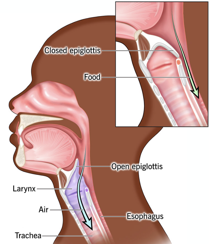
Mechanics of swallowing
Food is formed into a bolus of a tongue and then is swallowed.
Initiated voluntarily as food is swallowed and the tongue moves bolus towards the posterior portion of the pharynx.
Step 1: When food enters the back of mouth, the soft palate seals off the nasal cavity and breathing is temporarily paused. As the bolus passes the soft palate, the involuntary swallowing reflex is initiated.
Step 2: The elevation of the larynx, or voice box, and the subsequent folding of the epiglottis, keeps food out of the respiratory tract and directs it to the esophagus.
Step 3: After the bolus enters the esophagus, the larynx relaxes and the air passageway is restored.
Peristalsis
Coordinated muscle movement to direct food towards stomach regardless of gravity
Villi
Finger-like projections of the mucosal lining of the small intestine. They increase surface area and contain blood and lymphatic capillaries for nutrient transport.
Microvilli
Tiny extensions of the plasma membrane on epithelial cells that cover each villus. These form a structure called the brush border due to their brush-like appearance.
Brush Border
The name given to the wall of the small intestine because of the dense layer of microvilli. This border maximizes absorption and hosts digestive enzymes.
Brush Border Enzymes
Digestive enzymes embedded in the membranes of microvilli.
They assist in the final breakdown of carbohydrates, proteins, and fatty acids just before absorption into the cells.
Pancreas
A major accessory digestive organ located between stomach and duodenum. Secretes pancreatic juice through pancreatic duct into small intestine
Pancreatic juice
Contains:
Trypsin and Chymotrypsin – proteases stored in inactive form, activated in the duodenum.
Pancreatic Amylase – digests carbohydrates.
Lipase – digests fats.
Bicarbonate Ions
Neutralizes acidic chyme from stomach and create a suitable pH
Liver
Body’s largest internal organ and most metabolically active. Has four major functions:
Detoxification
Protein synthesis
Glycogen storage
Bile secretion
Detoxification
Modifies harmful substances like alcohol and carcinogens
Protein synthesis
Builds complex proteins from amino acids
Glyocgen storage
Assembles and stores carbohydrates as glycogen
Bile
A fluid that aids in fat digestion
Rectum
Final part of digestive tract that stores feces before expulsing them.
First sphincter: Smooth muscle, opens involuntarily with pressure.
Second sphincter: Voluntarily controlled.
Feces
Made of:
Undigested materials
Water
Electrolytes
Mucus
Bacteria
Color comes from bile pigments altered by bacteria.
Odor comes from bacterial byproducts like phenol, hydrogen sulfide, indole, skatole, and ammonia.
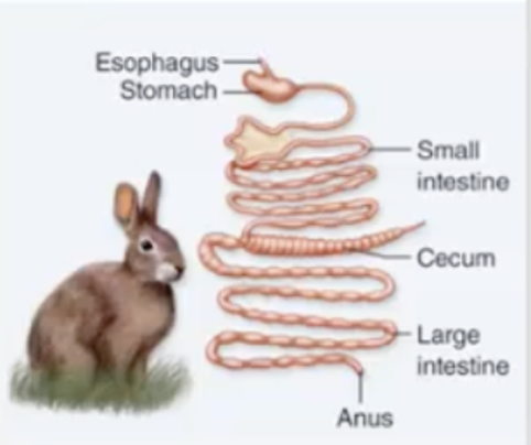
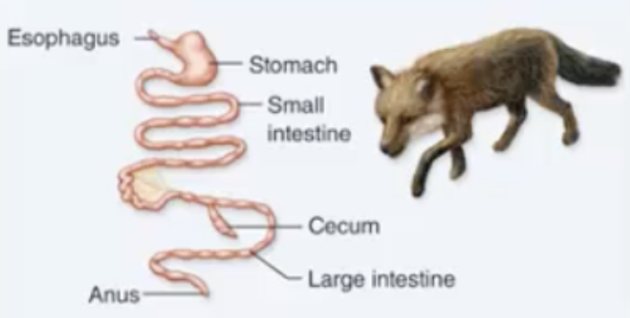
Nonruminant herbivore
Simple stomach, large cecum.
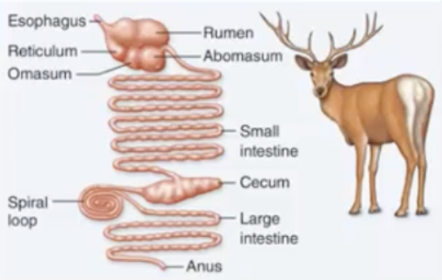
Ruminant herbivore
Four-chambered stomach with large rumen; long small and large intestine
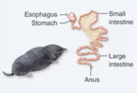
Insectivore
Short intestine, no cecum
Carnivore
Short intestine and colon; small cecum
Gastrin
A hormone that stimulates the stomach to produce acid (HCl) and enzymes, which help in the digestion of food. It also promotes the movement of food through the stomach.
Cholecystokinin (CCK)
Released in response to fatty chyme in the duodenum. It targets cells in the gallbladder, stimulating the release of bile to absorb fats. It also stimulates the pancreas to release additional digestive enzymes. Overall, it helps with digestion
Gastric Inhibitory Peptide (GIP)
Plays a role in regulating blood sugar levels and other metabolic processes. It reduces the production of stomach acid, which helps protect the stomach lining from damage.
Secretin
Released in response to acid
Stimulates pancreas to release bicarbonate which neutralizes acidic chyme
Circulatory system
Plays a very important role in sustaining life because it is responsible for the delivery of oxygen and nutrients to all cells as well as removing carbon dioxide and waste products. The maintenance of pH in the circulation of proteins and cells of the immune system
All organisms must have the ability to circulate nutrients, gases, and to rid their bodies of waste.
Purposes:
Transportation of all materials essential for metabolism, such as oxygen and nutrients, as well as the removal of metabolic wastes.
Regulation, by regulating body systems by carrying regulatory hormones throughout body and regulating body temperature
Protection, both for mechanical wounds and invading pathogens.
Erythrocytes
Red blood cells.
Hemoglobin in vertebrates (pigments that binds and transports oxygen)
Constitute approximately 45% of total volume of blood
Leuokocytes
White blood cells
Defend body against pathogens
Make up less than 1% of total blood volume
Platelets
Cell fragments that play an important role in blood clotting
Make up less than 1% of total blood volume
Hematopoiesis
Production of blood cells from bone marrow
Erythropoiesis
When oxygen availability in blood drops, the kidney converts plasma protein into a protein called erythropoietin (EPO) which stimulates the production of erythrocytes (red blood cells).
Since EPO accelerates erythrocyte production, it also increases oxygen carrying capacity in the blood. Some cheating athletes inject EPO to help with endurance.
erythropoietin
The glyocoprotein hormone made by kidneys that stimulates production of red low cells in the spongy bone marrow. More of this increases the number of red blood cells.
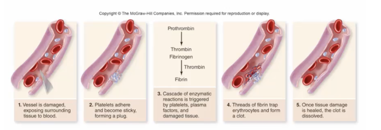
Blood clotting
First, when blood vessels are damaged from like a cut, the vessel walls will constrict.
Then, the platelets congregate to wound by sticking to one another and thus sealing off the wounded area
At this point, is a cascade of enzymatic reactions that the platelets initiate
Afterwards, the cascade of events ultimately produces protein threads of fibrillin that along with the platelets form a patch around wound
Finally, once the damage tissue is repaired, the patch will slowly dissolve.
Open circulatory systems
Type of circulatory system
No capillaries
Low or no blood pressure
Hemolymph
Long time to circulate
Examples
Insects 20-35 min
Lobster – 5-8 min
Crab – 1 min
Closed Circulatory Systems
Type of circulatory system
End-to-end Capillaries
Lymphatic circulation reenters
blood circulation.Higher blood pressure
Rapid circulation
Examples:
Human– 23 sec
Wolf & Dog – 16 sec
Rabbit – 7.5 sec
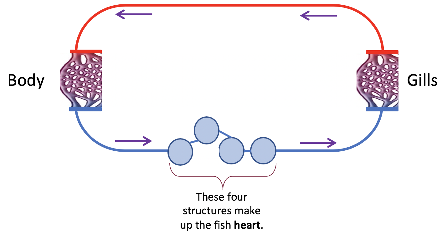
Fish circulatory system
Composed of four structures that form two pumping chambers. As blood moves from the heart via a(n) artery , it travels to the gills where the blood will release carbon dioxide and pick up oxygen . Thus, oxygenated blood is represented by the red color in the figure and deoxygenated blood is represented by the blue color in the figure. The blood travels from the gills, throughout the body of the fish, and once past arterioles, capillaries, and venules back to the heart via a(n)vein .
Amphibian circulatory system
Once blood is pumped by the heart and now must go through the pulmonary arteries to the lungs and is then returned to the heart by pulmonary veins. The blood is now ready to be circulated throughout the body through a process called double circulation
So the pulmonary circulation system transfers blood from the heart to the lungs and the systemic circulatory system from the blood from the heart to the rest of the body
They have a three-chambered heart which consists of two atria and one ventricle
Atria receive blodd
Ventricles pump blood out heart
Since there’s only ventricle, separation of the pulmonary and systemic circulation are incomplete in amphibians.
This means that there’s mixing of oxygenated and deoxygenated blood in ventricles
Reptile Circulatory system
Circulatory system that has adaptations that reduce the mixing of oxygenated and deoxygenated blood in the heart. Most have a ventricle partially divided by a septum, which helps separate the two blood types.
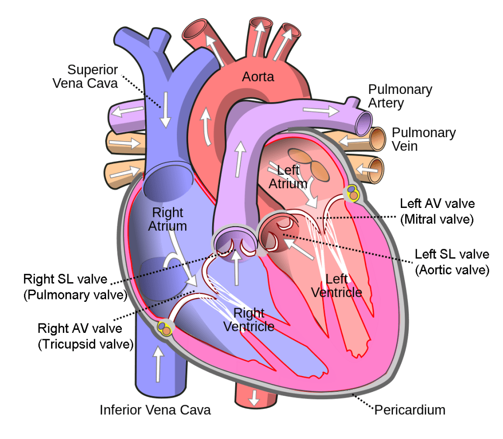
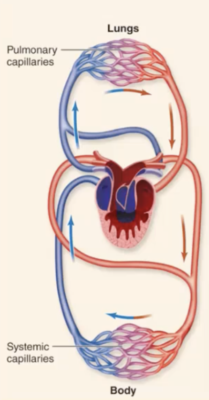
Mammalian circulatory system
The four-chambered heart supports a double circulatory system—pulmonary (to lungs) and systemic (to body)—which increases efficiency for high metabolism and endothermy.
Deoxygenated blood returns to the right atrium via the superior and inferior vena cava, moves to the right ventricle, and is pumped to the lungs through the pulmonary arteries.
Oxygenated blood returns to the left atrium via pulmonary veins, enters the left ventricle, and is pumped to the body through the aorta.
The heart beats ~75 times per minute: atria contract together first, followed by ventricles, maintaining rhythmic blood flow.
This efficient circulation is essential for sustaining warm-blooded life.
Study this. Watch video.