NPB: The Cardiovascular System Lecture Set #2
1/116
Earn XP
Description and Tags
Cardiac AP, ECG, Mechanical Events of the Heart The Vasculature
Name | Mastery | Learn | Test | Matching | Spaced |
|---|
No study sessions yet.
117 Terms
Cardiac AP vs Neuronal AP
shaped different
Longer lasting
T/F: A Cardiac AP is not stereotyped
False, it is
Depolarization
Influx:
Ca2+
Na+
Charge
Inside gets more electropositive
Repolarization
Efflux:
K+
Charge:
more electronegative inside

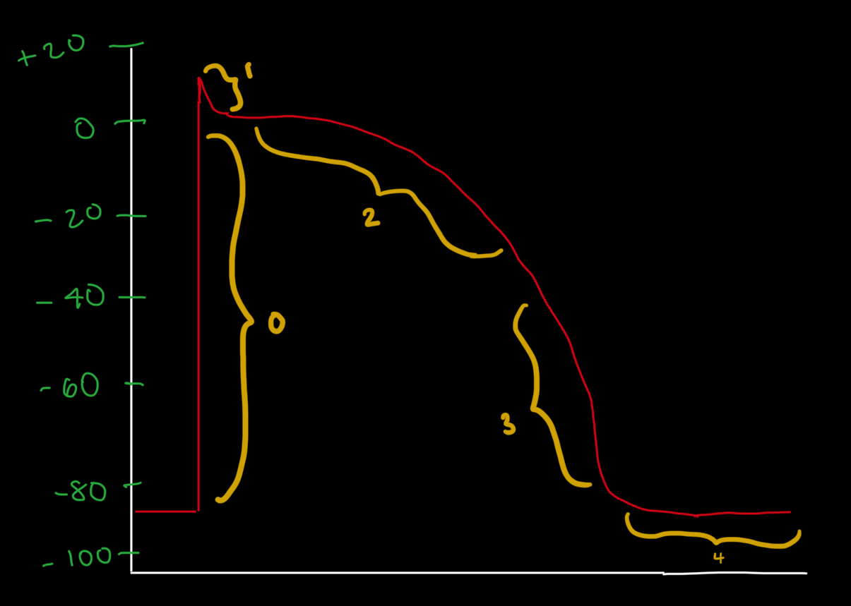
What Phase is A?
Phase 0
Phase 1
Phase 2
Phase 3
Phase 4
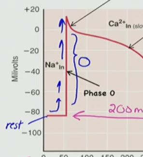
Phase 0
Phase 0
depolarizes quickly
Na+ influx via V-gated Na+ channel
tiny Ca2+ influx too via a V-gated Ca2+ channel
Na+ current quickly inactivates at peak like in neuron
Which of the following correctly describes Phase 0 of the cardiac action potential?
A) Ca²⁺ influx via T-type calcium channels
B) K⁺ efflux via delayed rectifier channels
C) Na⁺ influx via voltage-gated Na⁺ channels
D) Brief repolarization due to Cl⁻ influx
C) Na⁺ influx via voltage-gated Na⁺ channels
What ion is primarily responsible for the rapid depolarization seen in Phase 0 of the cardiac action potential?
A) K⁺
B) Ca²⁺ via T-type channels
C) Cl⁻
D) Na⁺ via voltage-gated Na⁺ channels
E) Mg²⁺
D) Na⁺ via voltage-gated Na⁺ channels

What Phase is B?
Phase 0
Phase 1
Phase 2
Phase 3
Phase 4
Phase 1

Phase 1:
membrane potential begins to repolarize
repolarizes through K+ Channel
via a Transient V-gated K+ Channel (IK) I=current/transient
K+ efflux
brief
During the cardiac myocyte AP, which phase is considered the brief repolarization?
A) Phase 2
B) Phase 4
C) Phase 0
D) Phase 1
E) Phase 3
D) Phase 1
Which of the following contributes to the brief repolarization immediately following the peak of depolarization in the cardiac AP?
A) Na⁺ influx
B) Ca²⁺ influx via L-type channels
C) Transient K⁺ efflux (Phase 1)
D) Cl⁻ influx
E) Slow Na⁺ leak channels
C) Transient K⁺ efflux (Phase 1)
What occurs during Phase 1 of the cardiac action potential?
A) Transient K⁺ efflux through voltage-gated channels (Iₖ,TO)
B) Rapid Na⁺ influx
C) Ca²⁺ influx through L-type channels
D) Sustained depolarization
E) Repolarization via chloride channels
A) Transient K⁺ efflux through voltage-gated channels (Iₖ,TO)
Which of the following statements is true about the shape of the cardiac action potential compared to the neuronal action potential?
A) Cardiac AP is shorter in duration
B) Cardiac AP lasts longer and has a different shape
C) They are identical in all phases
D) Neuronal AP is longer than cardiac
E) Cardiac AP does not involve voltage-gated ion channels
B) Cardiac AP lasts longer and has a different shape
What causes Na⁺ current to stop after the initial depolarization in Phase 0?
A) The Na⁺ channels are permanently open
B) Potassium influx overpowers it
C) Calcium inhibits Na⁺ entry
D) Na⁺ channels inactivate quickly at the peak
E) ATP is depleted
D) Na⁺ channels inactivate quickly at the peak

What Phase is C?
Phase 0
Phase 1
Phase 2
Phase 3
Phase 4
Phase 2

Phase 2: Plateau
flatten
Ca2+ current influx → muscle cell
L-type Ca2+ channel
hypertension patients block this channel
K+ leaving cell efflux via
delayed rectifier K+ Channel V-gated

What two ion movements contribute to the plateau phase (Phase 2) of the cardiac action potential?
A) Ca²⁺ influx via L-type channels and K⁺ efflux via delayed rectifier channels
B) Na⁺ influx and Cl⁻ influx
C) Only Ca²⁺ influx
D) Na⁺ and Ca²⁺ both efflux
E) K⁺ influx and Na⁺ efflux
A) Ca²⁺ influx via L-type channels and K⁺ efflux via delayed rectifier channels

Why does Phase 2 of the cardiac AP last ~200 msec?
A) Because Na⁺ influx continues the entire time
B) Due to a balance between Ca²⁺ influx and K⁺ efflux
C) Because Ca²⁺ channels are slow to open
D) Because K⁺ efflux stops completely
E) Because of a delay in depolarization
B) Due to a balance between Ca²⁺ influx and K⁺ efflux
There is slow influx of Ca2+ and slow efflux of K+

What Phase is D?
Phase 0
Phase 1
Phase 2
Phase 3
Phase 4
Phase 3
Phase 3
Repolarize the cell
Need L-Type Ca2+ to inactivate
Slowly activating V-gated K+ Channel
(same rectifying K+ Channels that were activated at Phase 2)
close off

What happens during Phase 3 of the cardiac action potential?
A) Plateau is maintained
B) Na⁺ influx returns
C) Ca²⁺ influx increases
D) V-gated Na⁺ channels reopen
E) L-type Ca²⁺ channels inactivate and K⁺ channels repolarize the membrane
E) L-type Ca²⁺ channels inactivate and K⁺ channels repolarize the membrane

Which channel is primarily responsible for repolarization in Phase 3?
A) Funny channel (Iₙ)
B) T-type Ca²⁺ channel
C) Voltage-gated K⁺ channel (delayed rectifier)
D) ATPase pump
E) Voltage-gated Na⁺ channel
C) Voltage-gated K⁺ channel (delayed rectifier)
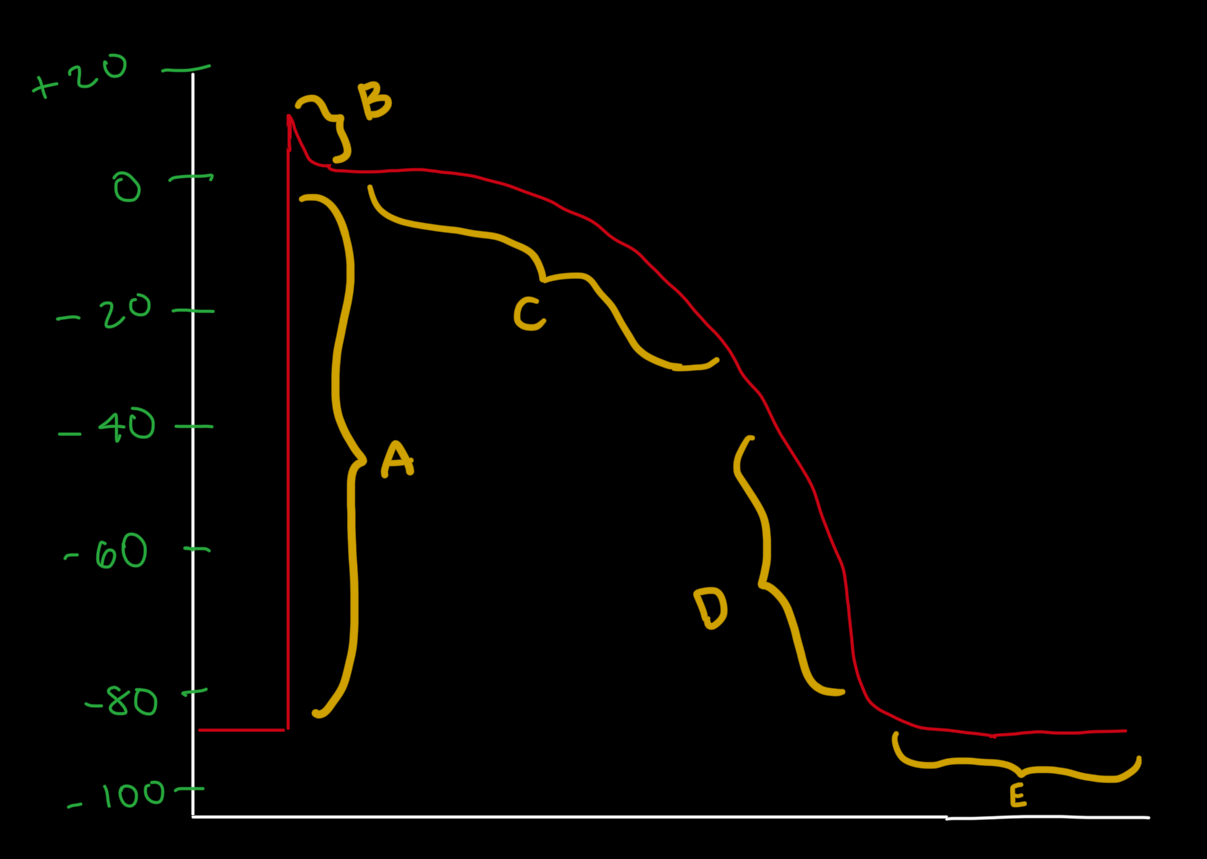
What Phase is E?
Phase 0
Phase 1
Phase 2
Phase 3
Phase 4
Phase 4

Phase 4
K+ continues effluxing
via dif K+ channel

What ion movement characterizes Phase 4?
A) Na⁺ continues leaking in
B) Ca²⁺ remains in the cytoplasm
C) Cl⁻ influx balances K⁺ efflux
D) No ion movement occurs
E) K⁺ continues to efflux via a different K⁺ channel
E) K⁺ continues to efflux via a different K⁺ channel
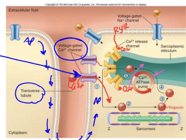
T/F: The DHPR was a true voltage sensor in skeletal muscle, so it does the same thing in cardiac muscle
False, the DHPR is a true channel in cardiac, but not in skeletal
What initially activates voltage-gated Ca²⁺ channels (DHPR) in the cardiac muscle cell membrane?
A. Calcium binding to troponin
B. The influx of sodium
C. An action potential (AP)
D. Activation of the sarcoplasmic reticulum
C. An action potential (AP)
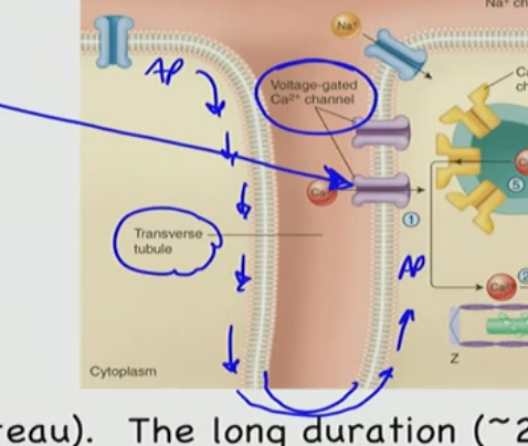
Which of the following best describes how ryanodine receptors (RyRs) are activated in cardiac muscle cells?
A. They are mechanically pulled open by DHPR
B. They are opened by ATP binding during the power stroke
C. They open in response to sodium influx during depolarization
D. They are activated by direct calcium binding (Ca²⁺)
D. They are activated by direct calcium binding (Ca²⁺)
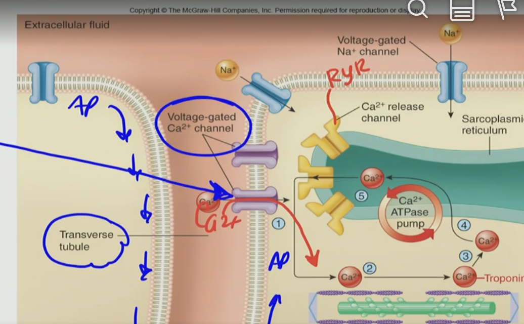
What is the result of calcium binding to the ryanodine receptors (RyRs) on the sarcoplasmic reticulum?
A. Closure of voltage-gated Ca²⁺ channels
B. Release of more Ca²⁺ from the sarcoplasmic reticulum
C. ATP production in the mitochondria
D. Inhibition of troponin binding
B. Release of more Ca²⁺ from the sarcoplasmic reticulum
What is the process called when Ca²⁺ triggers the release of more Ca²⁺ from the sarcoplasmic reticulum?
A. Calcium ATPase cycling
B. Excitation-Relaxation Coupling
C. Calcium-Induced Calcium Release (CICR)
D. Plateau Phase Discharge
C. Calcium-Induced Calcium Release (CICR)

When does CICR primarily occur during the cardiac action potential?
A. Phase 0 (rapid depolarization)
B. Phase 1 (initial repolarization)
C. Phase 2 (plateau phase)
D. Phase 3 (repolarization)
C. Phase 2 (plateau phase)
What causes the plateau phase (Phase 2) in the cardiac action potential?
A. Rapid sodium influx through voltage-gated Na⁺ channels
B. Simultaneous calcium influx and potassium efflux balancing the membrane potential
C. Continuous firing of action potentials from the SA node
D. Increased ATP production in the mitochondria
B. Simultaneous calcium influx and potassium efflux balancing the membrane potential
What happens during repolarization in cardiac excitation-contraction coupling?
A. RyRs open and Ca²⁺ floods into the cytosol
B. Voltage-gated Ca²⁺ channels inactivate, and Ca²⁺ is removed from the cytosol
C. Troponin releases Ca²⁺ and binds to actin
D. The sarcomere contracts and shortens
B. Voltage-gated Ca²⁺ channels inactivate, and Ca²⁺ is removed from the cytosol
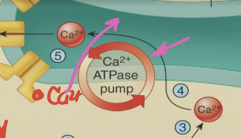
What role does the Ca²⁺ ATPase pump (seen in the diagram) play in this process?
A. It helps depolarize the membrane
B. It causes Ca²⁺ influx into the cytoplasm
C. It removes Ca²⁺ from the cytoplasm into the SR for relaxation
D. It opens RyR channels for CICR
C. It removes Ca²⁺ from the cytoplasm into the SR for relaxation
What happens to the L-type Ca²⁺ channels during repolarization of cardiac muscle cells?
A. They open wider to allow more calcium in
B. They activate ryanodine receptors again
C. They inactivate, stopping Ca²⁺ influx into the cytoplasm
D. They are mechanically blocked by the sarcomere
C. They inactivate, stopping Ca²⁺ influx into the cytoplasm;

What happens to intracellular Ca²⁺ during the repolarization phase of a cardiac action potential?
A. It continues binding to troponin to sustain contraction
B. It is pumped back into the sarcoplasmic reticulum (SR) by SERCA
C. It activates more L-type calcium channels
D. It binds to ryanodine receptors to trigger another release
B. It is pumped back into the sarcoplasmic reticulum (SR) by SERCA; happening constantly to contract & relax
Refractory Period
Time period
If you give an AP and try to stimulate the cell again, and can you get another AP
The absolutes refractory period of cardiac muscle
A) causes a tetanic contraction in the ventricles
B) is very short in duration
C) causes the AP to travel slowly in the conduction pathways
D) allows the atria to contract before the ventricles
E) is much longer than the refractory period in skeletal muscle
E) is much longer than the refractory period in skeletal muscle
What is a likely consequence of a premature contraction during the cardiac refractory period?
A. The heart will contract with greater force and eject more blood
B. The heart will fail to eject a proper volume of blood, reducing overall blood flow
C. The ventricles will enter tetanus, increasing cardiac output
D. The contraction will be delayed until the next normal heartbeat
B. The heart will fail to eject a proper volume of blood, reducing overall blood flow
T/F: Cardiac Muscle also does Tetany!
False, Cardiac Muscle does not do tetany because it doesn’t have the capacity to do so, since you would need a repeated stimuli and here we have a long refractory period
In cardiac muscle cells, what is the primary trigger for RyR activation on the sarcoplasmic reticulum?
A. Voltage change across the sarcolemma
B. Direct mechanical pull from DHPR
C. Binding of extracellular sodium
D. Binding of Ca²⁺ that entered through voltage-gated channels
D. Binding of Ca²⁺ that entered through voltage-gated channels
What is the name of the process where calcium entry causes further calcium release from the SR?
A. Calcium Pump Feedback
B. Calcium-Induced Calcium Release (CICR)
C. Voltage-Triggered Calcium Dump
D. Sarcomeric Calcium Excitation
B. Calcium-Induced Calcium Release (CICR)
During relaxation in cardiac muscle, what happens to the intracellular Ca²⁺?
A. It is broken down by enzymes
B. It continues to stimulate troponin
C. It is pumped back into the SR or out of the cell
D. It binds to ATP to fuel contraction
C. It is pumped back into the SR or out of the cell
Which statement is TRUE regarding excitation-contraction coupling in cardiac muscle (not skeletal muscle)?
A. RyRs are mechanically linked to DHPRs
B. Ca²⁺ does not play a role in contraction
C. RyRs are activated by Ca²⁺ binding, not by voltage directly
D. Ca²⁺ channels remain open throughout repolarization
C. RyRs are activated by Ca²⁺ binding, not by voltage directly
Which of the following sequences best describes the order of events in cardiac excitation-contraction coupling (including CICR)?
A. RyRs release Ca²⁺ → AP arrives → CICR begins → Ca²⁺ removed from cytosol → Relaxation
B. AP arrives → L-type Ca²⁺ channels open → Ca²⁺ influx triggers CICR via RyRs → More Ca²⁺ is released from SR → Ca²⁺ is removed → Relaxation
C. Ca²⁺ enters the cell → AP fires → CICR occurs → L-type Ca²⁺ channels inactivate → Depolarization
D. SR releases Ca²⁺ → CICR starts → AP spreads → Ca²⁺ channels stay open during repolarization
B. AP arrives → L-type Ca²⁺ channels open → Ca²⁺ influx triggers CICR via RyRs → More Ca²⁺ is released from SR → Ca²⁺ is removed → Relaxation
Why can't the heart go into tetanus like skeletal muscle?
A. It lacks voltage-gated Na⁺ channels
B. The refractory period is too short for summation
C. The refractory period lasts almost as long as contraction
D. The heart is not under voluntary control
C. The refractory period lasts almost as long as contraction
Approximately how long is the cardiac muscle refractory period?
A. 50 milliseconds
B. 100 milliseconds
C. 250 milliseconds
D. 500 milliseconds
C. 250 milliseconds
What is the main purpose of the long refractory period in cardiac muscle?
A. To allow for rapid, repeated contractions
B. To prevent the action potential from spreading
C. To ensure full ventricular filling before contraction
D. To ensure complete spread of AP and contraction before a new one starts
D. To ensure complete spread of AP and contraction before a new one starts
During which phase can another action potential not be generated in cardiac muscle?
A. Relative refractory period
B. Plateau phase only
C. Absolute refractory period
D. Repolarization
C. Absolute refractory period
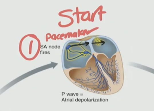
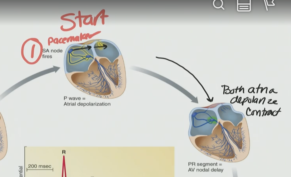
What is the first structure to initiate the cardiac action potential?
A. AV node
B. Bundle of His
C. Purkinje fibers
D. SA node
D. SA node/pacemaker
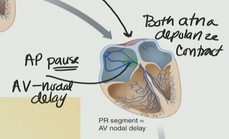
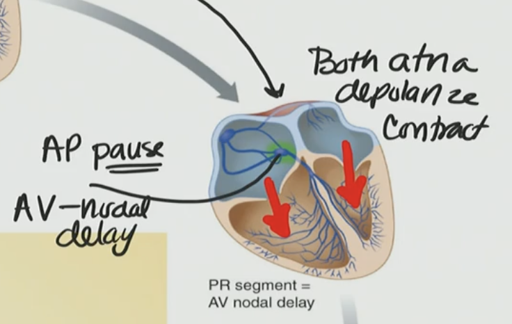
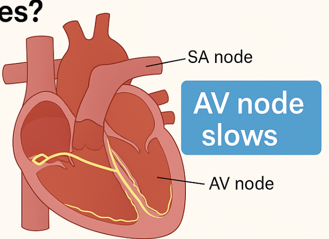
What is the function of the AV nodal delay?
A. To allow both ventricles to depolarize at the same time
B. To prevent the atria from contracting
C. To allow the atria to fully contract and fill the ventricles
D. To speed up conduction through the ventricles
C. To allow the atria to fully contract and fill the ventricles
The AV nodal delay ensures that:
A) The ventricles contract prior to atrial systole.
B) The atria contract and empty their contents into the ventricles prior to ventricular systole.
C) Ventricular diastole occurs before systole.
D) Tetanic contractions of cardiac muscle are impossible.
E) Atrial diastole occurs before atrial systole.
B) The atria contract and empty their contents into the ventricles prior to ventricular systole.
Which pathway spreads the pacemaker signal from the SA node to the atrial muscle?
A. Purkinje fibers
B. Interatrial and internodal pathways
C. Atrioventricular bundle
D. Vagal nerve
B. Interatrial and internodal pathways; because it spreads it to the AV from the SA, so SA → Interatrial and internodal pathways → AV
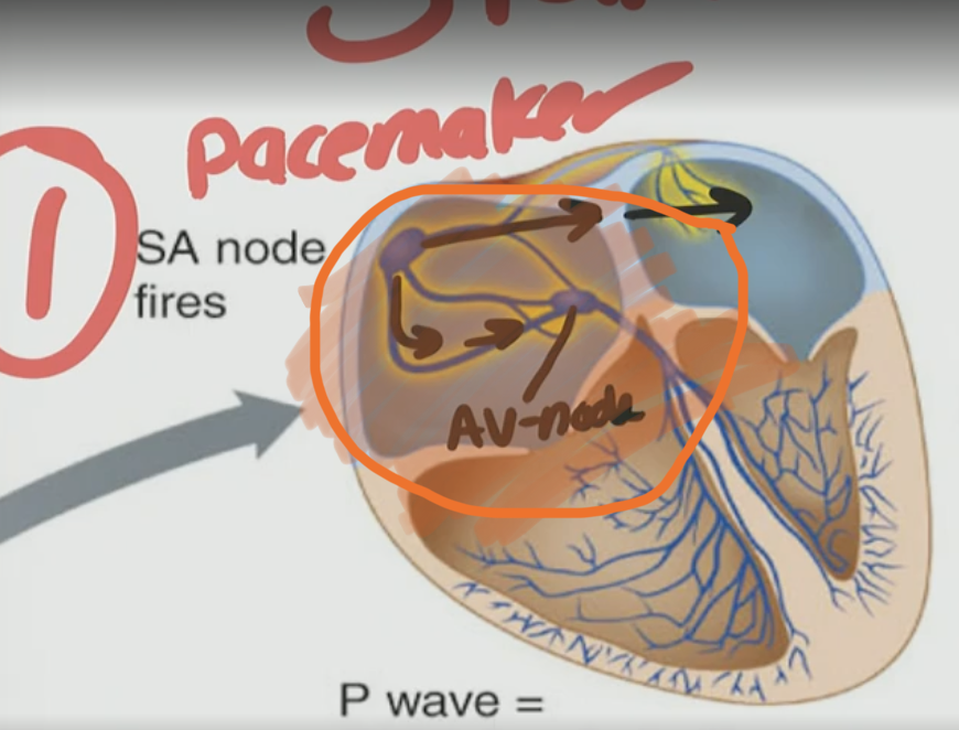
Where does the cardiac action potential pause briefly to allow the atria to contract before the ventricles?
A. SA node
B. Bundle of His
C. Purkinje fibers
D. AV node
D. AV node → AV nodal delay
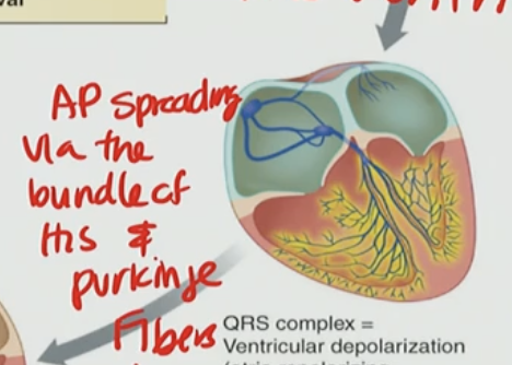
Which structures are responsible for rapid conduction through the ventricles?
A. AV node and SA node
B. Bundle of His and Purkinje fibers
C. Atrial muscle and interatrial pathways
D. T-tubules and myofibrils
B. Bundle of His and Purkinje fibers
What is the primary purpose of the AV nodal delay in the cardiac conduction system?
A. To allow both ventricles to contract at the same time
B. To increase the strength of atrial contraction
C. To ensure the atria have time to fully fill the ventricles before ventricular contraction
D. To speed up conduction through the atria
C. To ensure the atria have time to fully fill the ventricles before ventricular contraction
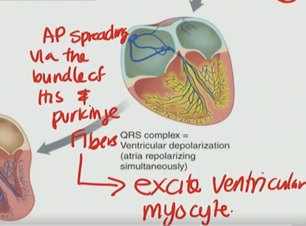
What is the purpose of the action potential spreading through the Bundle of His and Purkinje fibers?
A. To delay ventricular contraction until the next heartbeat
B. To excite the ventricular myocytes and trigger ventricular contraction
C. To depolarize the atrial myocytes
D. To repolarize the ventricles after contraction
B. To excite the ventricular myocytes and trigger ventricular contraction

For side A pick two:
A) Left Side
B) Right Side
C) Bicuspid
D) Tricuspid
For Side A, it will be B & D
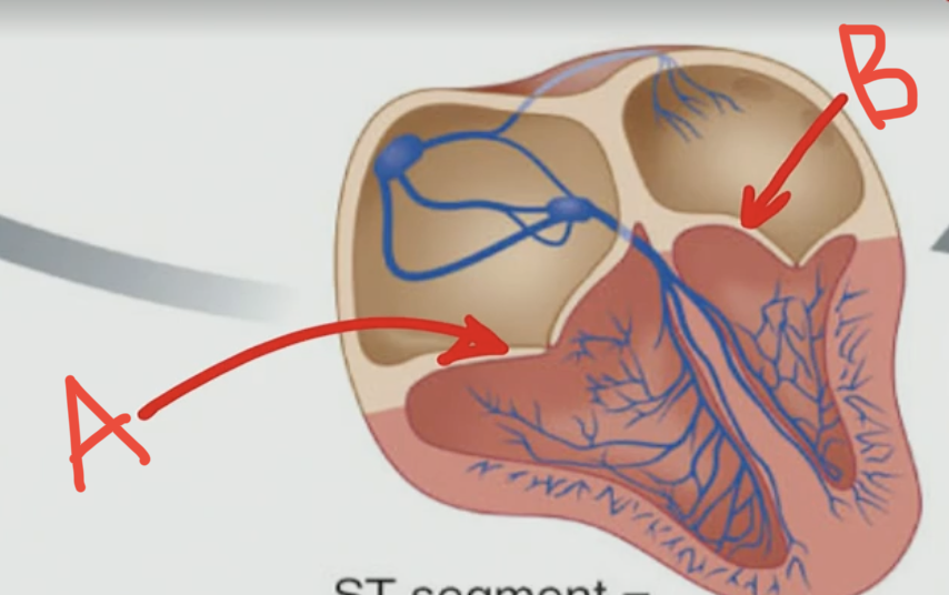
For side B pick two:
A) Left Side
B) Right Side
C) Bicuspid
D) Tricuspid
Side B is A & C

Is the Triscuspid & Bicuspid open or closed?
They are closed
What happens when the tricuspid and bicuspid (mitral) valves are closed during the cardiac cycle?
A. Blood flows freely from the ventricles into the atria
B. Blood is ejected into the atria to maintain pressure
C. Blood cannot flow backward into the atria from the ventricles
D. The ventricles are relaxed and passively filling
C. Blood cannot flow backward into the atria from the ventricles
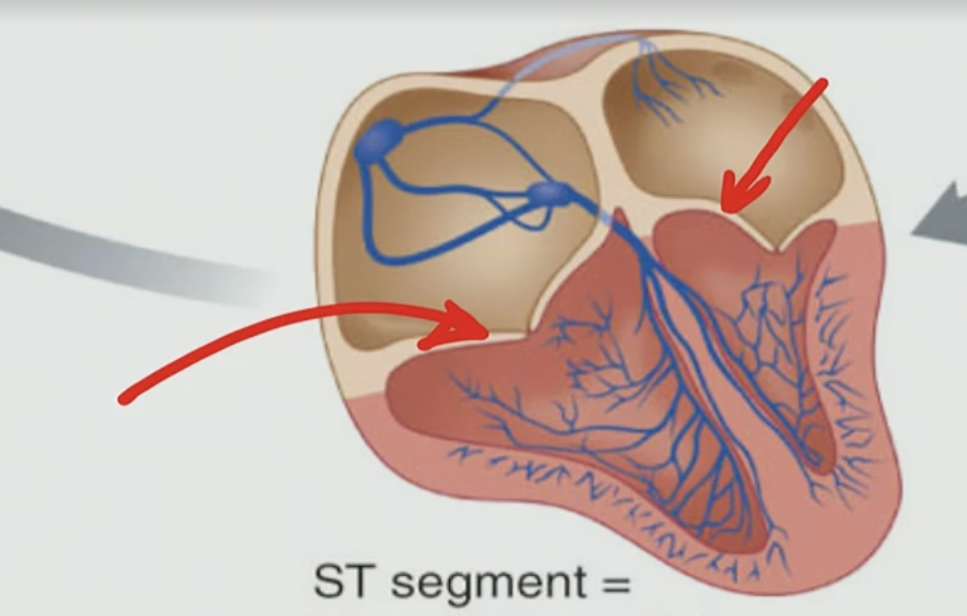
Semilunar Valves
Arterial Trunks
Pulmonary Valve
Aorta Valve
What happens when pressure in the ventricles becomes higher than the pressure in the aorta and pulmonary trunk?
A. The AV valves reopen to relieve the pressure
B. The atria contract again to help push blood
C. The semilunar valves open, allowing blood to be ejected into the arteries
D. The ventricles begin to relax and refill with blood
C. The semilunar valves open, allowing blood to be ejected into the arteries
Semilunar valves prevent backflow of blood from:
A) the arterial trunks to the ventricles
B) none of the above
C) the ventricles to the arterial trunks
D) the ventricles to the atria
E) the atria to the ventricles
A) the arterial trunks to the ventricles (up → down)
What occurs during repolarization of ventricular muscle cells in the cardiac cycle?
A. Voltage-gated Ca²⁺ channels open and cause another depolarization
B. The membrane becomes more positive due to Na⁺ influx
C. L-type Ca²⁺ channels inactivate, and K⁺ exits the cell to restore resting potential
D. Atria contract and push blood into the ventricles
C. L-type Ca²⁺ channels inactivate, and K⁺ exits the cell to restore resting potential

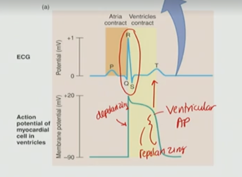
What is ECG/EKG for?
monitor your heart activity
looking at the echo of an AP
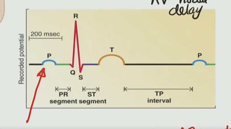
What does the P-wave on the ECG represent?
A. Ventricular depolarization
B. Atrial repolarization
C. Atrial depolarization
D. Ventricular repolarization
C. Atrial depolarization
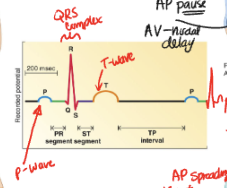
The QRS complex is like a ___ of AP
shadow
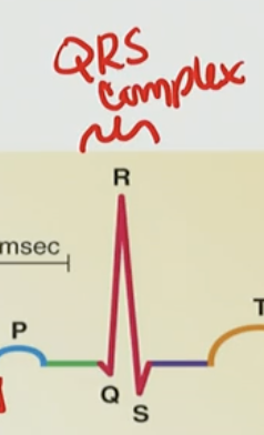

Which event corresponds to the QRS complex on the ECG?
A. Atrial depolarization
B. Atrial repolarization
C. Ventricular repolarization
D. Ventricular depolarization
D. Ventricular depolarization

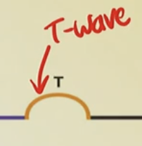
What event is represented by the T wave on the ECG?
A. Atrial depolarization
B. Ventricular depolarization
C. Atrial repolarization
D. Ventricular repolarization
D. Ventricular repolarization

The AV nodal delay appears as which segment on the ECG?
A. ST segment
B. PR segment
C. TP interval
D. QRS complex
B. PR segment
Which of the following correctly describes the sequence of events as they appear on a normal ECG?
A. T wave → P wave → QRS complex
B. P wave → T wave → QRS complex
C. P wave → QRS complex → T wave
D. QRS complex → P wave → T wave
C. P wave → QRS complex → T wave
Which of the following correctly describes the sequence of electrical events during a normal cardiac cycle as seen on the ECG?
A. Atrial repolarization → Atrial depolarization → Ventricular depolarization
B. Atrial depolarization → Ventricular depolarization → Ventricular repolarization
C. Ventricular depolarization → Atrial depolarization → Ventricular repolarization
D. Atrial depolarization → Ventricular repolarization → Ventricular depolarization
B. Atrial depolarization → Ventricular depolarization → Ventricular repolarization
Match each ECG wave to the correct electrical event:
ECG Wave | Event |
|---|---|
1. P wave | A. Ventricular repolarization |
2. QRS complex | B. Atrial depolarization |
3. T wave | C. Ventricular depolarization |
1 → B
2 → C
3 → A
Which part of the ECG waveform covers up atrial repolarization, making it not directly visible?
A. P wave
B. T wave
C. PR segment
D. QRS complex
D. QRS complex

What does an ECG measure?
A. Mechanical contraction of cardiac muscle
B. Sound of valve closures
C. Electrical activity from excitable tissues like the heart
D. Blood flow through coronary vessels
C. Electrical activity from excitable tissues like the heart
What does each waveform on an ECG correspond to?
A. A physical movement of the heart valves
B. A burst of calcium from the SR
C. A pressure change inside the ventricles
D. An electrical event such as depolarization or repolarization
D. An electrical event such as depolarization or repolarization
hat is the purpose of the electrode leads placed on the body in an ECG?
A. To stimulate heart contractions
B. To measure muscular contraction
C. To detect voltage differences caused by electrical activity in the heart
D. To monitor oxygen levels in the blood
C. To detect voltage differences caused by electrical activity in the heart
Which of the following ECG waves represents ventricular repolarization?
A) QRS complex
B) Ventricular repolarization occurs simultaneously with atrial depolarization and consequently cannot be recorded.
C) PR segment
D) P wave
E) T wave
E) T wave
Which of the following correctly matches ECG waves with their events?
A. P wave – Ventricular depolarization
B. QRS complex – Atrial repolarization (only)
C. T wave – Ventricular repolarization
D. P wave – Ventricular contraction
C. T wave – Ventricular repolarization
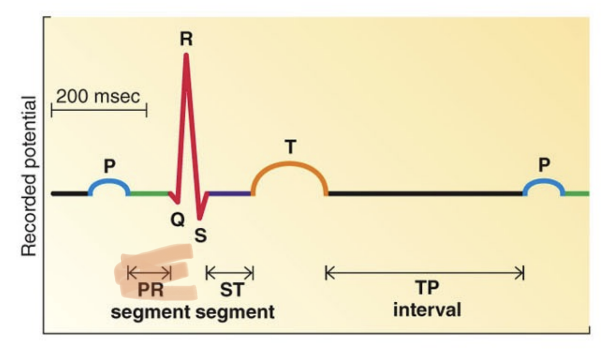
What does the PR interval on an ECG include?
A. Just the P wave
B. Only the QRS complex
C. The time from the start of the P wave to the start of the Q wave
D. The interval between the T wave and the next P wave
C. The time from the start of the P wave to the start of the Q wave

What does the PR interval represent?
A. Time from the start of atrial depolarization to the start of ventricular depolarization
B. Time from atrial repolarization to ventricular depolarization
C. Time between ventricular contraction and relaxation
D. Time from T-wave to the next P-wave
A. Time from the start of atrial depolarization to the start of ventricular depolarization
What might a longer-than-normal PR interval indicate?
A. Increased heart rate
B. Slow conduction through the AV node
C. Early ventricular repolarization
D. Stronger atrial contraction
B. Slow conduction through the AV node
What happens to the PR interval when heart rate increases?
A. The PR interval becomes longer due to more atrial filling
B. The PR interval remains the same regardless of heart rate
C. The PR interval shortens because the conduction system speeds up
D. The PR interval disappears completely
C. The PR interval shortens because the conduction system speeds up
What does the PR segment represent on an ECG?
A. The time for ventricular repolarization
B. The delay between the end of atrial depolarization and start of ventricular depolarization
C. The total time of one heartbeat
D. The duration of atrial repolarization
B. The delay between the end of atrial depolarization and start of ventricular depolarization
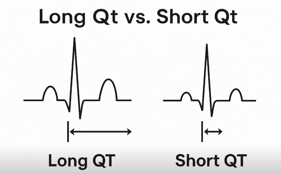
What is the QT interval measuring?
A. From the P wave to the end of the QRS
B. The total time for atrial and ventricular depolarization
C. The time between ventricular depolarization and repolarization
D. Only the time of atrial depolarization
C. The time between ventricular depolarization and repolarization
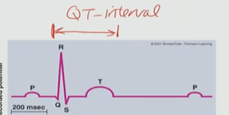
How does the QT interval change with an increase in heart rate (HR)?
A. It becomes longer to allow better filling
B. It stays constant due to pacemaker regulation
C. It shortens because the electrical events happen faster
D. It is unaffected by heart rate
C. It shortens because the electrical events happen faster
What part of the heart slows the AP before it enters the ventricles?
A. Gap Junctions
B. SA node
C. AV node
D. Internodal pathway
E. Bundle of His
C. AV node bc its important for AV nodal delay
The chordae tendineae:
A) contract when the ventricles contract
B) hold the right and left ventricles together
C) transmit the electrical impulse from the atria to the ventricles
D) keep the AV valves from everting during ventricular systole
E) hold the AV valves open during diastole
D) keep the AV valves from everting during ventricular systole
What is the purpose of the chordae tendinae?
A. They prevent the valve leaflets from everting backwards during ventricular contraction
B. They close the AV valve during ventricular contraction
C. They open the AV valve during ventricular relaxation
D. They prevent the the valve leaflets from closing during ventricular relaxation
E. They conduct the pacemaking AP in the ventricles
A. They prevent the valve leaflets from everting backwards during ventricular contraction
What cell do you find in the HCN Channel?
pacemaker cell
What allows the repeat of depolarization?
The pacemaker cell from HCN
What is a feature of Phase 2 of the cardiac muscle action potential?
A. The HCN channel is activated prior to Phase 2
B. This a period of Ca2+ through L-type channels
C. The cell repolarizes as K+ effluxes
D. This is the upswing of the cell’s AP
E. Na+ influxes into the cell during this phase.
B. This a period of Ca2+ through L-type channels
What initiates the cardiac cycle?
A. Ventricular contraction
B. T-wave generation
C. SA node firing
D. AV node delay
C. SA node firing
What does the PR segment represent in the ECG?
A. Atrial repolarization
B. AV nodal delay
C. Time between ventricular depolarization and repolarization
D. Time when ventricles are fully relaxed
B. AV nodal delay
In what order do the heart chambers contract during the cardiac cycle?
A. Ventricles contract first, then atria
B. Both atria and ventricles contract together
C. Atria contract first, then ventricles
D. Only ventricles contract
C. Atria contract first, then ventricles
What is the purpose of the delay at the AV node during the cardiac cycle?
A. To allow time for ventricular repolarization
B. To allow ventricles to eject blood
C. To allow atria to fully fill the ventricles
D. To stimulate the Purkinje fibers
C. To allow atria to fully fill the ventricles
What ECG wave corresponds with atrial depolarization?
A. T wave
B. QRS complex
C. P wave
D. PR interval
C. P wave
What electrical event does the QRS complex represent?
A. Atrial contraction
B. Ventricular repolarization
C. Ventricular depolarization
D. Atrial repolarization
C. Ventricular depolarization