Chapter 8 Mitosis and the Cell Cycle
1/103
There's no tags or description
Looks like no tags are added yet.
Name | Mastery | Learn | Test | Matching | Spaced |
|---|
No study sessions yet.
104 Terms
how long does the cell cycle take for mamallian liver cells?
more than one year
two sections of cell division
the miotic phase and interphase
cells spend more time in
interphase
Subsections of interphase
G0, G1, S, and G2
G0 phase
an offshoot from the cell cycle after division
-cells n this phase can choose not to divide but can also reenter the cell cycle
which types of cells remain in G0 and do not re-enter the cell cycle?
neurons
G1 Phase
the first growth phase
S phase
The synthesis phase of the cell cycle; the portion of interphase during which DNA is replicated.
G2 phase
The second growth phase of the cell cycle, consisting of the portion of interphase after DNA synthesis occurs.
-serves as a verification step to check everything was duplicated properly
M phase
when the cell divides
Where are the 3 checkpoints in the cell cycle?
G1, G2, M
What can G1 and G2 checkpoint do?
check if the cell is large enough (only G1)
-if environment is right
-if there is any damage to the DNA
Heterokaryon
formed by fusing a M phase cell with an interphase cell
-DNA from interphase begins to condense, meaning that the M phase is capable of inducing condensation
-used to be called the MPF
MPF
a miner of cyclin and cyclin dependent Kinase
-CDK is a serine +a theronine so it can phosphorylate either on other proteins
how does active CDK control the cell cycle
by phosphorylating other proteins
why are they called cyclins
because the cyclin part of the dimer goes through a cycle of synthesis and degradation
-every new cell gets new cyclins
the core of the cell cycle control system (CDK)
1. cyclins are made and degraded by proteosomes cyclically
2.CDK protein levels remain constant throughout the cycle
3.Presence/absence of cyclin determines whether or not CDK becomes activated (cyclin is needed to phosphorylate things)
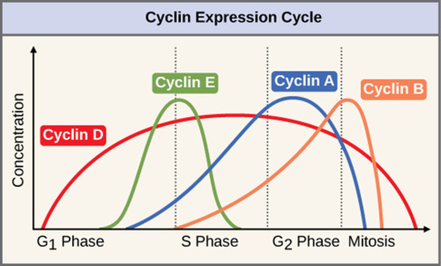
G1 cyclins
Cyclin D (CDK4 or 6)
also Cyclin E
cyclin E
control the G1/S checkpoints and controls movement to the S phase
S cyclins
also Cyclin E and Cyclin A (CDK2)
M cyclins
Only Cyclin A using CDK1
Cyclin A
control the G2/M checkpoint. They remain active until destroyed by APC
Cyclin B
Prevalent during M phase
-drives nuclear envelope breakdown and condensation of chromosomes
cyclin expression cycle
Varying levels of different cyclins in each stage
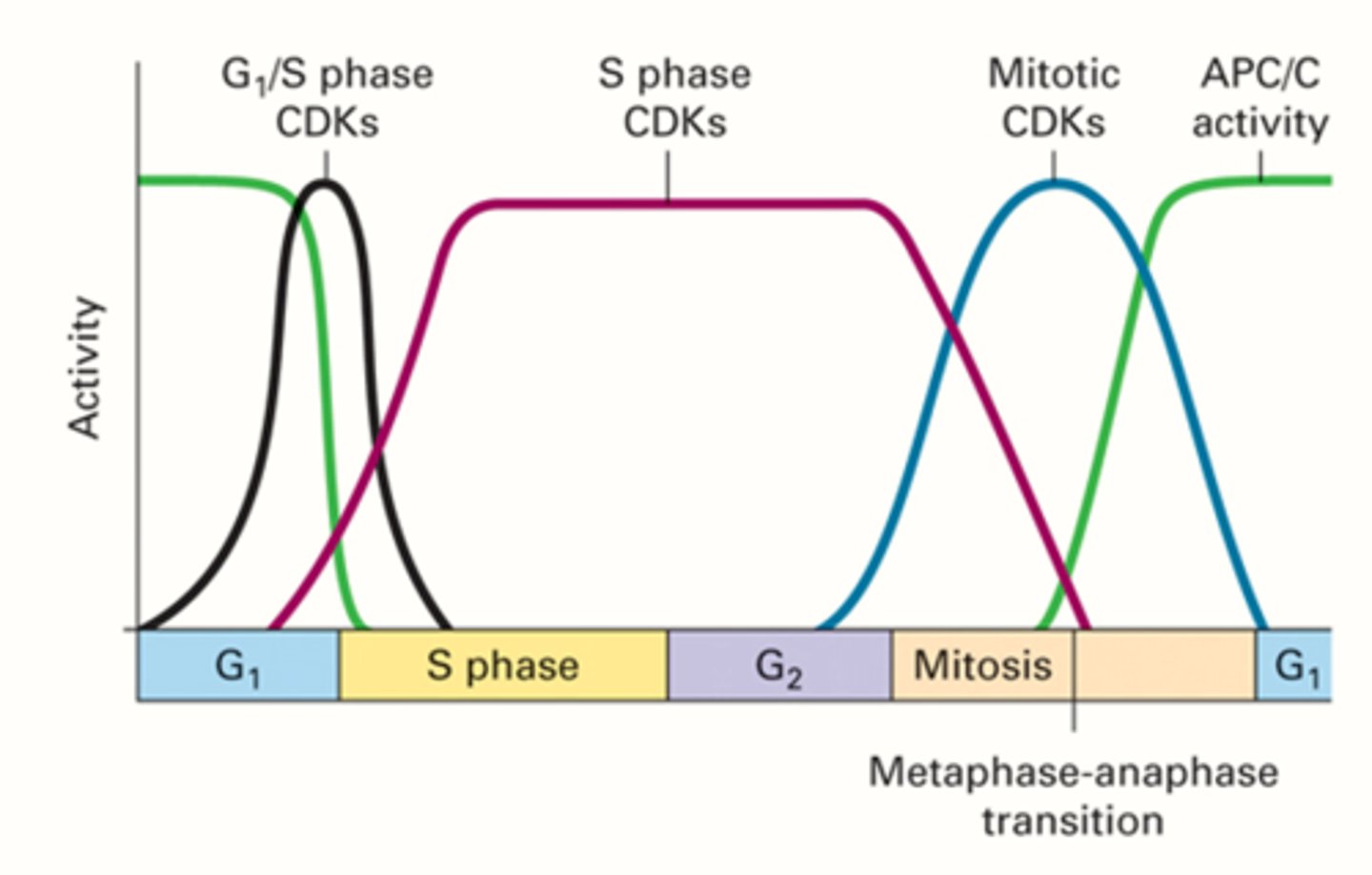
Activity of CDKs APC/C in each phase
the values of each cyclin will change, but the overall amount of cyclin is constant, specific cyclins only around when they have to be
what controls the signaling cascade that determines proliferation or apoptosis?
CDK2 and CDK4 (E and D)
step one of the cell cycle
the cell is in G1 phase, DNA prereplication complexes begin to assemble (multiple per chromosome)
-DNA replication will start from these places
step 2 of the cell cycle
G1 cyclin CDK inactivates CDH1
step 3 cell cycle
G1 cyclin CDK activates the S phase but they remain inactive until activation of a nearby inhibitor by cyclin CDK
step 4 of the cell cycle
G1 cyclin CDK phosphorylate the inhibitor, activating the S phase cyclins
-also signals for ubiquinone to be added by SCF ub ligase
step 5 cell cycle
once SCF ubiquinates the S phase inhibitor, it gets degraded by the proteasome
step 6 cell cycle
the cell enters S phase once SCF is degraded
-the cell will then progress to G2 after DNA replication
step 7 cell cycle
in G2, miotic Clyclin CDKs are activated, the cell enters mitosis and during metaphase, all chromosomes line up at the center of the cell
step 8 cell cycle
during metaphase, chromosomes attach to the microtubules
-when anaphase begins, APC-CDC20 activates to degrade the securin holding the chromosomes together. They separate and move to the cell poles
step 9
after anaphase, another proteosome will degrade any M cyclins left over and the cell will proceed to telophase and into cytokinesis
RB
retinoblastoma
-connects to transcription factor E2F and when activated by Cyclin D CDK, it phosphorylates RB leading to E2F release, which can bind to genes and determine gene expression
if a mutation occurs in the RB protein, what can happen
a tumor may develop in the eye and become cancerous
Retinoblastoma is always diagnosed before the age of 3 because
a child with this mutation cannot make RB proteins
Does RB mutation halt the cell cycle?
no because regardless, E2F is still being made however cell division will become uncontrolled
which transcription factor is always active?
E2F
S phase cyclin are only needed during S phase but can begin to accumulate before. Inactivation of this is done by
Sic1
how does Sic1 become removed for S phase cyclin activation
it gets phosphorylated by G1 cylins and becomes a target for ubiquitone and get degraded
-once degraded, S cyclins are activated
once an active S phase cyclin is present
it phosphorylates several components causing conformational changes, allowing for proteins to detach from the replication orgin which is what must occur for DNA to unwind
-phosphates are added back after, directly onto the components allowing for DNA replication to begin
unreplicated DNA checkpoint
before G2; prevents mitosis before completion of synthesis
spindle assembly checkpoint
check for chromosome attachment to spindle before mitosis
-if not, MAD2 prevents separation of the chromosomes until ready
segregation checkpoint
occurs during mitosis before telophase
-if the chromosome is not at the poles, CdC14 prevents movement to telophase
if any issues occur at the checkpoints,
P53 comes in and halts cell replication to prevent further damage
how is P53 important in cancer?
stopping damaged DNA from being replicated stops replication of cancer cells
the four stages of mitosis
1. Prophase
2. Metaphase
3. Anaphase
4. Telophase
Summary of interphase
chromosomes are duplicated, cohesion via cohesins and centrosomes are duplicated
-during G2, DNA duplication occurs
-during S phase, chromosomes are replicated
prophase
spindle poles migrate to the poles of the cell
-chromosome condensation via condensin 1 and 2 begins
-some cohesin is removed, some keep the chromatids together until anaphase
-the kinectochore assembles on the centromere
where is the location for kinetochore assembly and where sister chromatids sit when conjoined?
the centromere
-acts as the attachment point for microtubules during cell division
prometaphase
chromosomes are captured by microtubules
-the nuclear envelope begins to break down
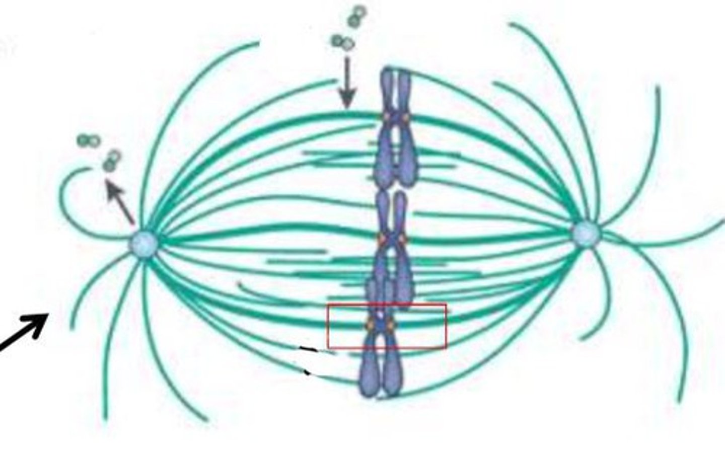
metaphase
chromosomes attached to their spindles are lined up at the metaphase plateq
anaphase
chromatids are separated and pulled to opposite poles via shortening of the kinetochore tubules
process for separating chromatids during anaphase
APC/C ubiquirin ligase degrades securin and activates the separase protease
-activated separase eats the rest of the cohesin and the chromatids can separate freely
telophase
the nuclear envelope begins to reform
-chromosomes decondense and the mitotic spindle disappears
cytokinesis
divides the cytoplasm into two separate cells
cytokinesis in animal cells is done via
cleavage furrow by the contractile ring in actin remodeling
cytokinesis in plant cells is done via
a cell plate
mitotic apparatus
The collective term for all the spindle fibers that form during mitosis
the mitotic apparatus is made up of
the spindle and astral microtubules
three types of microtubules
astral, polar, kinetochore
kinectochore microtubules
Attach to the kinectochore on the centromere of each chromosome
(direct interaction)
chromosome biorientation
where kinectochores attach to microtubules on the opposite spindles during mitosis in order to ensure equal separation of chromatids
if too many chromatids were attached to a spindle
a cell could end up with an extra chromosome or without one
ex: down syndrome
polar microtubules
interact with other polar microtubules to push poles apart
astral microtubules
form tufts at the end of the mitotic apparatus to hold spindle poles in place
what are the two ways chromosomes are captured in prometaphase?
end capture or side capture
end capture
when a microtubule by chance comes into contact with the chromosome as it grows, it can capture it
-very random, does not always occur
side capture
if the microtubule does not make it to the kinetochore region, kinetochore proteins can interact to bring them to the microtubule
chromosome congression
alignment of chromosomes at metaphase plate
steps to chromosome congression
1. kinetochores attach to the microtubule using end or side capture
2. if end, the chromosome is drawn to the spindle pole by a dynein-dynactin motor protein complex (walks to the - end)
3. the microtubule on the opposite pole picks up the other chromatid, allowing for bi-orientation
congression
-if bi-oriented chromosomes are attached to several microtubules, they locate to a central point between them
following congression, chromosomes experience
a "tug of war" until all but one kinetochore tubule shorten (oscillation)
on the shortening sides of a oscillated chromatid,
kinesin 13 stimulates disassembly at the + end
the dynein-dynactin motor moves the chromosome
toward the spindle pole (-)
on the lengthening side of the oscillated chromatid
kinesin 7 maintains its connection and grows the tubule
NCD80
a sleeve like protein complex form around the KMT (kinetochore microtubule) and attach to the kinetochore of the chromosome
how many NCD80 per chromosome
2
the site of dynein or kinesin motor cargo binding
Ncd80
mechanism of Ncd80
it is pulled by the dynein dynactin motor while the MT depolymerizes behind to make space for chromosomal movement
-also connects the microtubule to the chromosome
CEN-PA
a special histone exclusively found at the kinetochore region of chromosomes
-marks the kinetochore for the mitotic apparatus
-associated with the inner kinetochore
CPC
chromosomal passenger complex
-contains aurora B kinase
Aurora B kinase
phosphorylates Ndc80 at the kinetochore in the presence of low tension, releasing these attachments
when biorientation occurs, kinetochore regions bind to opposing microtubules, causing tension and a change in confirmation, causing pulling away from the CPC. Because of this:
Aurora B can no longer phosphorylate those proteins, resulting in a strong attachment of ncd80 and the microtubule
-this tension increases stabillity
if chromosomes are organized wrong
weak association and Aurora B kills Ncd80
cohesin is degraded by
separase
securin is an inhibitor of ______. Securin is degraded by
separase, anaphase ubiquitin ligase and APC/C and cdc20
once securin is degraded, what is activated
separase
separase degrades the _____ holding chromatids together
Cohesin
Anaphase A and anaphase b occur
simultaneously
the force from anaphase A comes from
the kinetochore tubules interacting with the chromosomes
the force from anaphase B comes from
the polar microtubules interacting with eachother at the spindle
anaphase A
rapid shrinking of the microtubules at the + end, leading to depolymerization
-bc of this, the tubules shrink towards the spindle pole and the sister chromatids are pulled there as well
anaphase B
polar microtubules push against eachother causing the poles to move apart, resulting in the elongation of the spindle
-astral tubules help as well
kinesin motors
attached to 2 different polar microtubules and the heads walk toward the +
-this movement causes a pushing in the opposite direction, allowing for polymerization of the new MT as they stretch
spindle elongation mechanism in anaphase b
using polar and astral mt aided by kinesins and dynein's
polar MT in spindle elongation
in the overlapping regions of polar MT, kinesins are present, and the heads walk toward the + end of the mT
-this causes pushing and elongation
astral MT in spindle elongation
they interact with dynein's bound to the plasma membrane
-their heads walk to the - end and pull, causing elongation