Hemostasis pt 2
 Call Kai
 Learn
Learn Practice Test
 Spaced Repetition
 Match
Match Flashcards
 Knowt Play
1/49

There's no tags or description
Looks like no tags are added yet.
Name | Mastery | Learn | Test | Matching | Spaced | Call with Kai | Chat |
|---|
No analytics yet
Send a link to your students to track their progress
50 Terms
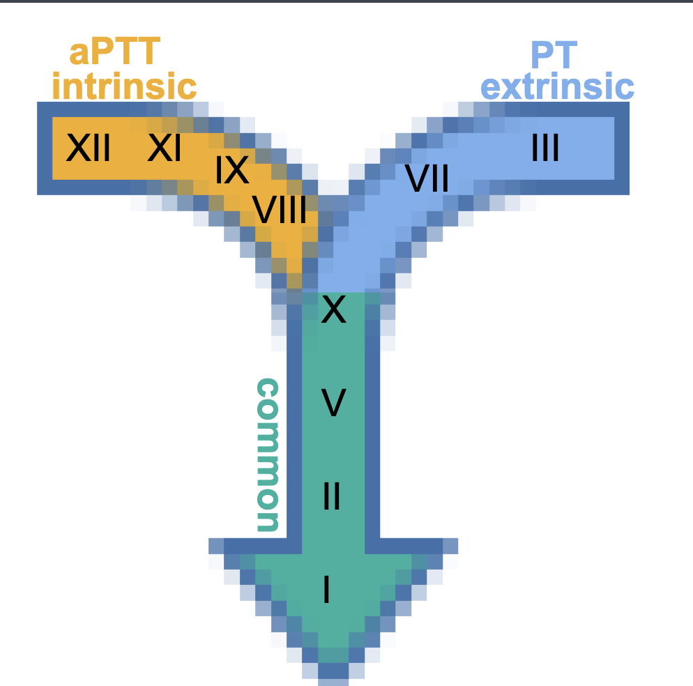
Intrinsic (aPTT)
Time required for plasma to clot after adding:
⚬ contact activator
⚬ phospholipid- will not work without phospholipid; must be bound to a cell
⚬ Ca- needed to activate the next thing
Extrinsic (PT)
(prothrombin time)
Time required for plasma to clot after adding tissue factor (III) and Ca
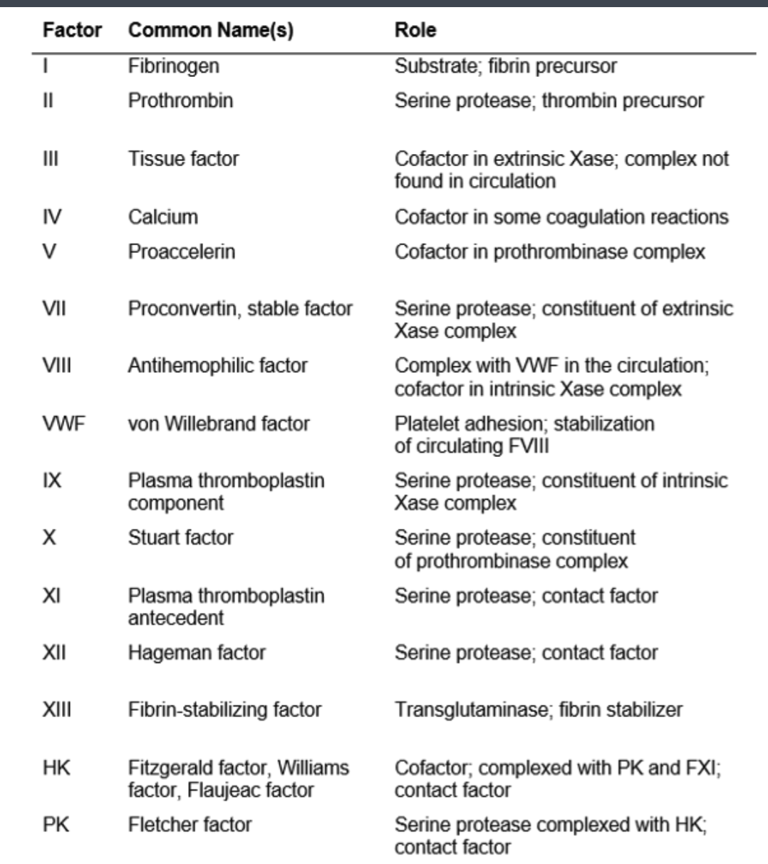
Coag proteins
most circulate as zymogens
⚬ named in order of discovery, NOT rxn sequence
⚬ roman numeral followed by “a” indicates activated
form
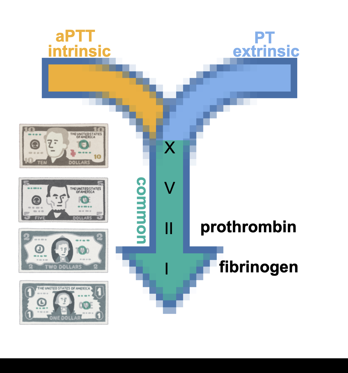
Common pathway
X marks the spot
• V fits into X = V is cofactor for X
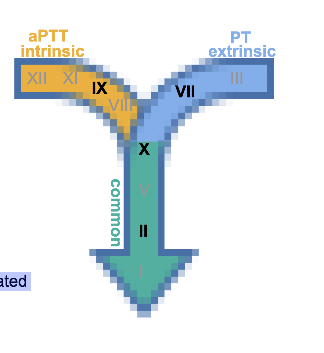
Intrinsic (TENENT)
the aPTT is a basic TENENT of coagulation studies
⚬ twelve
⚬ eleven
⚬ nine
⚬ eight
⚬ ten (beginning of common)
extrinsic
3 + 7 = 10
• “PT” has less letters than “aPTT”, shorter pathway,
fewer steps to remember, that’s lucky, lucky number 7
• III = tissue factor
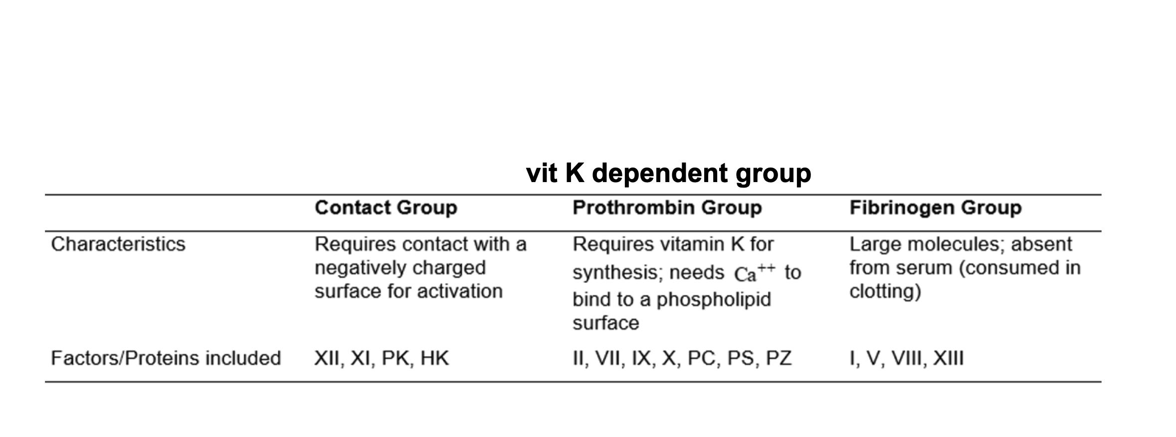
vit K dependent group
PROTIENS THAT NEED VIT K
II, VII, IX, X
• Protein C, Protein S
⚬ (inhibit coag)
• synthesized by liver
• all found in serum EXCEPT II (prothrombin)
• Vitamin K enables Ca²⁺-dependent binding to activated
platelet membranes
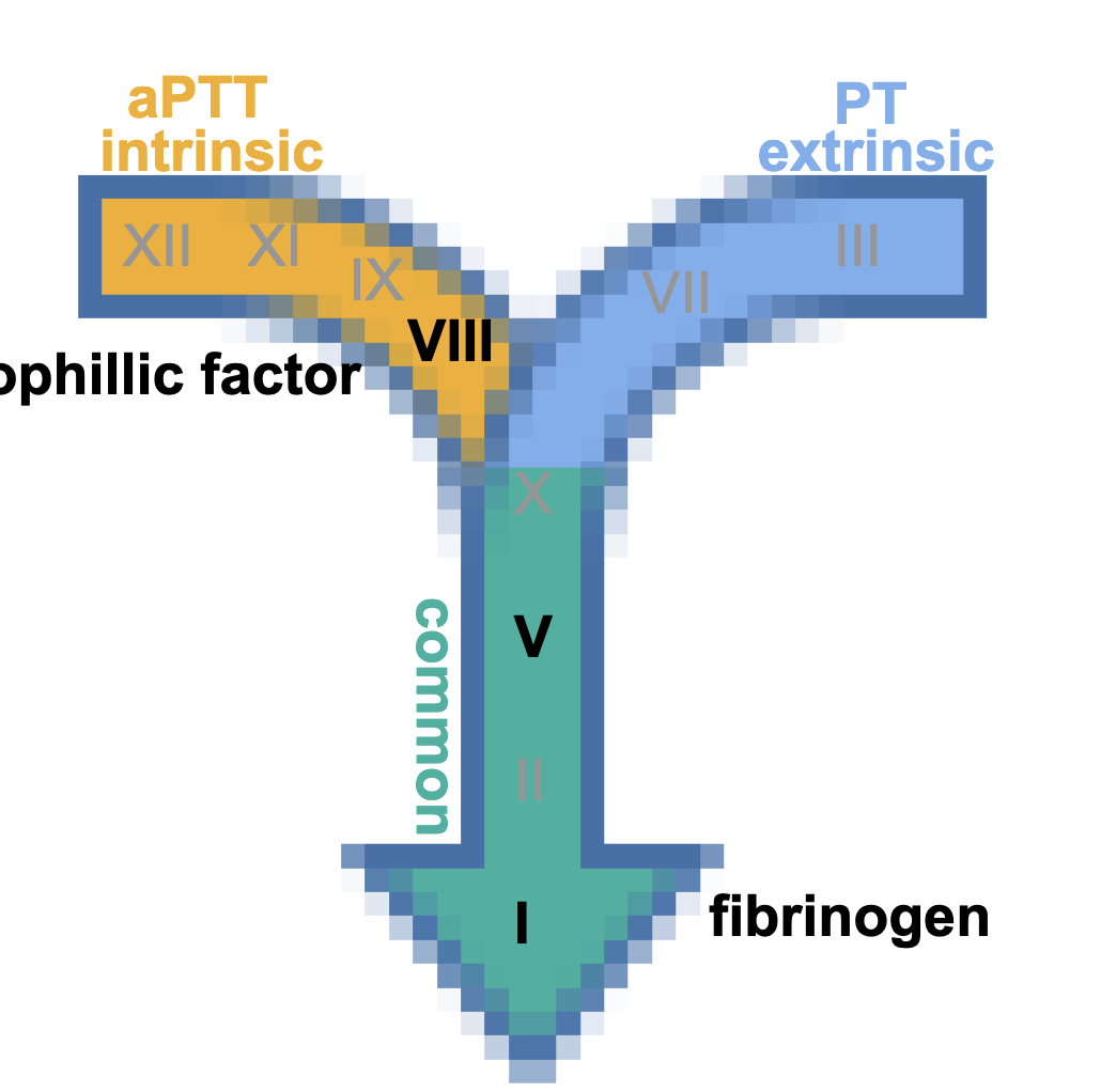
fibrinogen group
PROTIENS THAT NEED FIBRINOGEN
thrombin-sensitive
• I, V, VIII, XIII
⚬ I (fibrinogen) → fibrin
⚬ XIII = fibrin stabilizer
• not found in serum
contact group
activated by contact with negatively charged surfaces
⚬ collagen in basement membrane
⚬ initiate intrinsic pathway (aPTT)
• XII, XI, PK, HK
⚬ PK = prekallikrein
⚬ HK = high-molecular- weight-kininogen
⚬ deficiencies in all except XI are asymptomatic but cause prolonged
PTT
• found in serum in reduced amount
• also play role in fibrinolysis, activation of complement, inflammation
Summary of contact group, Vit K and fibrinogen
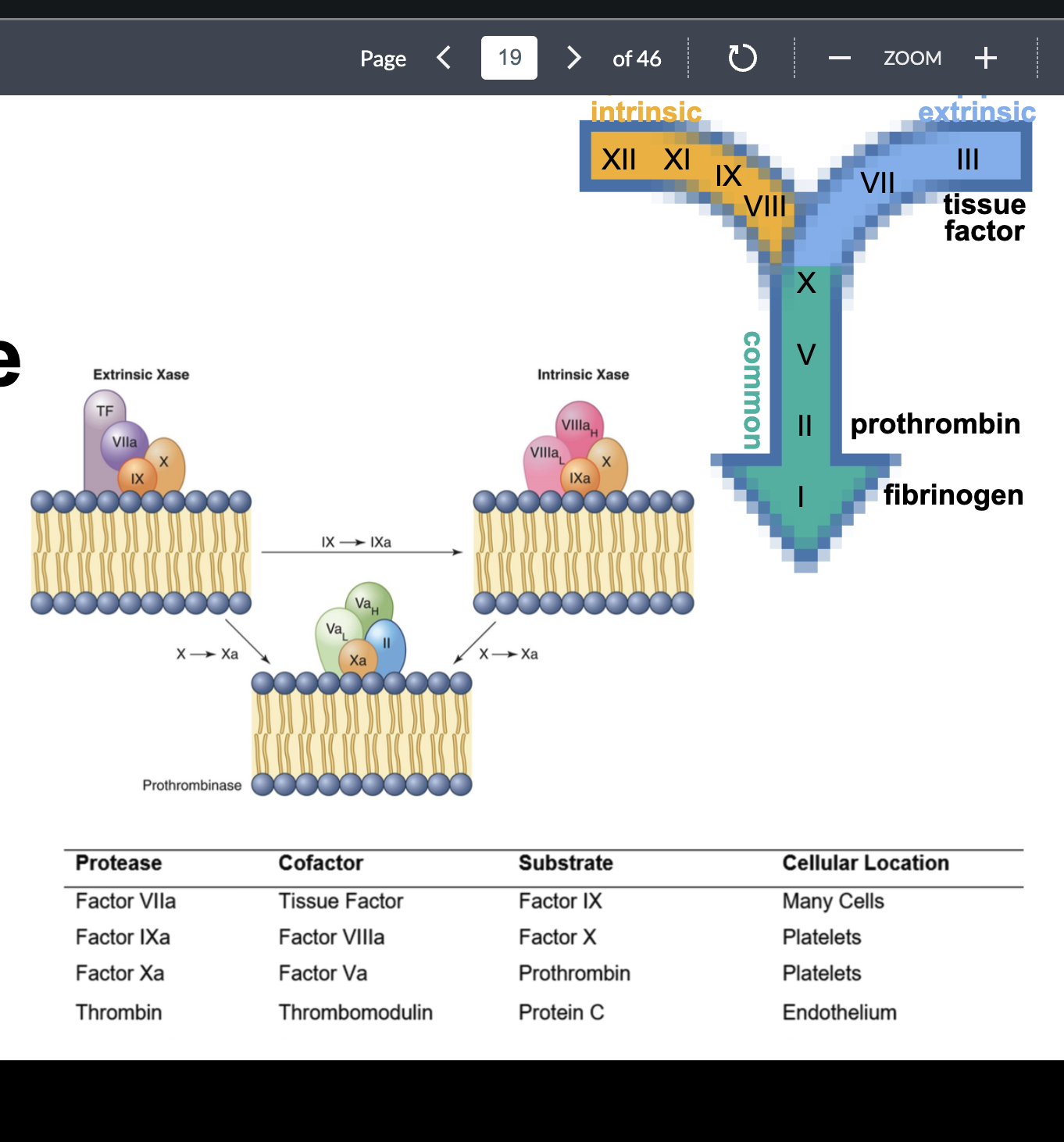
coagulation cascade
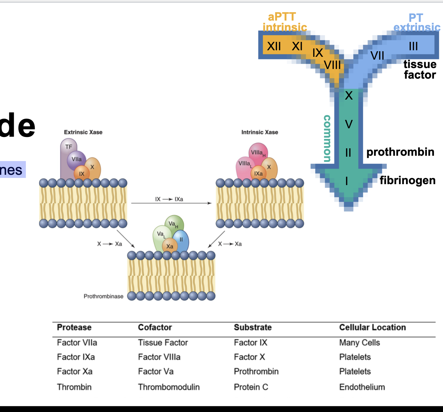
complex formation on phospholipid membranes
⚬ localizes clotting to site of injury
⚬ clotting factors bind to membrane of activated plts
■ complex = enzyme + cofactor + substrate
coagulation cascade complex formation
on phospholipid membranes
⚬ extrinsic Xase
■ starts coagulation
⚬ intrinsic Xase
■ amplifies factor X activation
⚬ prothrombinase
■ receives Xa from either complex
■ converts prothrombin to thrombin
coagulation cascade intrinsic pathway - aPTT
contact factors
■ XII, XI, PK, HK
■ activated when exposed to negatively
charged surfaces
• glass, celite (silica), ellagic acid
• does not require Ca
deficiency in XII, PK, HK
■ prolonged aPTT
■ no bleeding phenotype
⚬ deficiency in XI
■ prolonged aPTT
■ variable bleeding phenotype
coagulation cascade intrinsic pathway - aPTT (FACTOR DETAILS)
XII = Hageman factor
■ activated by collagen in vivo
■ activated by glass, silica in vitro
⚬ XI
■ only contact factor associated with abnormal
bleeding (variable)
⚬ IX = Christmas factor
■ vit K dependent
⚬ VIII = antihemophilic factor
■ deficiency = hemophilia A
■ circulates as complex with vWF
coagulation cascade extrinsic pathway - PT
vessel injury → nonvascular cells such
as fibroblasts, smooth muscle cells with
tissue factor (TF, III) are exposed to
blood
⚬ TF (III) binds VII in the presence of Ca
■ complex activates X and IX
coagulation cascade extrinsic pathway - PT FACTOR DETAILS
TF (III) = thromboplastin
■ transmembrane glycoprotein
■ cofactor for VII
⚬ VII
■ vit K dependent
coagulation cascade common pathway
begins with factor X
■ vit K dependent
■ activated via extrinsic or intrinsic Xase
⚬ V
■ 20% in plt alpha granules
■ positive feedback loop with thrombin:
• Va helps make thrombin, thrombin
activates more V
coagulation cascade common pathway FACTORS
prothrombin (II)
■ vit K dependent
■ cleaved by prothrombinase complex to
⚬ thrombin (IIa)
■ procoagulant
■ anticoagulant
■ antifibrinolytic
coagulation cascade common pathway thrombin (IIa)
procoagulant
• Fibrinogen → fibrin
• Activates V, VIII, XI, XIII
• Activates platelets
• Stimulates endothelial release of vWF and
expression of TF
(coagulation cascade common pathway) fibrinogen (I)
■ acute phase reactant
■ most abundant coag protein
⚬ 3 stages → fibrin
proteolysis = cleaved by thrombin into fibrin
monomers
polymerization = self-assemble into fibrin polymers
fibrin polymers stabilized by XIIIa
• creates covalent bond crosslinks between fibrin
• fibrin mesh stabilizes primary plt plug
• = secondary hemostasis
fibrinolytic system
digesting and removing fibrin
• activated in response to
⚬ coag cascade activation
⚬ fibrin formation
• limited to area of fibrin formation
• excessive fibrinolysis = bleeding
• inadequate fibrinolysis = excessive clotting
(fibrinolytic system) plasminogen (PLG)
synthesized by liver
⚬ activated by plasminogen activators (PA) to plasmin (PLN)
⚬ plasmin substrates=breakdown:
■ fibrin (Ia)
■ fibrinogen (I)
■ V, VIII, vWF
■ complement
■ plt surface receptors-stops receiving signals
(fibrinolytic system) 2 major PAs in circulation
⚬ tPA = tissue plasminogen activator(t for tissue)
■ main activator for dissolving fibrin clots inside vessels
• most released by endothelial cells
■ binds to fibrin => much more effective
• = localizes/concentrates fibrinolysis to clot
⚬ uPA = urokinase-type plasminogen activator
■ fibrin- independent
■ functions mainly in tissues (tissue remodeling/cell movement)
• other activators produced during contact phase- start plasminogen
⚬ kallikrein- don’t care about
⚬ XIIa
⚬ XIa
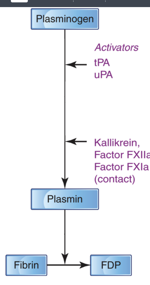
plasmin (fibrinolytic system)
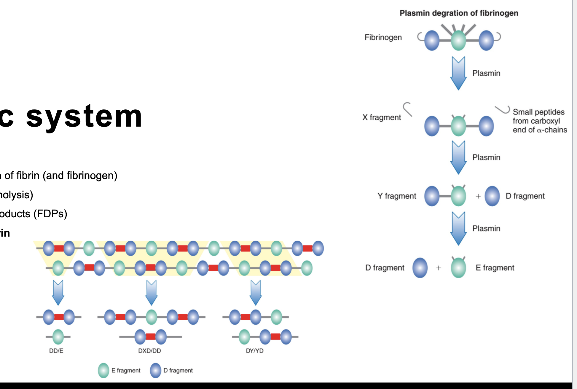
⚬ responsible for degradation of fibrin (and fibrinogen)
■ = fibrinolysis (fibrinogenolysis)
■ → fibrin degradation products (FDPs)
■ DDimer specific for fibrin
D (DDimer) and E fragments
Plasmin cannot break down D fully
Plasmin breaks down fibrinogen, which then can break down D
Elevated D-dimer most likely indicates a clot but does not confirm it
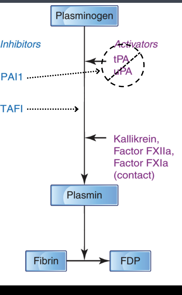
fibrinolytic system inhibitors of fibrinolytic system
prevent systemic proteolysis
⚬ action:
■ at the PLG activation step
• PAI1 = plasminogen activator inhibitor 1
⚬ primary inhibitor of tPA and uPA
• TAFI = thrombin-activatable fibrinolysis inhibitor
⚬ activated by thrombin/thrombomodulin complex
⚬ inhibits plasminogen → plasmin
■ directly on plasmin
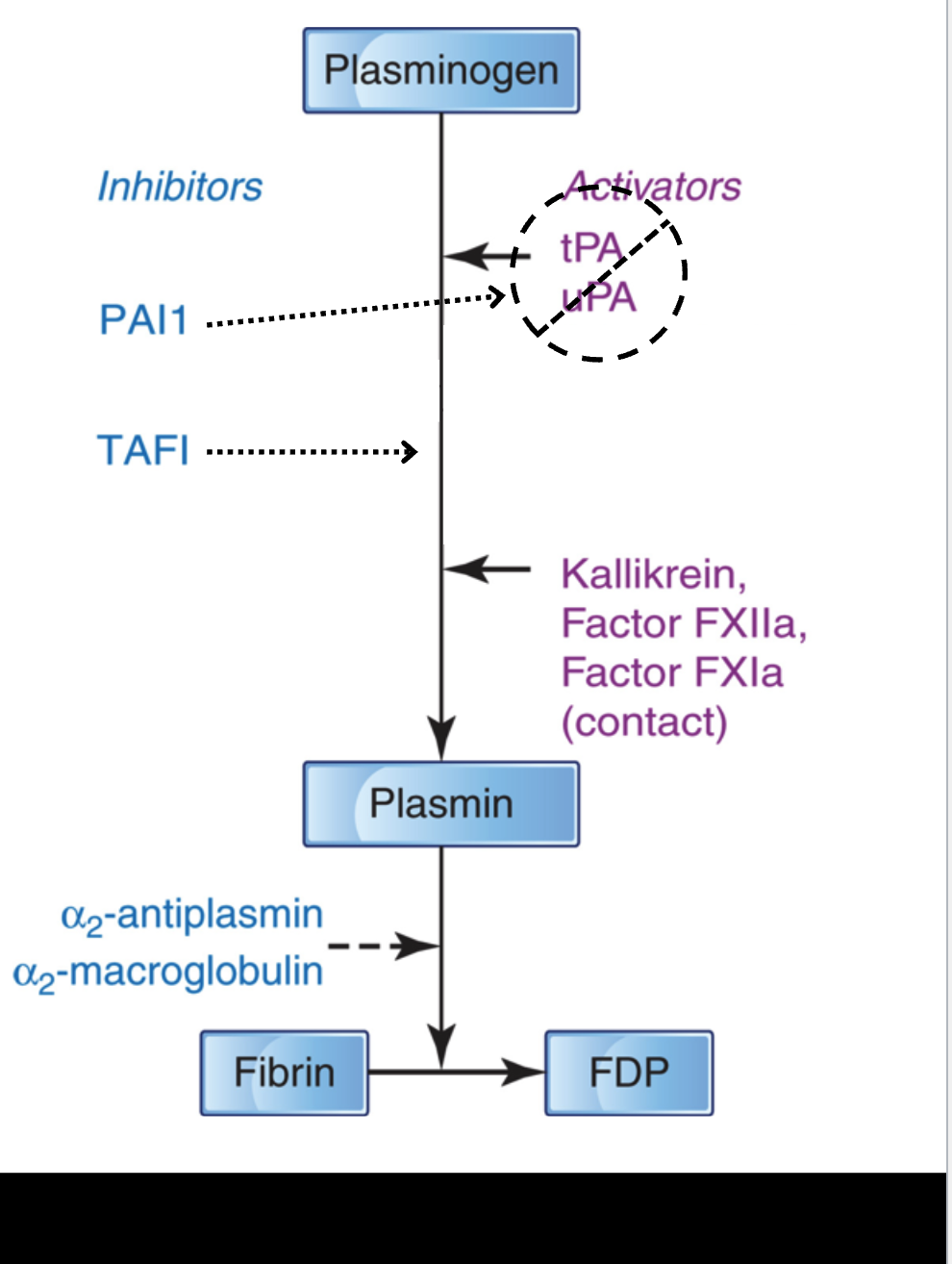
fibrinolytic system inhibitors of fibrinolytic system PT2
prevent systemic proteolysis
⚬ action:
■ at the PLG activation step
• PAI1
• TAFI
■ directly on plasmin
• α₂-antiplasmin
⚬ principle inhibitor of plasmin
⚬ cross-linked to fibrin by XIIIa(13)
• α₂-macroglobulin
⚬ backup inhibitor for plasmin
control of hemostasis
physical control = blood flow
⚬ vasoconstriction
⚬ return to normal blood flow → limits coagulation by
dilution of activated factors
physical control: liver clearance
⚬ liver produces many clotting factors
⚬ removes activated factors complexed with inhibitors,
and FDPs
⚬ liver disease may cause hemorrhage
■ reduced production of coag factors
■ if unable to remove activated proteases =contribute to systemic fibrinolysis or thrombosis
control of hemostasis feedback control positive
positive feedback amplification of coagulation
■ thrombin(II)-activates fibrinogen to fibrin(I to Ia)
• major activator of plts
• promotes release of plt Va
• activates Va, VIIIa, XIa, XIIIa
■ Xa
• activates VIIa and VIIIa
control of hemostasis negative feedback inhibition
control of hemostasis
• feedback control
⚬ negative feedback inhibition
■ thrombin
• activate OR inactivate Va and VIIIa
■ fibrin
• has strong affinity for thrombin, limiting amount of
thrombin available to convert fibrinogen to fibrin
■ FDPs = inhibit fibrin formation
control of hemostasis biochemical inhibitors pt1
⚬ antithrombin
■ most important inhibitor of serine proteases
■ inactivates:
• thrombin (IIa)
• Xa
• IXa, XIa, XIIa
■ activity enhanced by
• endothelial heparan sulfate
• pharmaceutical heparin
control of hemostasis biochemical inhibitors pt 2
biochemical inhibitors
⚬ antithrombin
⚬ protein c system = protein c and protein s
■ vit K dependent
■ synthesized primarily by liver
■ thrombin - thrombomodulin activates protein c
■ activated protein c + protein s
• inactivate Va and VIIIa
control of hemostasis biochemical inhibitors pt 3
antithrombin
⚬ protein c system = protein c and protein s
⚬ thrombomodulin
■ binds thrombin on intact endothelial cells
■ complex activates protein c
■ (Free thrombin promotes clotting;
thrombomodulin-bound thrombin helps limit
clotting)
control of hemostasis biochemical inhibitors pt 4
antithrombin
⚬ protein c system = protein c and protein s
⚬ thrombomodulin
⚬ TFPI = tissue factor pathway inhibitor
■ inhibits extrinsic/tissue factor pathway
■ binds Xa
■ TFPI-Xa complex inhibits TF-VIIa
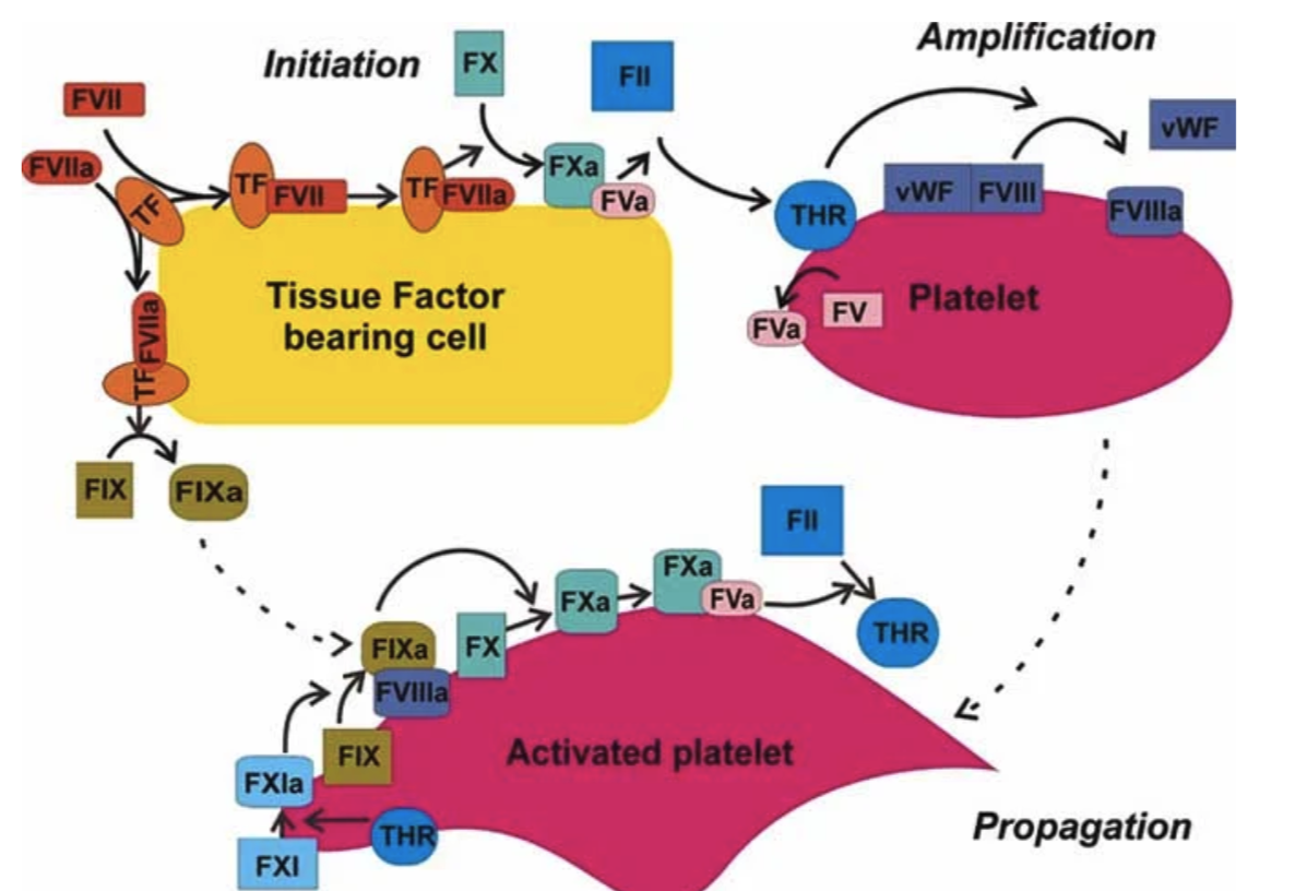
physiologic pathway = cell-based model
initiation
⚬ vascular injury exposes cells expressing TF
⚬ TF-VIIa activates X and IX
⚬ small amount of thrombin generated
• amplification
⚬ thrombin activates plts and factors V, VIII, XI
• propagation
⚬ coagulation complexes assemble on activated plts
⚬ thrombin burst → fibrin formation
⚬ XIIIa stabilizes fibrin
• termination
⚬ natural anticoagulants limit coagulation to injury site
Vit K required
C. XI, XII, PK, HK
1972
Which proteins are Vitamin K-dependent anticoagulants?
Protein C and Protein S
Vitamin K allows clotting factors to bind to:
Platelet phospholipid membranes
Fibrinogen Group
I
V
VIII
XIII
1,5,8,13
VIII deficiency
→ Hemophilia A
IX deficiency
→ Hemophilia B
Contact Group
XII
XI
Prekallikrein (PK)
High molecular weight kininogen (HK)
Activated by:
Collagen
Glass
Silica
Negatively charged surfaces
Deficiency of:
XII
PK
HK
Produces:
✔ Prolonged aPTT
❌ No bleeding
Only XI deficiency may produce bleeding.
Common Pathway
Starts at:
Factor X
↓
Xa + Va
↓
Prothrombinase
↓
II → IIa
↓
Fibrinogen
↓
Fibrin
↓
XIIIa stabilizes clot
Plasmin
Digests:
Fibrin
Fibrinogen
Factor V
Factor VIII
vWF
Produces:
FDPs
Main plasmin inhibitor
α₂-antiplasmin
Main coagulation inhibitor
Antithrombin