Brain and Behavior exam 1
 Call Kai
 Learn
Learn Practice Test
 Spaced Repetition
 Match
Match Flashcards
 Knowt Play
1/111

Earn XP
Description and Tags
Ch 0-2
Name | Mastery | Learn | Test | Matching | Spaced | Call with Kai | Chat |
|---|
No analytics yet
Send a link to your students to track their progress
112 Terms
three major types of experiments are
-correlation
-somatic intervention
-behavioral intervention
correlation experiments
measures how much a behavioral measure varies with a biological measure
somatic intervention
manipulate the body and see how that affects behavior
behavioral intervention
affect behavior and measure resulting physiological change
level of analysis
A concept to describe what level you are reducing a problem to, from social to cellular or even molecular.
Give a brief description (one or two sentences) of a situation that might not strictly be a within or between subjects experiment. (Hint: Some variables can't be a within-subject experiment)
A correleational study that studies an individual's stress levels and their hours of sleep is neither a within or between subjects experiment.
The modern field of neuroscience using the scientific method and objective experimentation:
Started approximately a century ago.
Epigenetics is:
Changes in gene expression that do not involve changes to the genome.
What is ontogeny?
Change in an individual during it's life time.
within-subjects experiment
every subject experiences both the experimental and control conditions
Computer science advances have helped neuroscience by:
Allowing the simulation of various biological functions within the brain, from neurons to circuits and Providing the ability to process datasets too large for humans to do by hand.
The idea that different regions of the brain control different aspects of thought is
localization of function
what is behavioral neuroscience
study of the biological bases of psychological processes and behavior
modern era uses..
the scientific method and objective experimentation, only about 100 years old
interdisciplinary neuroscience
neuroscience involves many different sciences
epigenetics
studies factors that affect gene expression w/o altering the genes themselves
levels of analysis
scope of experimental approaches, from social interaction down to the molecular level
within subjects
control group is the same set of subjects tested before any alteration
between subjects
experimental group is compared to a separate control group that has been treated identically except for the manipulation
somatic intervention
alteration of a brain structure or function to see how behavior is altered
behavioral intervention
intervention in a behavior to see how brain structure or function is altered
correlation
measures how much a body measure varies w/a behavioral measure
neurons
the basic units of the nervous system
four zones of the neuron
input: dendrites
integration: the cell body(soma)
conduction: axon or nerve fiber
output: axon terminals
axon
conducts output info away from the cell body as an electrical impulse
dendrite
receives info from other cells
bipolar neurons
one axon, one dendrite; sensory receptors(vision, smell, hearing)
unipolar neurons
a single extension branches in two directions, forming an input zone and an output zone; touch from body to spinal cord
multipolar neurons
one axon, many dendrites; most common type; most common neuron
nissal stains
outline all cell bodies because the dyes are attracted to RNA distributed within the cell
golgi stains
fill the whole cell and includes details, only stain a small proportion of neurons
immunocytochemistry
an antibody binds to the protein and chemical treatments make the antibody statement
in situ hybridization
uses radioactively labeled lengths of RNA or DNA to identify neurons in which a gene is being expressed
tract tracers
reveal the axonal projections of neurons( anterograde and retrograde tracers)
the brainbow
visualized different types of neurons simultaneously using fluorescent proteins inserted into cell DNA
5 types of techniques to visualize cell
-nissal stains
-golgi stains
-immunochemistry
-tract tracers
-the brainbow
anatomy of the synapse
-pre-synaptic membrane
-post synaptic membrane
-synaptic cleft
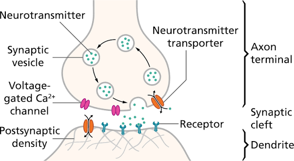
pre-synaptic membrane
on the axon terminal of the presynaptic neuron
post synaptic membrane
on the dendrite or cell body of the post synaptic neuron
synaptic cleft
a gap 20-40 nanometers that separates the membranes
mechanism of synaptic release
action potential arrival
calcium influx
vesicle mobilization and docking
fusion and exocytosis
post-synaptic action and recycling
what is the first step of the mechanism of synaptic release
action potential arrival, causes cell membrane to depolarize
second step of synaptic release
calcium influx, depolarization opens voltage gated calcium channels, which causes ca2+ to rush into the cell
third step of mechanism of synaptic release
vesicle mobilization and docking, synaptic vesicles store the neurotransmitters
fourth step of mechanism of synaptic release
fusion and exocytosis; cell membranes merge causing exocytosis
5th step of mechanism of synaptic release
post-synaptic action and recycling, Neurotransmitters cross the cleft and bind to specific receptors on the next cell, creating a new electrical or chemical response
layers of the cortex
neocortex(6 layers), mesocortex(6 layers), aliocortex(5 layers), periallocortex
types of glia
-myelin cell(oligodendrocytes and schwann cells)
-astrocytes
-microglial cells
oligodendrocytes
form the myelin sheath in the brain and spinal cord(CNS)
schwann cells
provide myelin to cells outside the brain and spinal cord (PNS)
astrocytes
star shaped cells with many processes thar stretch around and between neurons and blood vessels
microglial cells(microglia)
small cells that remove debris from injured cells
what is myelination
process in which specialized glial cells wrap around a neuron’s axon to form a fatty insulation layer known as the myelin sheath
how does myelination affect the action potential
myelin acts as a fatty insulating layer along the axon as electrical impulses travel down toward the axon terminals and the nodes of ranvier interrupt this insulation at regular intervals, leaving specific gaps where the axon is directly exposed
cranial nerves
connected to the brain; innervate the head neck and visceral organs directly from the brain
cranial nerve 1
olfactory: smell
cranial nerve 2
optical: vision
cranial nerve 3
occulomotor: eye movement
cranial nerve 4
trochlear: eye movement
cranial nerve 5
trigeminal: facial sensation chewing muscles
cranial nerve 6
abducens: eye movement
cranial nerve 7
facial: taste sensation, facial muscles
cranial nerve 8
vestibulocochlear: hearing and balance
cranial nerve 9
glossopharyngeal: throat sensation, throat muscles
cranial nerve 10
vagus: innervates the heart, liver, and intestines
cranial nerve 11
spinal accessory:neck muscles
cranial nerve 12
hypoglossal: tongue
cranial nerve mnemonic
on old olympus’ towering top a friendly viking grew vines and hops
sympathetic chain
innervates the sympathetic chain and innervates the sympathetic nervous system(fight or flight)
spinal nerves : DAVE
dorsal = afferent, ventral = efferent
parasympathetic nervous system
rest and recovery
afferent
carries info into a region of interest( A for arrive)
efferent
carries info away from the CNS( E for exit)
four lobes of the brain
frontal, parietal, temporal, occipital
grey matter
contains more cell bodies and dendrites which lack myelin
white matter
consists mostly of axons w/ white myelin sheaths
limbic system
structures important for learning and memory
amygdala
emotional regulation and perception of odor
structures of the limbic system
amygdala, hippocampus and fornix, cingulate gyrus, olfactory bulb, thalamus and hypothalamus
hippocampus and fornix
learning
cingulate gyrus
attention
olfactory bulb
sense of smell
thalamus
cluster of nuclei that relay sensory info
hypothalamus
contains nuclei w/ many functions, also controls the pituitary gland
brain stem consists of:
medulla, pons, and midbrain
midbrain sensory systems consist of:
superior and inferior colliculi
superior colliculi
process visual info
inferior colliculi
process auditory info
basal ganglia includes:
striatum, caudate nucleus, putamen, and nucleus accumbens, globus pallidus, substantia nigra, and subthalamic nucleus
striatum
includes caudate nucleus, putamen, and nucleus
function of the basal ganglia
help coordinate voluntary movement, learning, habits, and emotions
Meninges
protective membrane that surrounds the brain and spinal cord: consists of 3 layers: dura mater, pia mater, arachnoid membrane
dura mater
tough most outermost layer of meninges
pia mater
delicate most innermost layer of meninges
arachnoid membrane
lies between the other two layers, filled w cerebrospinal fluid(CSF)
meningitis
acute infection of the meninges
meningiomas
tumors formed in the meninges
ventricular system
series of chambers filled with cerebral spinal fluid(CSF), contains lateral ventricle, choroid plexus, third ventricle, and fourth ventricle
lateral ventricle
in each hemisphere of the ventricular system, extend into all four lobes and is lined with the choroid plexus
choroid plexus
a membrane that produces CSF