Chapter 2: Neurons and Glial Cells
 Call Kai
 Learn
Learn Practice Test
 Spaced Repetition
 Match
Match Flashcards
 Knowt Play
1/60

Earn XP
Description and Tags
Vocabulary flashcards generated from lecture notes covering neuronal structures, glial cell types, action potential phases, and key neurotransmitters.
Name | Mastery | Learn | Test | Matching | Spaced | Call with Kai | Chat |
|---|
No analytics yet
Send a link to your students to track their progress
61 Terms
Neuron
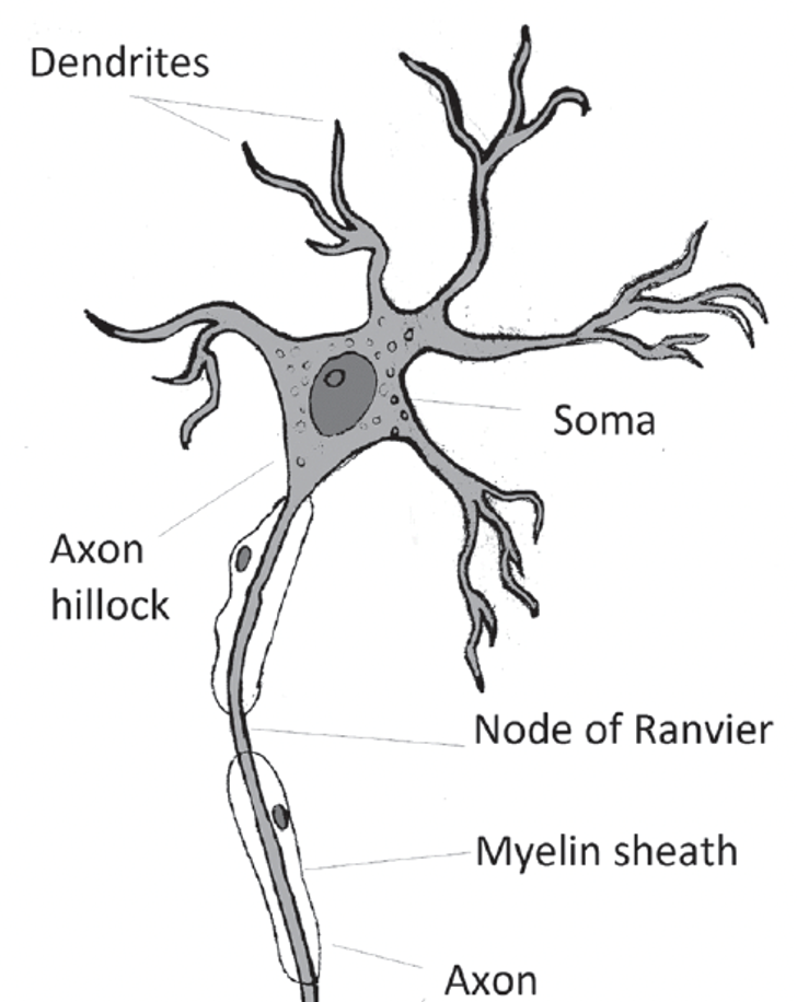
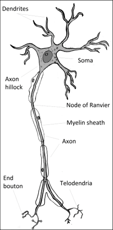
The smallest functional unit of the nervous system, also known as the nerve cell. Each neuron consists of a soma (cell body) and cell processes, including dendrites and an axon.

Soma
The cell body of a neuron containing organelles such as the nucleus, nucleolus, endoplasmic reticulum, ribosomes, Golgi apparatus, lysosomes, mitochondria, and neurofibrils. It is the metabolic center of the cell.

Nucleus
A large spherical structure within the soma containing DNA, which controls protein and enzyme synthesis.
Nucleolus
Located within the nucleus, it is the site of ribosomal RNA synthesis.
Endoplasmic Reticulum (ER)
A network of membranes within the cytoplasm responsible for protein and lipid synthesis. Rough ER has ribosomes for protein production, while Smooth ER synthesizes steroid hormones and processes toxins.
Ribosomes
Organelles responsible for protein synthesis, found on the rough ER or floating freely in the cytoplasm.
Golgi Apparatus
An organelle involved in collecting, packaging, and releasing proteins and lipids produced by the ER.
Lysosomes
Organelles containing enzymes responsible for breaking down and recycling cellular waste and organelles.
Mitochondria
Double-membrane organelles that generate energy for the cell through metabolism. Critical for neuronal function due to high energy demand.
Neurofibrils
Threadlike structures in the cytoplasm that facilitate communication among organelles within the cell body.
Dendrites
Highly branched processes that serve as the input system of the neuron, receiving information from other neurons.
Axon
The sole output structure of a neuron that transmits electrical impulses away from the soma.
Axon Hillock
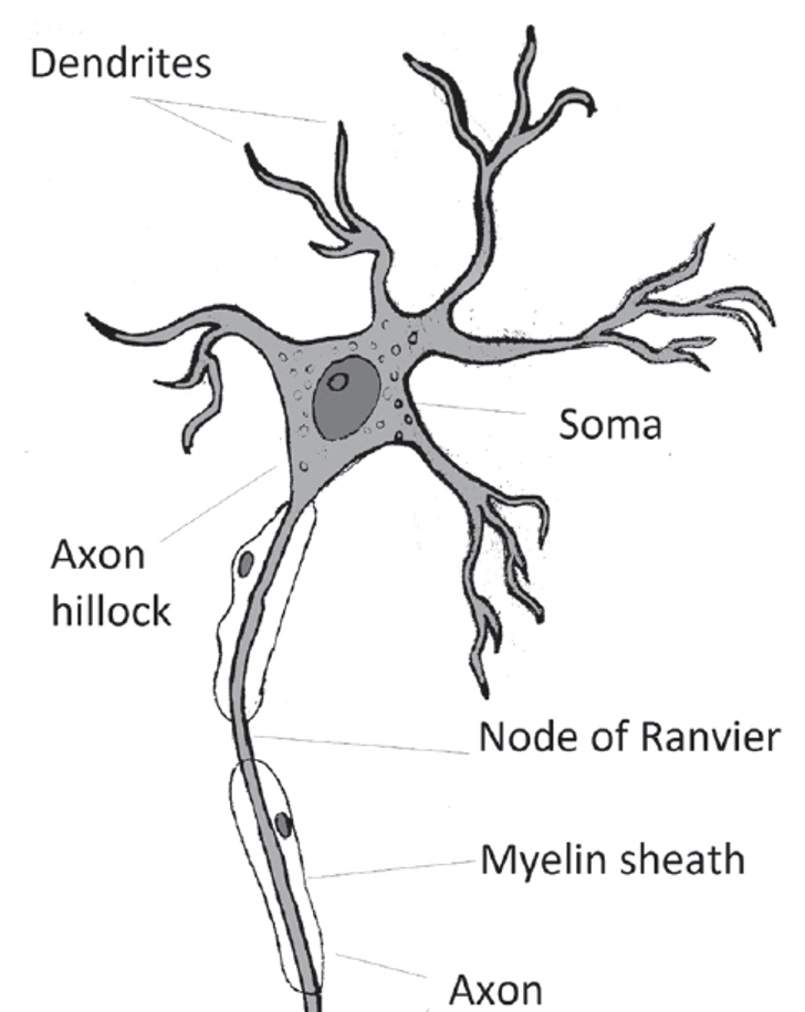
The site of action potential generation located at the root of the axon.

Myelin Sheath
An insulating covering of axons that greatly enhances the speed of neural impulse conduction.

Nodes of Ranvier
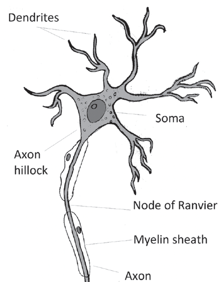
Gaps in the myelin sheath that allow for saltatory conduction, greatly increasing conduction velocity.
Telodendria
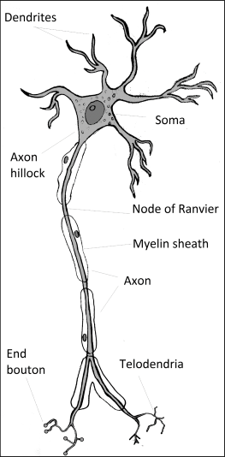
Terminal branches of the axon that end in end boutons.
End Boutons
Specialized terminals at the end of telodendria that contain synaptic vesicles filled with neurotransmitters.
Synapse
The point of communication between two neurons, consisting of the presynaptic end bouton, synaptic cleft, and postsynaptic receptors.
Unipolar Neuron
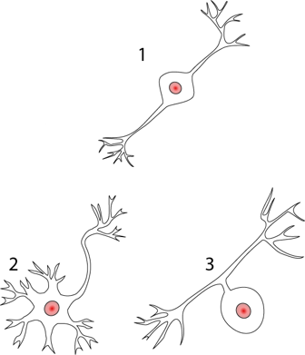
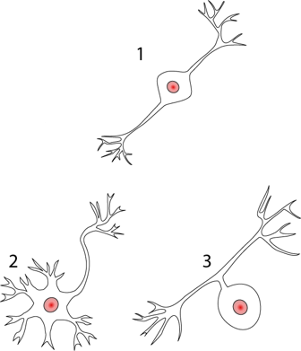
A neuron with one process extending from the soma, primarily found in sensory systems.
Bipolar Neuron
A neuron with one axon and one dendrite, commonly found in sensory systems such as the retina and auditory pathways.
Multipolar Neuron
A neuron with many dendrites and one axon, the most common type in the central nervous system.
Golgi Type I Neuron
A neuron with a long axon that communicates across distant regions of the nervous system.
Golgi Type II Neuron
A neuron with a short axon that communicates locally, often called interneurons.
Afferent Neuron
A neuron that transmits information to the central nervous system, generally sensory in nature.

Efferent Neuron
A neuron that carries information from the central nervous system to the periphery, generally motor in function.

Interneuron
A neuron that provides communication between other neurons, the most common type in the nervous system.

Glial Cells
Non-neuronal cells that provide structural and metabolic support to neurons, as well as play active roles in information processing.
Microglia
Specialized immune cells in the CNS that act as macrophages, removing waste products and defending against infection.
immune defense/cleanup
Astrocytes
Glial cells that provide structural support, regulate neurotransmitters, ions, and blood flow, and maintain the blood-brain barrier.
Blood-brain barrier + synapse/ion regulation

Oligodendrocytes
Glial cells in the CNS that generate myelin for axons.

Schwann Cells
Glial cells in the PNS that generate myelin and assist in axon repair.

Radial Glia
Glial cells that act as scaffolds for neuron migration during development and regulate synaptic plasticity.
Guide neuron migration in development
Satellite Cells
Glial cells in the PNS that surround neurons in ganglia and regulate the chemical environment.
Enteric Glia
Glial cells in the digestive tract involved in autonomic regulation.
Ependymal Cells
Glial cells lining the ventricles of the brain that secrete cerebrospinal fluid (CSF).
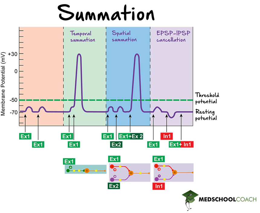
Excitatory Postsynaptic Potential (EPSP)
A depolarizing postsynaptic potential that increases the likelihood of an action potential.
Inhibitory Postsynaptic Potential (IPSP)
A hyperpolarizing postsynaptic potential that decreases the likelihood of an action potential.
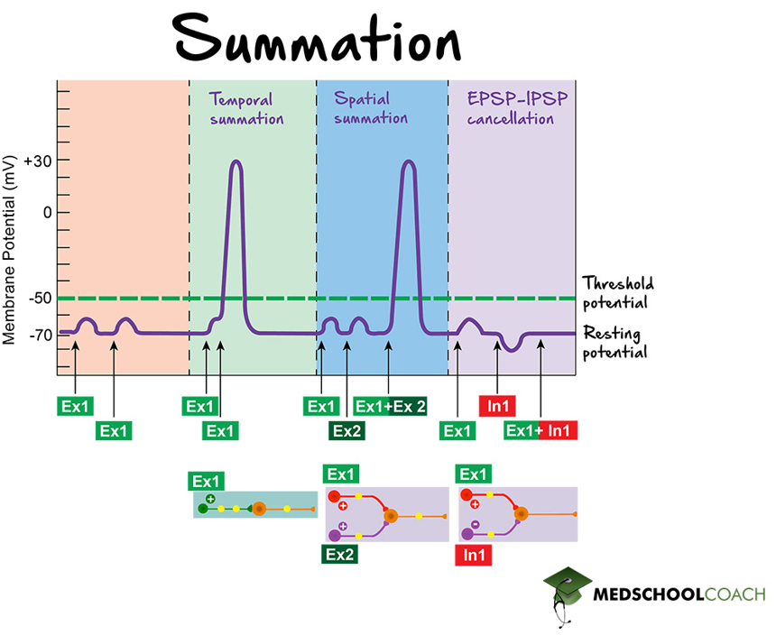
Action Potential
An all-or-nothing electrical event generated at the axon hillock that propagates along the axon to transmit information.
Saltatory Conduction
The process by which action potentials jump from node to node along a myelinated axon, increasing conduction speed.

Neurotransmitters
Chemical messengers released from presynaptic neurons into the synapse to excite or inhibit postsynaptic neurons.
Glutamate
The most prevalent excitatory neurotransmitter in the CNS.
Aspartate
An excitatory neurotransmitter that stimulates receptors similar to glutamate.
Gamma-Aminobutyric Acid (GABA)
The most prevalent inhibitory neurotransmitter in the CNS.
Glycine
An inhibitory neurotransmitter primarily active in the spinal cord.
Acetylcholine (ACH)
A neurotransmitter responsible for muscle activation, memory, and regulation of consciousness.
Dopamine (DA)
A monoamine neurotransmitter involved in reward, emotion, movement regulation, and inhibition in specific brain areas.
Norepinephrine (NE)
A monoamine neurotransmitter and hormone involved in stress response, arousal, and attentional focus.
Serotonin
A monoamine neurotransmitter involved in mood regulation, appetite, memory, and sleep.
Neuropeptides
A diverse class of neurotransmitters and hormones involved in regulating pain, emotion, and social behavior.
Adenosine Triphosphate (ATP)
An energy molecule that also functions as a neurotransmitter involved in pain signaling and neural communication.
Synaptic Vesicles
Small membrane-bound sacs located within the end boutons of the presynaptic neuron. They contain neurotransmitters that are released into the synaptic cleft during synaptic transmission.

Post-synaptic Neuron
The neuron that receives the signal at the synapse. It has receptor sites on its membrane that bind neurotransmitters released from the presynaptic neuron.

Pre-synaptic Neuron
The neuron that sends the signal at the synapse. It contains synaptic vesicles that release neurotransmitters into the synaptic cleft.

Synaptic Cleft
The small gap (approximately 20nanometers wide) between the presynaptic and postsynaptic neurons where neurotransmitters are released to transmit signals.

Membrane Potential
The electrical potential difference across a cell’s membrane, resulting from the distribution of ions inside versus outside the cell.
Depolarization
The process during which the inside of a neuron becomes less negative relative to the outside, usually due to the influx of sodium (Na+) ions. It is the initial phase of an action potential.

Repolarization
The process of returning the membrane potential back to its resting negative state after depolarization, primarily due to the efflux of potassium (K+) ions.
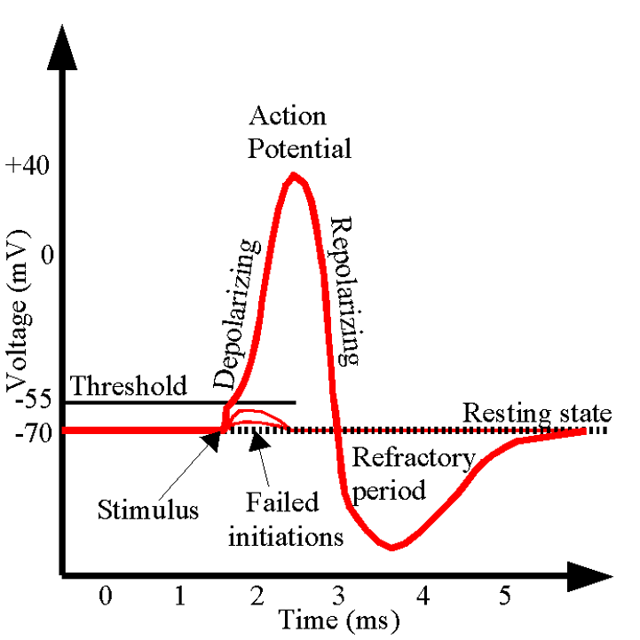
Hyperpolarization
A state where the inside of the neuron becomes more negative than the resting potential, making it less likely for the neuron to fire another action potential immediately.

5 Ways to Classify Neurons
Number of processes: How many “arms” leave the soma. Bipolar (1 dendrite + 1 axon; retina, auditory, olfactory) and multipolar (1 axon + many dendrites; e.g., spinal motor neurons) are the human types. Pseudo-unipolar = our sensory nerves.
Dendrite shape/arborization: The branching pattern of the dendritic tree (e.g., fan, spindle, spherical). Just know that dendrite shape varies a lot by neuron type.
Axon length/Golgi type: Golgi type 1 = long axons for long-distance signaling (e.g., a motor neuron from your back to your toe). Golgi type 2 = short axons; interneurons connecting nearby cells.
Function: Afferent (sensory, signal goes IN to CNS), efferent (motor, signal goes OUT from CNS), and interneurons (connect the two).
Conduction velocity: How fast the signal travels; thicker myelin = faster.
Neuromuscular junction
The neuromuscular junction (NMJ) is the synapse between a motor neuron and a muscle fiber.
A motor unit = one neuron + all the muscle fibers it activates. When the neuron fires, ACh is released, the muscle’s receptors are activated, and the muscle fiber contracts (all-or-none, like the neuron).
Innervation ratio = neurons-to-fibers. Low ratio (1 neuron : many fibers) → powerful, slow-twitch work; high ratio (1 neuron : few fibers) → fine, fast-twitch control. Speech muscles use both. (Big-picture only.)

Blood-brain barrier (BBB)
Astrocycles build and maintain
Key protective mechanism of the brain
Filtering system to prevent infectious agents, damaging molecules and certain classes of immune cells from having unrestricted access to our signaling neurons
