Concepts of Care for Patients with Dysrhythmias (NUR 411)
 Call Kai
 Learn
Learn Practice Test
 Spaced Repetition
 Match
Match Flashcards
 Knowt Play
1/27

Earn XP
Description and Tags
Ch. 28
Name | Mastery | Learn | Test | Matching | Spaced | Call with Kai | Chat |
|---|
No analytics yet
Send a link to your students to track their progress
28 Terms
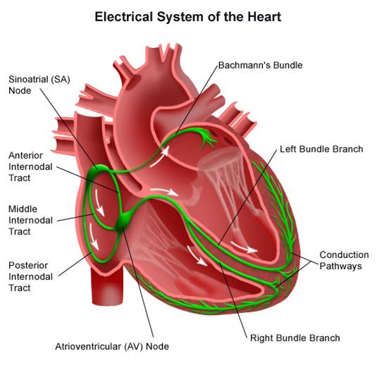
Cardiac Conduction System
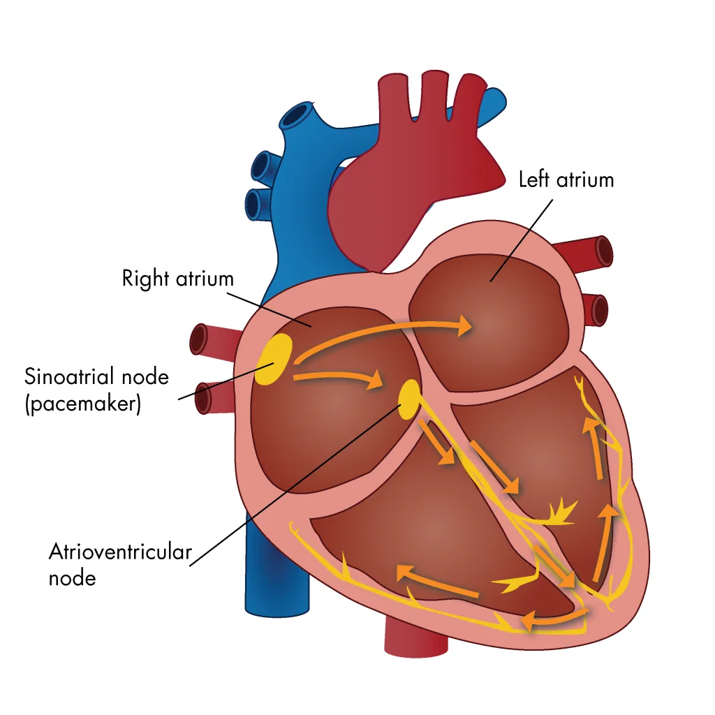
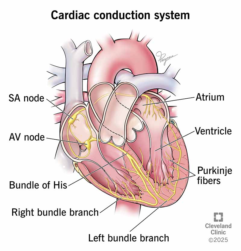
1.) Sinoatrial (SA) Node → primary pacemaker located near the junction of the right atrium & SVC
2.) Atrioventricular (AV) Node → delays impulses before proceeding to ventricles; located beneath the right atrial endocardium (between the tricuspid valve & coronary sinus)
3.) Bundle of His → part of the AV junction that carries impulses; continues through the interventricular septum via bundle branches
4.) Right Bundle Branch → travels down the right side of the interventricular septum; carries impulses to the Purkinje network
5.) Left Bundle Branch → extends down the left side of the interventricular septum; carries impulses to the Purkinje network
6.) Purkinje Fibers → network located on the endocardial surface of both ventricles; responsible for rapid conduction of electrical impulses throughout ventricles
Sinoatrial (SA) Node
The heart’s primary pacemaker; located close to the surface of the right atrium (near the junction with the SVC); spontaneously/rhythmically generates electrical impulses at 60-100 beats/min; changes in HR influenced by the sympathetic & parasympathetic nervous systems

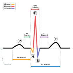
P Wave
Reflects atrial depolarization caused by impulses from the SA node moving through atrial muscle
Atrioventricular (AV) Junction
Consists of…
1.) A transitional cell (T cell) zone
2.) The AV node
3.) The bundle of His

Atrioventricular (AV) Node
Lies beneath the right atrial endocardium (between the tricuspid valve & coronary sinus); T cells (transitional cells) cause impulses to slow down before proceeding to the ventricles; controlled by the sympathetic & parasympathetic nervous systems
Transitional Cells (T Cells)
Cells of the AV junction that are specialized to slow down or delay electrical impulses in the AV node; enable the atria to contract (atrial kick) & the ventricles to fill
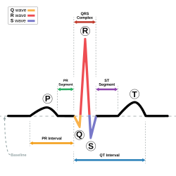
PR Segment
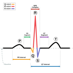
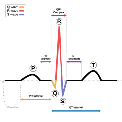
Reflects the delay of electrical impulses in the AV node before proceeding to the ventricles
Bundle of His
Connects with the distal portion of the AV node & continues through the interventricular septum; extends as a right & left bundle branch; composed of Purkinje cells that enable rapid conduction of electrical impulses throughout the ventricles (ventricular depolarization)

PR Interval
Reflects the time required for atrial depolarization & impulse travel through the conduction system (Bundle of His → Purkinje network); measured from the beginning of the P wave to the end of the PR segment (normally 0.12 to 0.20 s)
Purkinje Fibers
Located at the ends of both the right & left bundle branch systems; interweaving network located on the endocardial surface of both ventricles (apex → base); composed of Purkinje cells

QRS Complex
Reflects ventricular depolarization; measured from the beginning of the Q wave to the end of the S wave
Purkinje Cells
Compose the bundle of His, bundle branches, & terminal Purkinje fibers; responsible for the rapid conduction of electrical impulses throughout the ventricles; triggers ventricular depolarization & subsequent ventricular muscle contraction
ECG Components
1.) P Wave → atrial depolarization
2.) PR Segment → impulse travel through the AV node and through the ventricular conduction system to Purkinje fibers
3.) PR Interval → time for atrial depolarization, impulse delay, and travel to Purkinje fibers
4.) QRS Complex → ventricular depolarization
5.) ST Segment → early ventricular repolarization
6.) T Wave → ventricular repolarization
7.) U Wave → slow repolarization of ventricular Purkinje fibers; may suggest electrolyte abnormality
8.) QT Interval → total time for ventricular depolarization & repolarization; prolonged QT may lead to torsades de pointes

Automaticity
Pacing function; ability of cardiac cells to generate an electrical impulse spontaneously & repetitively; disturbances may involve either an increase or decrease in pacing function
Excitability
Ability of non-pacemaker heart cells to respond to an electrical impulse that begins in pacemaker cells
Depolarization
Normally negatively charged cells within the heart muscle develop a positive charge
Conductivity
Ability to send an electrical stimulus from cell membrane to cell membrane; excitable cells depolarize in rapid succession until all have undergone depolarization
Contractility
Mechanical activity of the heart; ability of atrial & ventricular muscle cells to shorten their fiber length in response to electrical stimulation, causing sufficient pressure to push blood forward
Normal Sinus Rhythm (NSR)
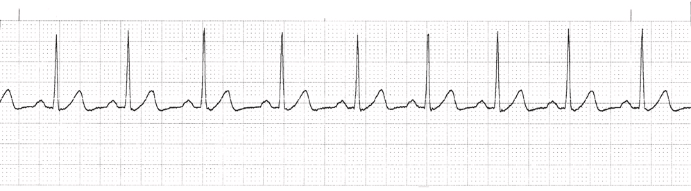
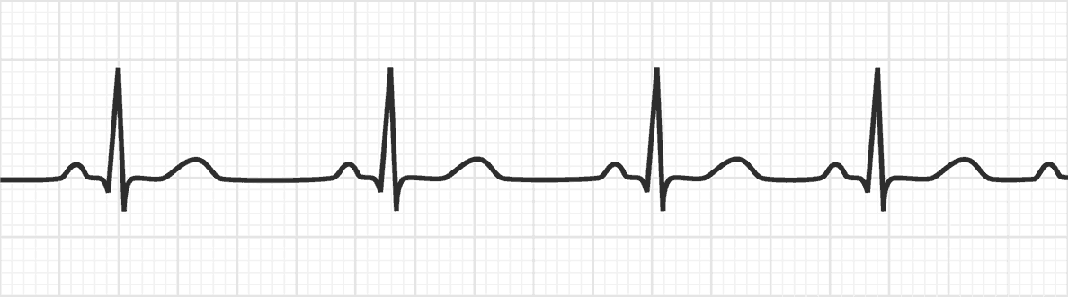
Rhythm originating from the SA node that meets the following ECG criteria:
i.) Rate → Atrial and ventricular rates of 60 to 100 beats/min
ii.) Rhythm → Atrial and ventricular rhythms regular
iii.) P waves → Present, consistent configuration; one P wave before each QRS complex
iv.) PR interval → 0.12 to 0.20 second and constant
v.) QRS duration → 0.06 to 0.11 second and constant
Sinus Arrhythmia
Variant of NSR that results from changes in intrathoracic pressure during breathing & meets the following ECG criteria:
i.) Rate → Atrial and ventricular rates between 60 and 100 beats/min
ii.) Rhythm → Atrial and ventricular rhythms irregular, with the shortest PP or RR interval varying at least 0.12 second from the longest PP or RR interval
iii.) P waves → One P wave before each QRS complex; consistent configuration
iv.) PR interval → Normal, constant
v.) QRS duration → Normal, constant
Dysrhythmia
Any disorder of the heartbeat
3 Categories of Common Dysrhythmias
1.) Premature Complexes → early rhythm complexes
2.) Bradydysrhythmias → HR slower than normal (typically < 60 beats/min)
3.) Tachydysrhythmias → HR faster than normal (typically > 100 beats/min)
Premature Complexes
Cardiac cell or cell group (other than SA node) becomes irritable & fires before the next sinus impulse is produced; pause between the premature & normal complex creates an irregularity in rhythm (palpitations)
3 Rhythmic Premature Complexes
1.) Bigeminy → normal complexes followed by a premature complex and a pause; two-beat pattern
2.) Trigeminy → two sequential normal complexes are followed by a premature complex and a pause; three-beat pattern
3.) Quadrigeminy → three sequential normal complexes followed by a premature complex and a pause; four-beat pattern

Bradydysrhtyhmias (Bradycardias)
Dysrhythmias that occur with HR < 60 beats/min…
i.) Generally well-tolerated if the BP is adequate (↓ myocardial oxygen demand, ↑ diastole, adequate coronary perfusion time)
ii.) Symptomatic if BP is inadequate (↓ coronary perfusion, ↓ CO, myocardial ischemia, MI, hypotension)

Tachydysrhythmias (Tachycardias)
Dysrhythmias that occur with HR > 100 beats/min…
i.) Shorten diastolic filling time → less coronary perfusion time
ii.) Initially ↑ CO & BP but prolonged tachydysrhythmias ↓ diastole & SV (↓ CO, BP, coronary perfusion)
iii.) Work of the heart & myocardial oxygen demand subsequently ↑
iv.) May lead to HF and associated signs/symptoms (dyspnea, lung crackles, JVD, fatigue/weakness)
Key Features of Sustained Tachydysrhythmias & Bradydysrhythmias
i.) Chest discomfort, pressure, or pain (which may radiate to the jaw, back, or arm)
ii.) Restlessness, anxiety, nervousness, confusion
iii.) Dizziness, syncope
iv.) Palpitations (in tachydysrhythmias)
v.) Change in pulse strength, rate, and rhythm
vi.) Pulse deficit
vii.) Shortness of breath, dyspnea
viii.) Tachypnea
ix.) Pulmonary crackles
x.) Orthopnea
xi.) S3 or S4 heart sounds
xii.) Jugular venous distention
xiii.) Weakness, fatigue
xiv.) Pale/ash gray, cool, skin; diaphoresis
xv.) Nausea, vomiting
xvi.) Decreased urine output
xvii.) Delayed capillary refill
xvii.) Hypotension