Biochem: Nitrogen Metabolism 2
 Call Kai
 Learn
Learn Practice Test
 Spaced Repetition
 Match
Match Flashcards
 Knowt Play
1/49

There's no tags or description
Looks like no tags are added yet.
Name | Mastery | Learn | Test | Matching | Spaced | Call with Kai | Chat |
|---|
No analytics yet
Send a link to your students to track their progress
50 Terms
Porphyrin
-5 ring molecule
A=acetate
P=propionate
M=methyl
V=Vinl
requires iron to be functional
eg Heme side chian = MVMVMPPM
Biosynthesis of heme 1st step
glycine + succinyl CoA (from TCA)
δ-aminolevulinic acid (ALA) by enzyme δ-ALA synthase.
-inhibited by hemin and heme (product inhibition)
-hemin-drug, reduces heme production
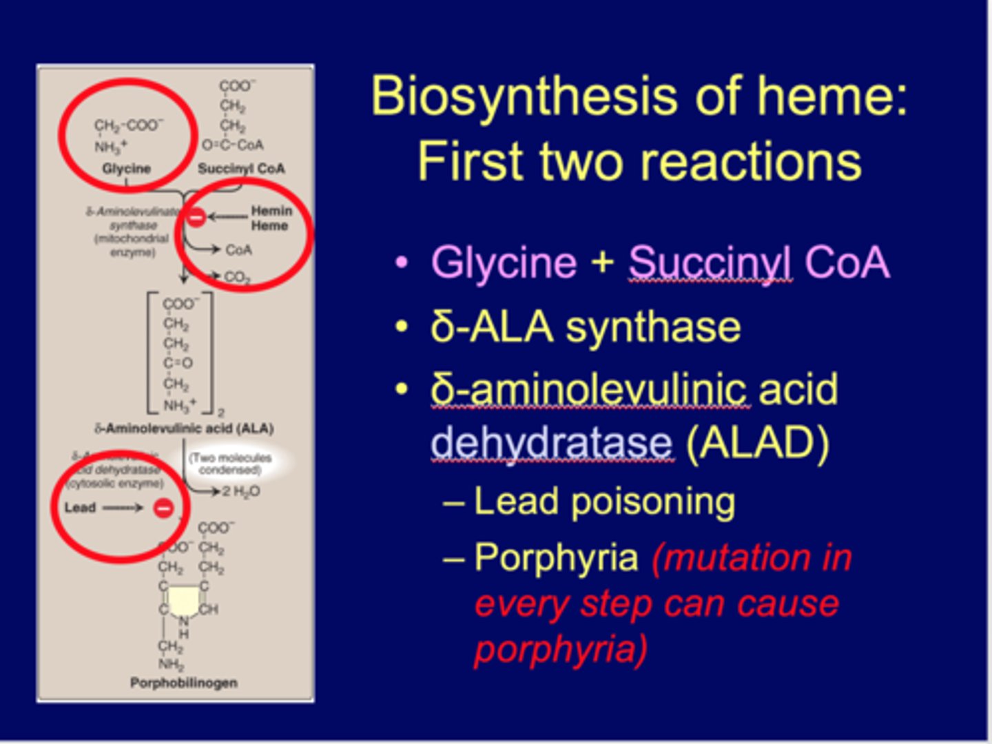
The 2nd step of heme biosynthesis
forms prophobilinogen by enzyme δ-aminolevulinic acid dehydratase (ALAD)
-inhibited by lead (lead also inhibits last step)
-leads to lead poisoning or porphyria
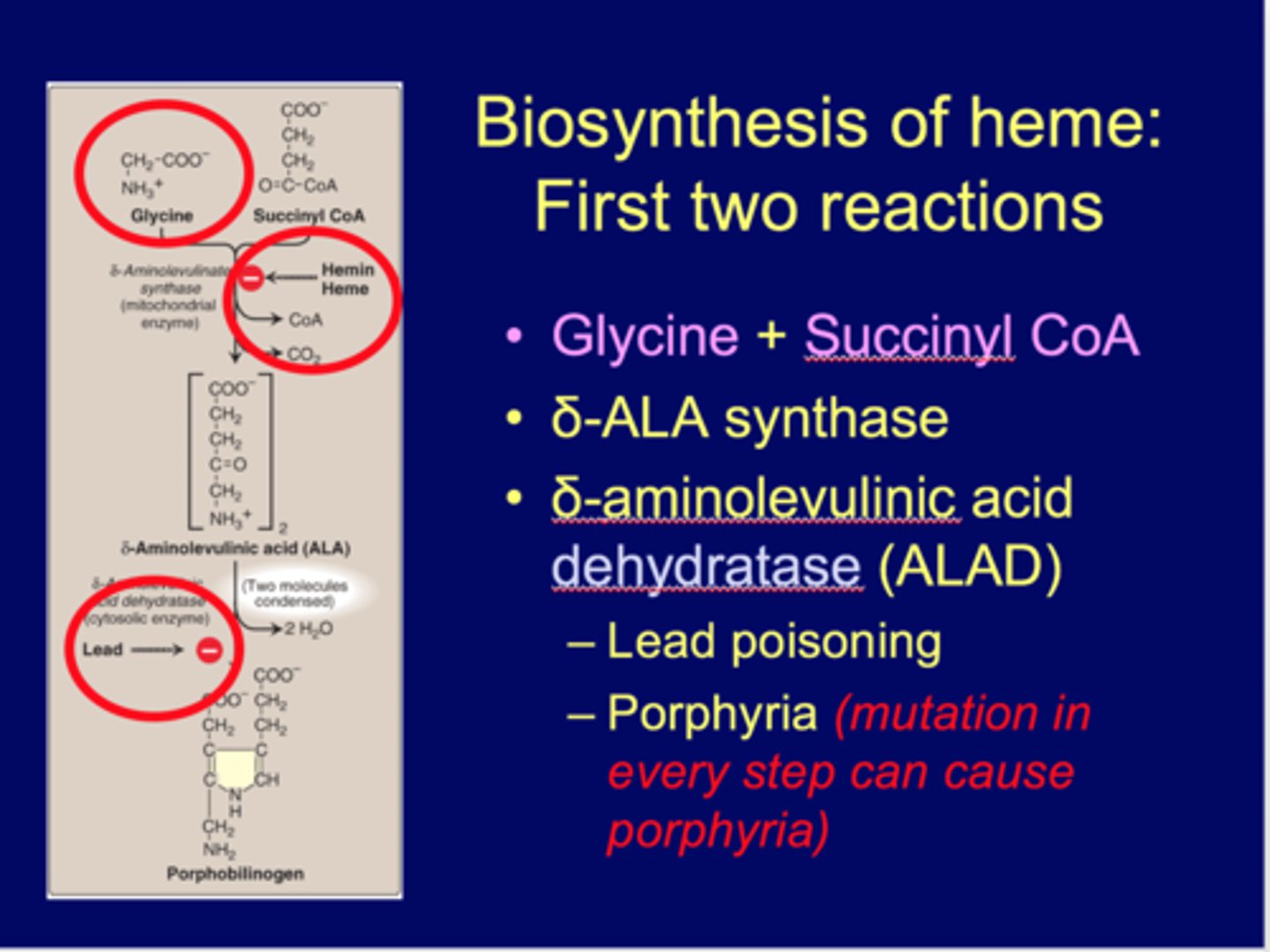
Lead poisoning (Pb replaces Fe) inhibits what steps/enzymes of heme biosynthesis?
1. δ-aminolevulinic acid dehydratase (ALAD) (2nd step)
2. Ferrochelatase (last step), inhibits moving of iron into the ring of heme
-porphyria cutanea tarda-chronic and most common
- causes brownish urine
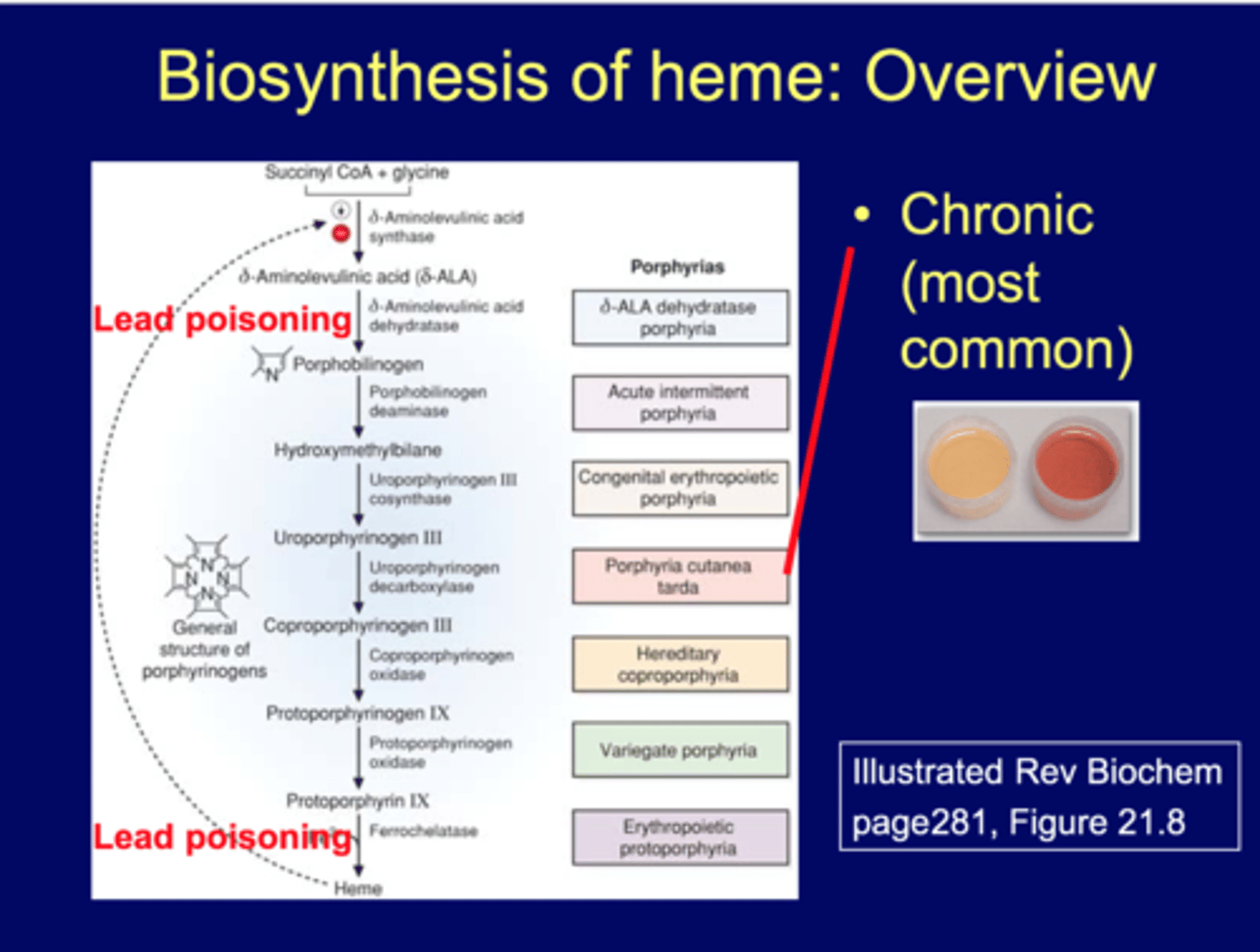
Porphyrias
- deficiency: any enzyme in heme biosynthesis pathway
-accumulation& increase excretion of porphorins or porphorin precursors
Clinical manifestations:
Sensitive to sun light (photosensitivity)
Skin itches/burns
sunlight produces ROS, start to burn->oxidative stress
Chronic hepatic porphyria: *MOST COMMON
Acute hepatic porphyria: liver type
Erythropoietic porphyria: RBC type
Degradation of Heme
In Macrophages:
Heme ---- Biliverdin ----Bilirubin (B) (unconjugated, UC)
In Liver:
Bilrubin -----Bilirubin diglucuronide (BG; direct bilirubin/conjugated)
In Intestine:
BG--- Bile---- Urobilinogen/Sterocobillin (by intestinal bacteria)
B= "inactive bilrubin" (billirubin = uncojugated billirubin )
BG = direct bilirubin (cojugated billirubin)
*more water- soluable, bilirubin diglucuronide by adding 2 glucoric acid molecules, catalyze by UDP-glucuronosyltransferace
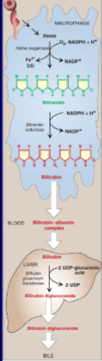
Hyperbilirubemia (high bilirubin in blood) is also called ____.
juandice
UDP glucuronosyltransferase deficiencies
UGT1A1 gene
Crigler-Najjar syndrome Type 1: No enzyme function
Crigler-Najjar syndrome Type 2: 20% of the function
-High levels of unconjugated bilirubin in the blood (unconjugated hyperbilirubinemia) and yellowing of the skin and eyes (jaundice).
Gilbert syndrome: 30% of the function
-Mild unconjugated hyperbilirubinemia, which rarely leads to episodes of jaundice
Crigler-Najjar syndrome Type 1
No enzyme function
Crigler-Najjar syndrome Type 2
20% of the function
-High levels of unconjugated bilirubin in the blood (unconjugated hyperbilirubinemia) and yellowing of the skin and eyes (jaundice).
Gilbert syndrome
30% of the function
-Mild unconjugated hyperbilirubinemia, which rarely leads to episodes of jaundice
UDP glucuronosyltransferase deficiencies also called
bilirubin-UGT, Bilirubin UDP glucuronosyltransferase, bilirubin uridine diphosphate glucuronosyltransferase.
***Reaction: In liver, more water- soluable, bilirubin diglucuronide by adding 2 glucoric acid molecules, catalyze by
UDP-glucuronosyltransferace
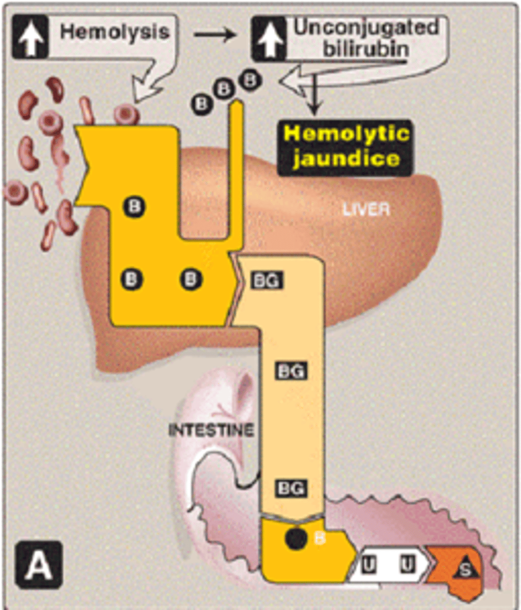
Hemolytic Jaundice
B (uncojugated billirubin) high, BG higher but liver may not catch up with conjugation->unconjugated hyperbilirubinemia
lab test? HIGH TOTAL BILIRUBIN
- result from extensive hemolysis (sickle cell anemia, pyruvate kinase or G-6-P deficiency) (hemoglobin overload)
- bilirubin is produced faster then it can be conjugate
(B = bilirubin/ UCB) (CB/BG= conjugated bilirubin) (U= urobilinogen) (S= stercobilin; UDP = uridine diphosphate)
Hepatocellular Jaundice
Liver damage/disease
-BG (conjugated bilirubin) higher, leaked from liver, conjugated hyperbilirubinemia
lab test? HIGH DIRECT BILIRUBIN
HIGH ALA & AST
Enzyme deficiency
-B high, BG low, unconjugated hyperbilirubinemia
lab test? HIGH TOTAL BILIRUBIN
AST/ALT normal unless liver injury involved
(B = bilirubin/ UCB) (CB/BG= conjugated bilirubin) (U= urobilinogen) (S= stercobilin; UDP = uridine diphosphate)
Obstructive Jaundice
-B and BG high in blood, conjugated and unconjugated hyperbilirubinemia
lab test? HIGH DIRECT BILIRUBIN and TOTAL BILIRUBIN
AST/ALT may be high, not always (if liver problem will be high)
- results from obstruction in common bile duct (by cancer, gallbladder stone etc)
-pain in abdomen, pale/clay colored stool, BG high in urine
S: hyperbilirubinemia (liver regurgitates CB into bb), "urinary bilirubin" (CB eventually excrete into urine)
(B = bilirubin/ UCB) (CB/BG= conjugated bilirubin) (U= urobilinogen) (S= stercobilin; UDP = uridine diphosphate)
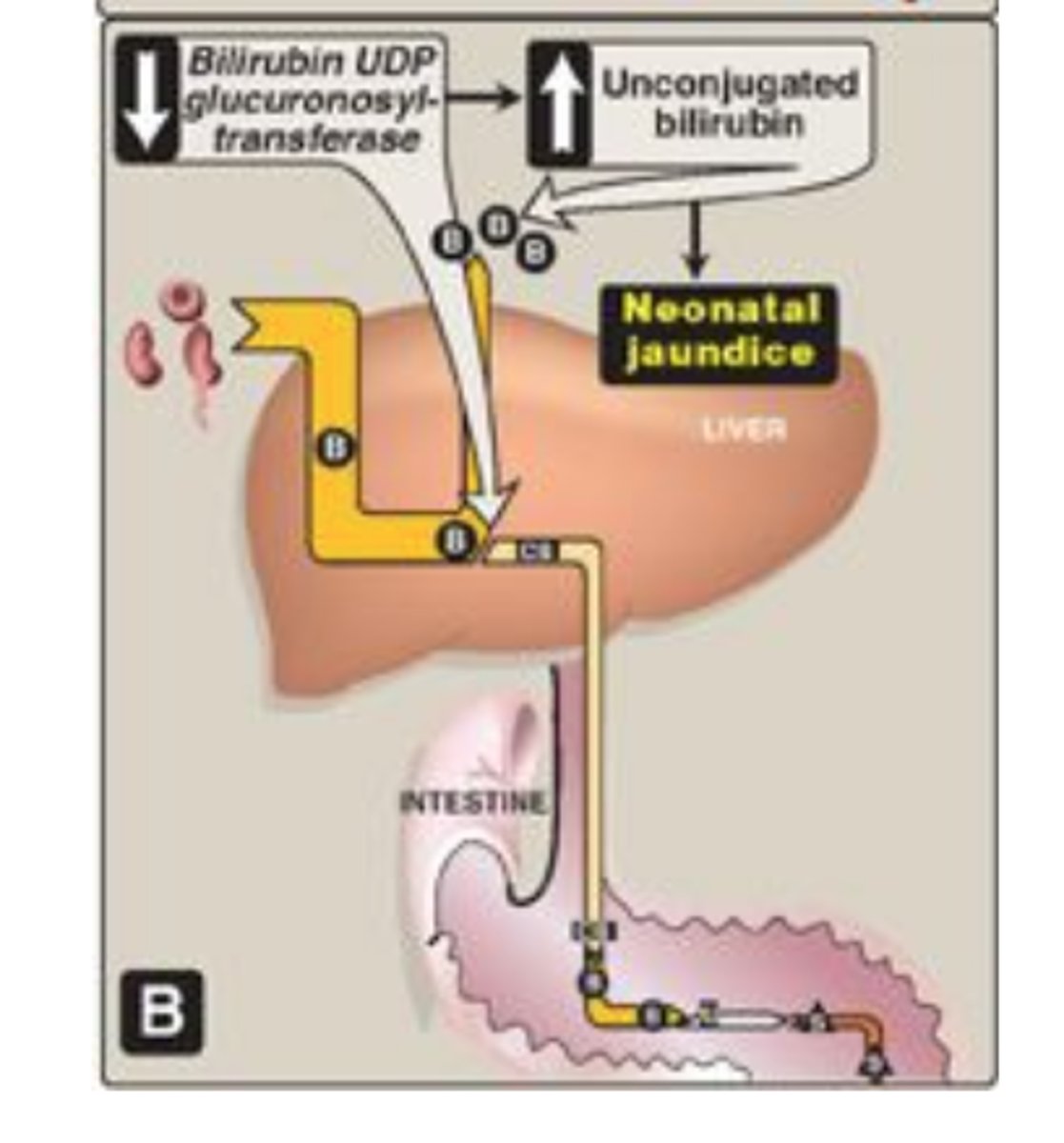
Neonatal Jaundice
Similar to enzyme deficiency
-B (unconjugated bilirubin)
-UGT enzyme not made in first postnatal week
-unconjugated hyperbilirubinemia
(B = bilirubin/ UCB) (CB/BG= conjugated bilirubin) (U= urobilinogen) (S= stercobilin; UDP = uridine diphosphate)
Lab test for bilirubin
Total and direct bilirubin are directly measured
The van den Bergh reaction:
-Colorimetric estimation of serum bilirubin by coupling with diazotized sulphanilic acid.
1. Direct bilirubin = Bilirubin diglucuronide
(water-soluble bilirubin glucuronides/conjugated form) react rapidly so that is called "direct".
2. Total bilirubin
is measured after solubilized with methanol. (unconjugated bilirubin)
What does this mean?
ALT 20 (normal range, 7-30 IU)
AST 25 (normal range, 9-32 IU)
Bilirubin, total: 10 (normal range 0-1.0 mg/dL)
Bilirubin, direct:.5 (normal range 0-.04 mg/dL)
Hemolytic Jaundice
Alcohol dehydrogenase (ADH)
enzyme for alcohol intoxication and metabolism
(ADH1A, 1B, 1C, 4-7)
-found in liver and stomach
-first step of alcohol metabolism
1B*2->half east asians
1b*3->15-25% of african americans
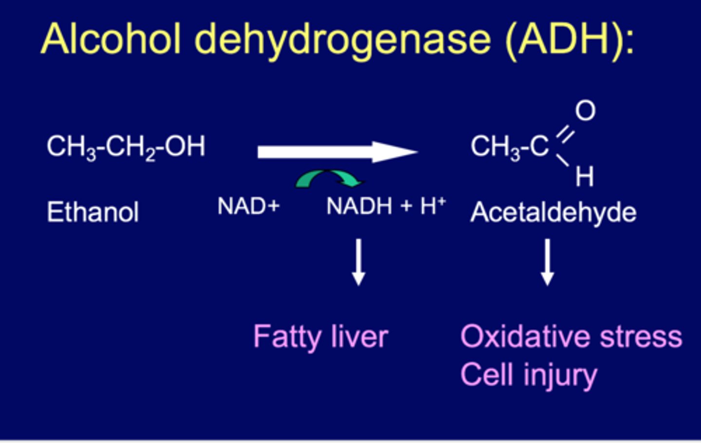
ADH Reaction
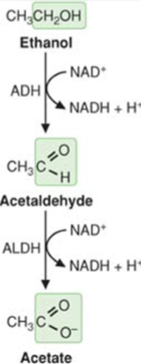
ethanol + NAD+ <---->acetaldehyde + NADH
NADH->fatty liver
acetaldehyde->oxidative stress (ROS), cell injury
Which of the following would be expected to occur after acute alcohol ingestion?
An increase in the NAD+/NADH ratio
A decrease in the NAD+/NADH ratio
decrease NAD+/ NADH ratio
*acute alcohol ingestion results in increase in cytoplasmic and mitochondrial NADH
Ethanol metabolism
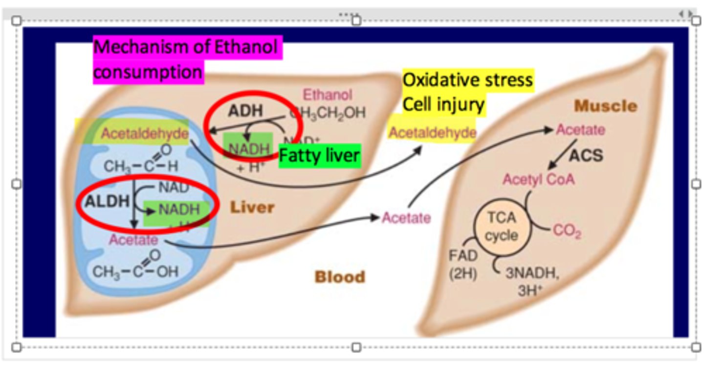
ethanol --> acetaldehyde --> acetate (liver)
acetate leaks out to muscle->makes Acetyl CoA using ACS
acetaldehyde can also leak out into blood (toxic)
ethanol-->acetaldehyde uses ADH
acetaldehyde-->acetate uses ALDH
Ethanol metabolism 3 steps
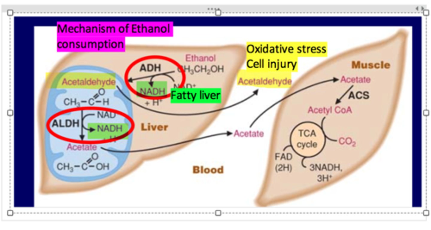
1. In cytoplasm, Ethanol oxidized to produce NADH and Acetaldehyde by alcohol dehydrogenase (ADH)
2. In mitochondria, Acetaldehyde oxidize to produce Acetate and NADH by acetaldehyde dehydrogenase (ALDH)
3. Fate of acetate mediated by ACS (acetyl-CoA synthetase)
Ethanol can also be oxidized by
1. CYP2E1 in microsomes (MEOS: microsomal enzyme oxidation system)
2. Catalase in peroxisomes
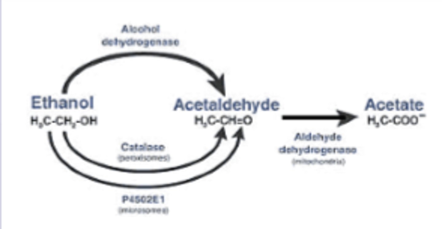
Acute effects of ethanol ingestion are due to the elevated NADH/NAD+ ratio, which leads to
- hyperlipidemia (high FA synthesis, inhibition of fatty acid oxidation->fatty liver)
-hypoglycemia (impaired GNG, pyruvate convert to alternate rxn pathways)
- lactic acidosis (high lactate)
- Ketogenesis (break down of FA & ketogenic AA)
- Hyperuricemia (high uric acid)
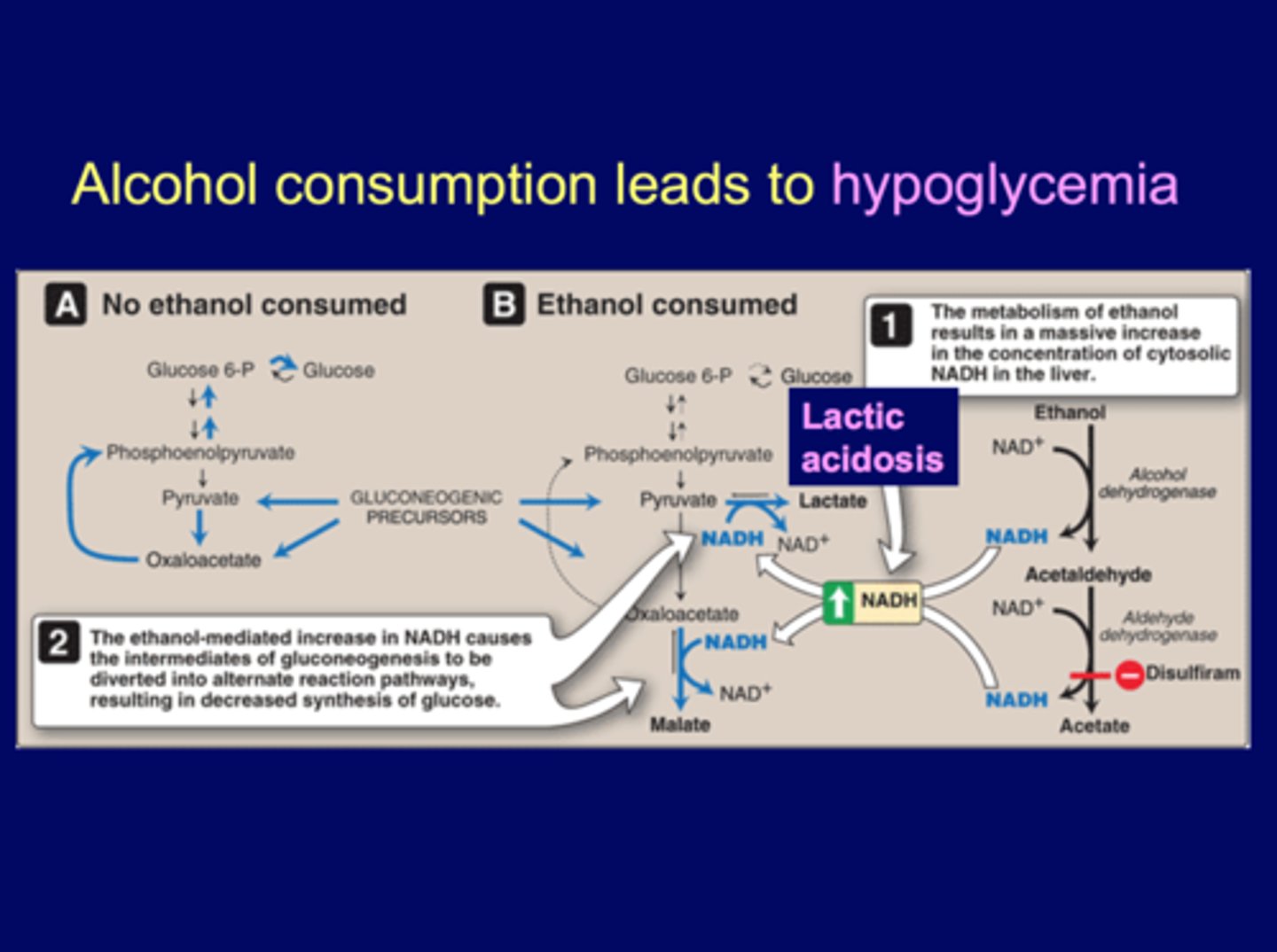
Alcohol consumption leads to hypoglycemia (mechanism)
The ethanol-mediated increase in NADH causes intermediates of gluconeogenesis to be diverted to alternate rxn pathways:
pyruvate to OAA-->malate (driven by NADH)
pyruvate to lactate (lactic acidosis)
decreases precursors for glucose
-exhaust pyruvate and OAA (increase lactate and malate)
-HIGH levels of cytosolic NADH → Impaired gluconeogenesis (pyruvate convert to lactate, OAA convert to malate) →
Hypoglycemia (decrease glucose)
Which of the following would be expected to occur after acute alcohol ingestion?
The activation of fatty acid oxidation
Lactic acidosis
Deprivation of malate
lactic acidosis
DNA and RNA
DNA and RNA Purines
-Adenine (A)
-Guanine (G)
-can't open/recycle the rings, final metab->uric acid
DNA and RNA Pyrimidines
-Thymine (T) (DNA)
-Cytosine (C)
-Uracil (U) (RNA)
-can recycle pyrimidines
Nucleotide vs. Nuecleoside
nucleotide= base + pentose sugar + P
nucleoside = base +pentose sugar
Phosphate provides energy
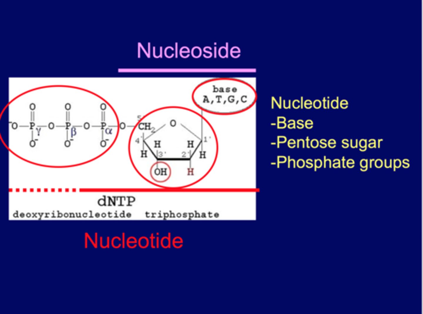
Ribose (RNA) vs 2-Deoxyribose (DNA)
ribose = OH @C2
deoxyribose = H @C2 (deoxy=no oxygen)
OH unstable-RNA harder to deal with
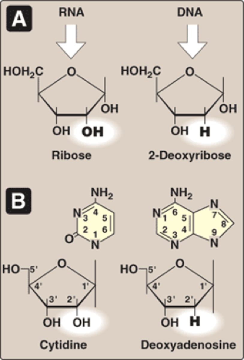
Purines and pyrimidines
-dietary uptake low
-synthesized by de novo or salvage pathways
-made from simple molecules (de novo)
-salvaged from existing bases (salvage pathways)
De novo purine synthesis
made from:
- Amino acids (Glutamine, Glycine, and Aspartate)
- Folic acids (N10-formyltetahydrofolate) ....contributes 1 C
- CO2
*purine ring is costructed in LIVER, in series of rxns that donate Carbon and Nitrogen to ribose 5- phosphate (from pentsoe phosphate pathway)
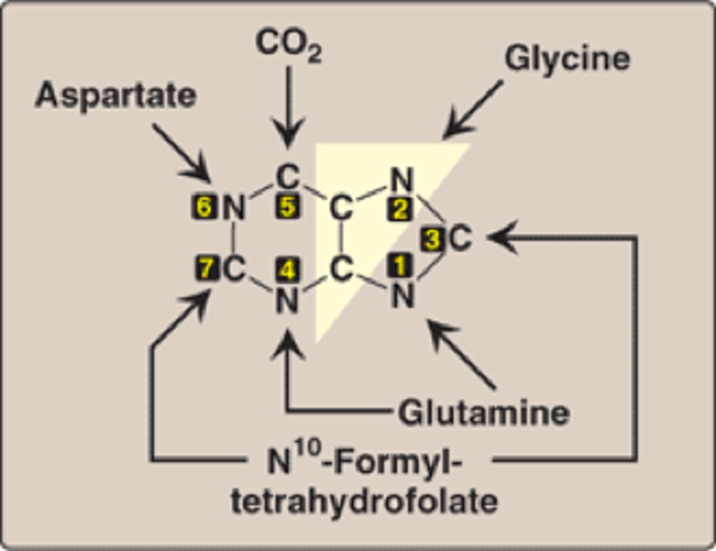
De novo pathway
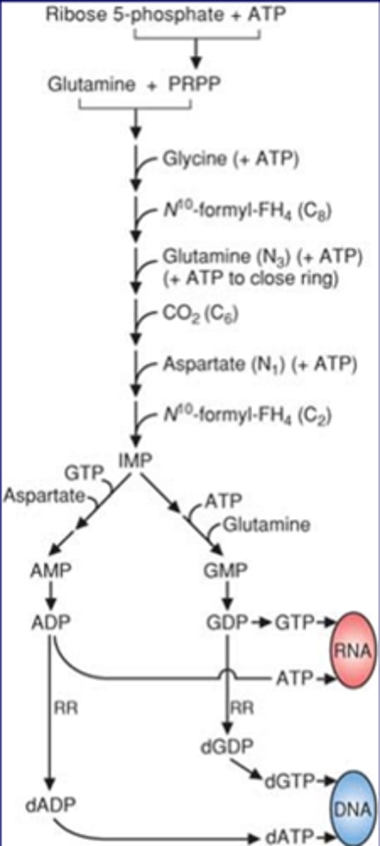
produces 6 ATP (5 for dATP)
comes from ribose 5 phosphate, from pentose phosphate shunt
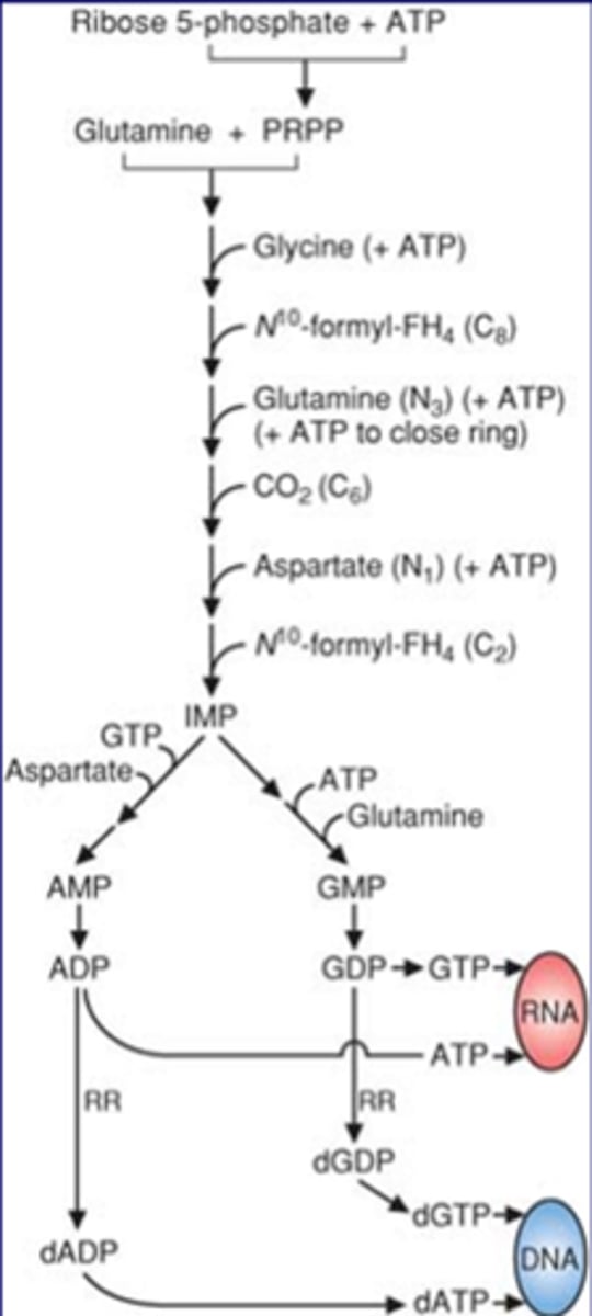
-glutamine + PRPP ---->IMP
-folic acid (N10 formyl FH4) important in 2 places
-if folic acid deficient->deficient in G and A nucleotide
-A can be supplemented
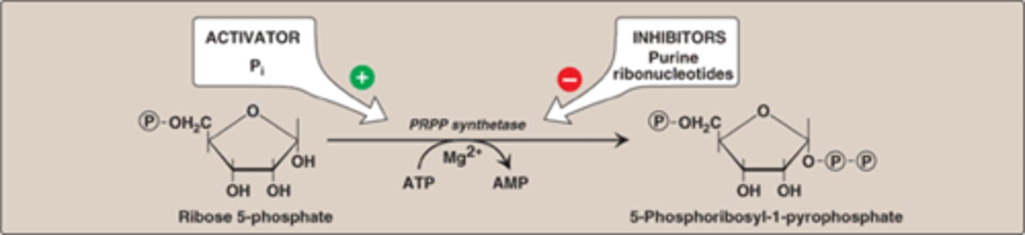
First reaction of de novo
R5P to PRPP (first step of de novo synthesis)
RRPP synthase used
inhibit: purine ribonucleotides
activator: Pi (phosphate)
The 2nd reaction of purine de novo synthesis
PRPP to 5-phosphoriboylamine by ...aminotransferase
ACTIVATOR: PRPP
INHIBITOR: AMP, GMP, IMP
N10 formyl FH4 at 2 places
2 drug groups
-PABA analog
-Folic acid analog
PABA
-sulfoamides->antibiotics, inhibit folic acid biosynthesis in bacteria, does not effect humans.
Folic Acid
-used on folic acid, inhibits reaction to block N10 formyl FH4 in 2 places, blocks formyltransferase at both locations, blocks A and G biosynthesis
IMP (Inosine) precursor for A and G
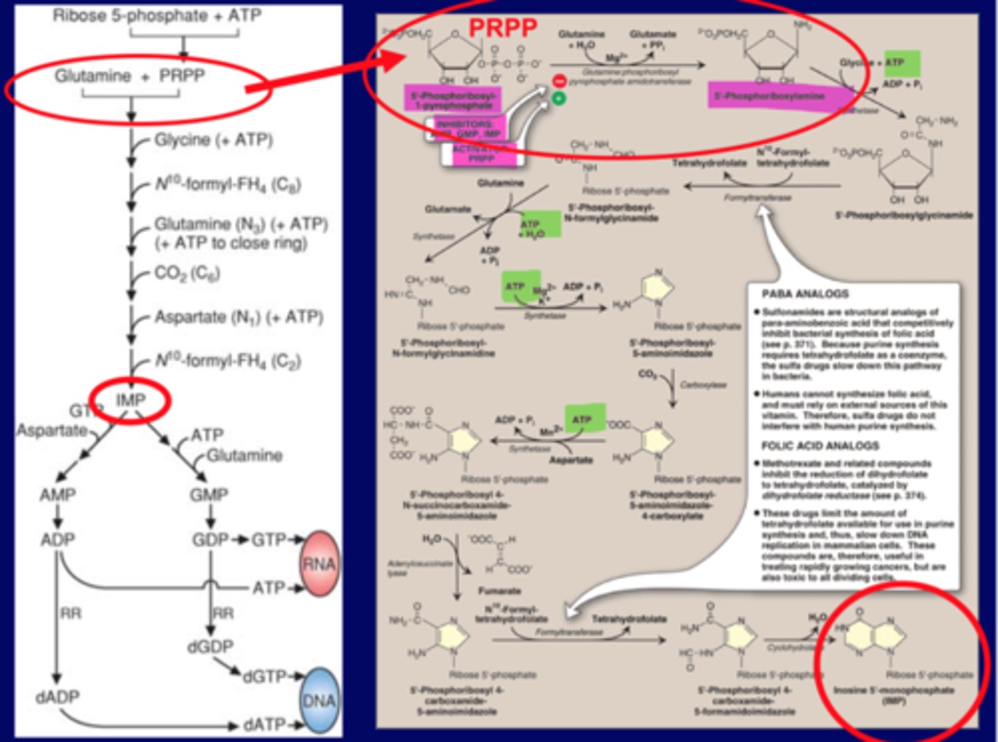
Purine synthesis inhibitors (PABA)
antibiotics
No effect on humans
PABA analogs inhibit bacterial synthesis of folic acid
PABA: para aminobenzoic acid
sulfonamides
inhibits sulfonamides
inhibits N10 formyl FH4 in two placesN10 formyl FH4 in two places
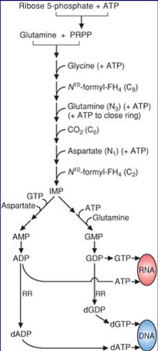
Purine synthesis inhibitors (folic acid analog)
Folic acid analogs
-Methotrexate
-inhibits two transferase reactions
-stops growing cells
-useful in treating cancers, rheumatoid arthritis
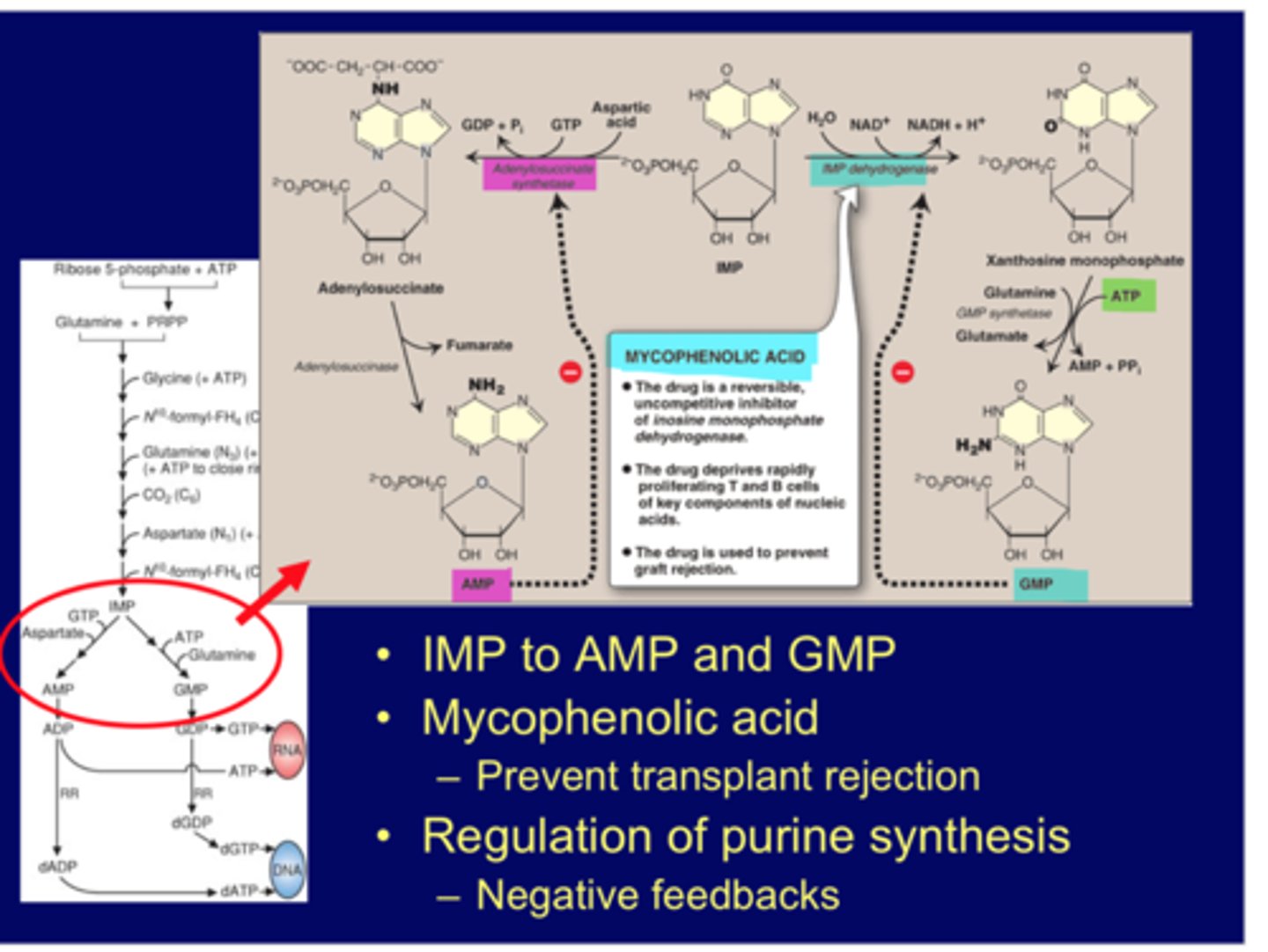
IMP reaction
IMP to AMP and GMP
-uses adenylosuccinate synthase and IMP dehydrogenase respectively
-inhibited by AMP and GMP respectively (neg. feedback)
drug mycophenolic acid also inhibits (reversible, non-comp)
-inhibits guanine pathway only and immune system, used to prevent transplant rejection
-when inhibit nucleotide pathway, any proliferation cell is impacted (stem cells, bone marrow, immune cells-T and B)
-if give inhibitor you suppress the immune system and other recycling system (skin, hair, GI side effects)
-main place is to stop cancer
De novo pyrimidine synthesis
Glutamine + CO2 + 2 ATP ---> carbamoyl phosphate
Inhib: CP2, UTP
stim: PRPP
Made from
-Amino acids (Aspartate, Glutamine)
-CO2
FH4/folic acid deficiency-impact T nucleo
if deficient will increase orotic acid if enzyme is inhibited
carbamoyl phosphate comes from urea cycle, increased by UCD, if happens will leak out and also contribute to orotic acid
CPS1-urea cycle deficiency
CPS2-pyrimidine biosynthesis
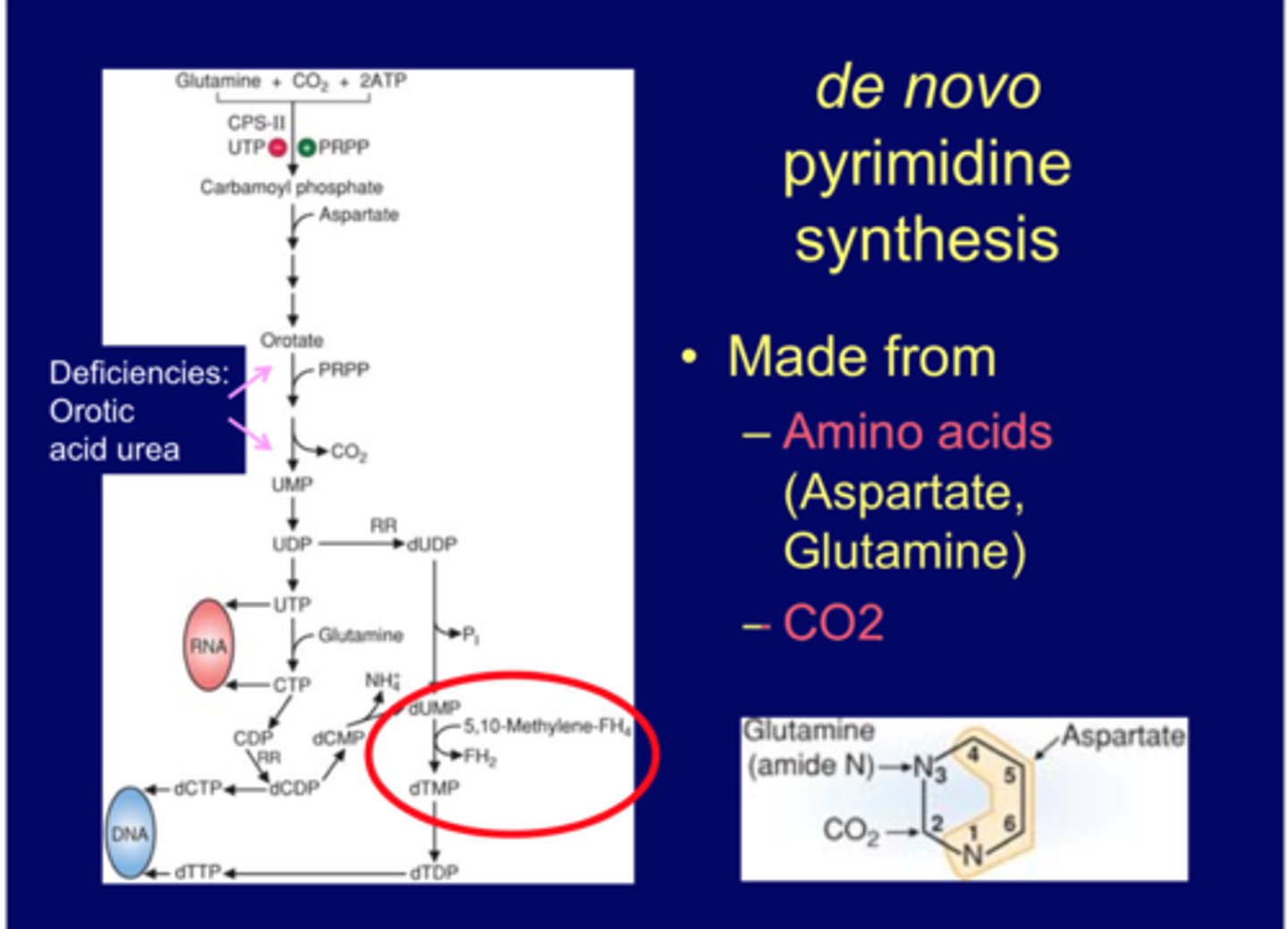
De novo pyrimidine synthesis (OAU)
Orotic acid urea caused by:
-UMP synthase def->OA high, ammonia normal
-UMP-synthase inhibitors->Gout treatment (Allopurinol) or chemotherapy (6-azauridine)
-OTC def (UCD)->OA high, ammonia high, CP high vs
-CP1 def->OA low, ammonia high, CP low
UMP inhib most common, know the 2 drug treatments above
OTC 2nd leading cause->look at ammonia, if high UCD problem, if OA and CP low then its CPS1 issue not OTC
UMP synth def 3rd cause (not ammonia related, enzyme issue)
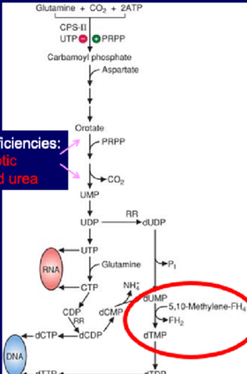
Anti-cancer drug: 5-FU (FU cancer)
-block thymidylate synthetase
-dUMP--->dTMP reaction is coupled with folic acid cycle (5, 10 methylene FH4)
-enzyme makes T nucleotide
-uses UMP
5-FU inhibits T synthesis, inhibits cell proliferation
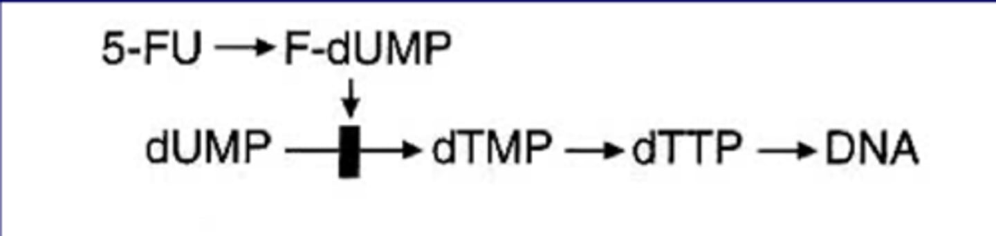
Salvage of bases (purines)
Salvage pathway for Purines
Purines that result from the normal turnover of nucleic acids or small amount obtained from diet can be converted to nucleoside triphosphates and used by the body.
*important for brain
focus on IMP, can make AA (makes AMP or GMP)
Hypoxanthine-make IMP, circle reaction, uses HGRPT (transferase type), if problem causes Lesch Nyhan (same issue for Guanine-->GMP also uses HGPRT)
Adenosine->Inosine using ADA, if def causes SCID
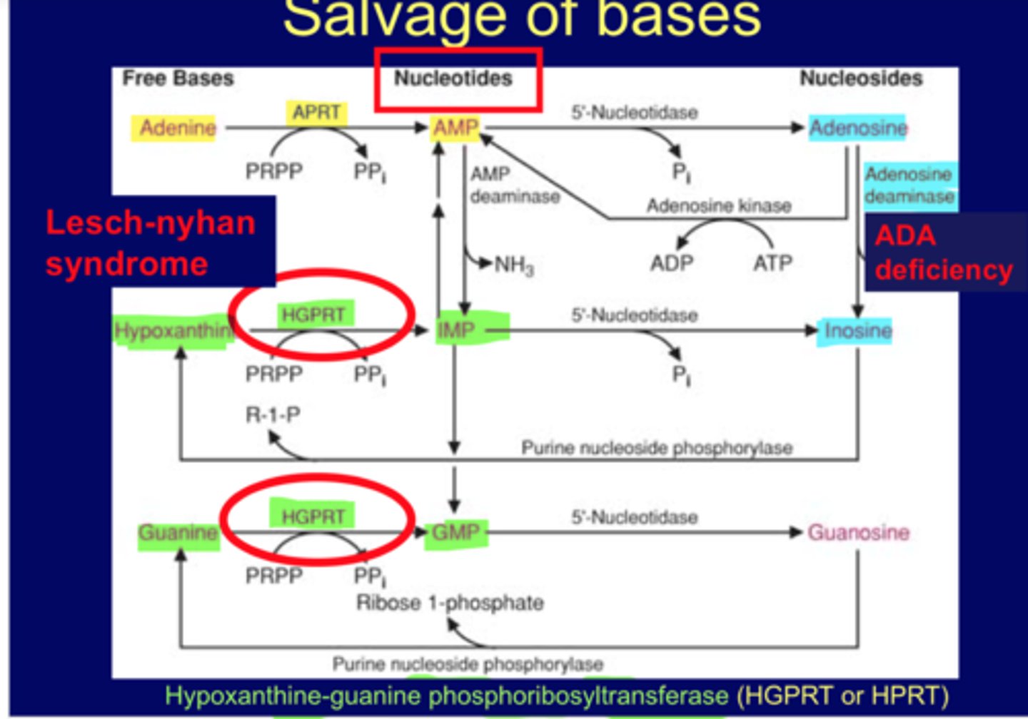
Lesch-Nyhan syndrome
X linked recessive
defect in salvage pathways (enzyme def) (cause)
increased de novo purine synthesis (result not cause)
nucleotide go down, compensate from increase in de novo synthesis
excessive uric acid and neurologic def:
-self mutilation, involuntary movements
HGPRT issue
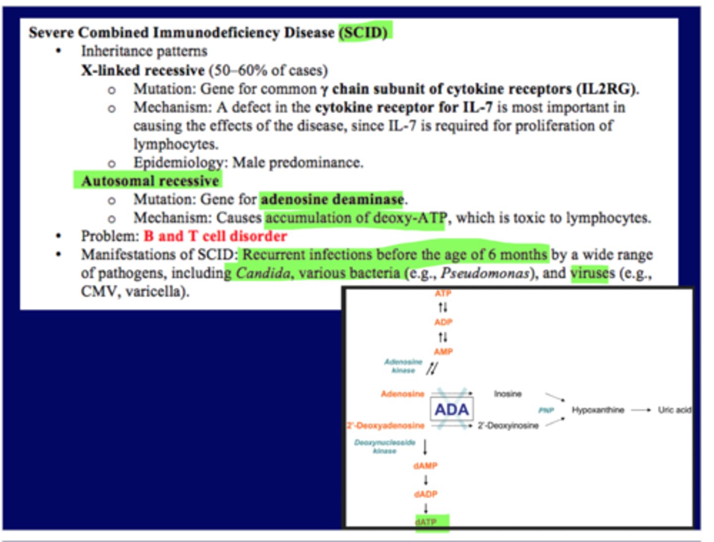
Severe combined immunodeficiency disease (SCID)
X linked recessive (major cause, 50-60%) leading death in babies
-immune system problem, mutation in y chain subunit of -cytokine receptors (IL2RG)
-defect in cytokine receptor for IL7, can't bind and activate immune reaction
Autosomal recessive (2nd type)
-adenosine deaminase (ADA) problem
-deoxynucleotide (dATP) accumulates, toxic to lymphocytes, decreases B and T cells
-ADA used for degradation pathway to make uric acid, since ADA def only goes to the dATP pathway instead
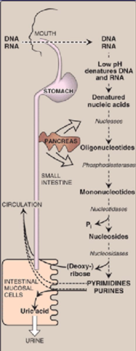
Degradation in the small intestine
-DNA/RNA to oligonucleotides to purine/pyrimidine
-ribonucleases and deoxyribonucleases (pancreas)
-nucleotidase
(low dietary uptake) we most throw away nucleotides
-we cut them into pieces, can cut nucleases more
-purine is thrown away by uric acid, little bit is used
some plant sources with DNA/RNA can make gout worse and contribute to uric acid
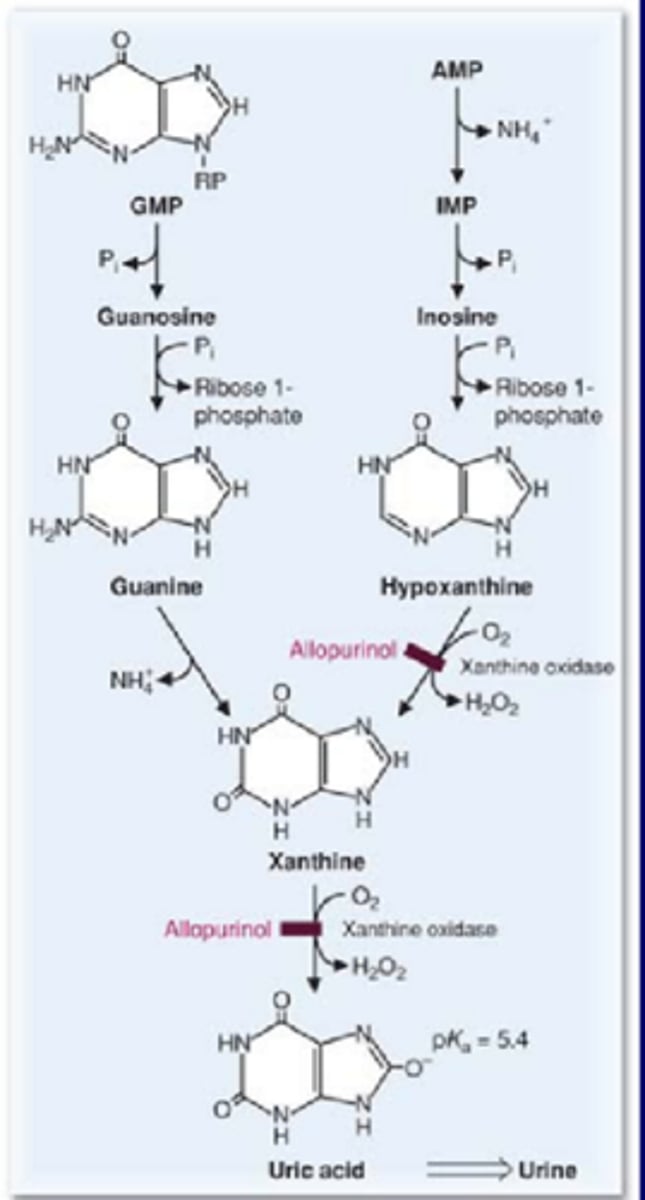
Degradation of purines
-mainly in liver
-AMP/GMP to Xanthine to Uric acid
Allopurinol
-inhibitor of xanthine oxidase
-treat Gout with overproducers of uric acid
can't open purine ring so we partially degrade
two reactions, both use xanthine oxidase->degrades AMP portion->to help form uric acid
Gout
High uric acid: multiple causes (inflammation problem)
-under excretion
-overproduction
acute: use anti inflammatory drugs (first)
long-term: uric acid lowering drugs/diet (after)
Degradation of pyrimidines
pyrimidine ring is opened (purine rings can't be opened)
degraded to highly soluble, b-alanine and b-aminoisobutyrate
Overview
Two different ways to get nucleotides
they can either be made (de novo)
or recycled (salvage pathway)
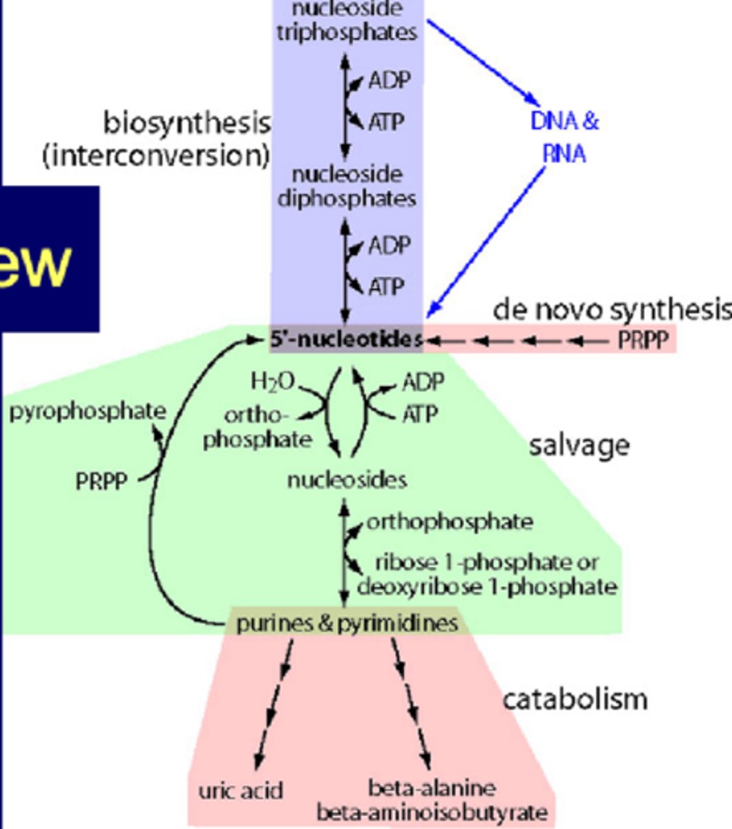
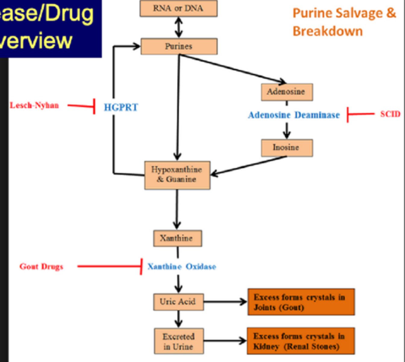
Disease/Drug Overview
Purines->Adenosine->Inosine w/ ADA (SCIDS if inhibited)
Inosine or Purine->Hypoxanthine & Guanine->Purines w/HGRPT (Lesch-Nyhan if inhibited)
Hypoxanthine & Guanine->Xanthine->Uric acid w/Xanthine oxidase (Gout drugs to inhibit)
Uric acid->excess forms crystals in joints (gout)
excreted in urine->excess forms crystals in kidney (rental stones)