Ch. 10 Neurons
 Call Kai
 Learn
Learn Practice Test
 Spaced Repetition
 Match
Match Flashcards
 Knowt Play
1/148

There's no tags or description
Looks like no tags are added yet.
Name | Mastery | Learn | Test | Matching | Spaced | Call with Kai | Chat |
|---|
No analytics yet
Send a link to your students to track their progress
149 Terms

What is the historical perspective of understanding brain function?
•Aristotle believed the heart was the seat of the soul
•Today, the brain is recognized as the source of human
uniqueness

What is the scientific challenge of understanding brain function?
Decoding how neural circuits produce complex behaviors
(e.g., speech, creativity, emotion)

What are the three levels of study in understanding the brain?
Reductionist: Individual neuron responses
•Integrative: Neuron networks and circuits
•Behavioral: Start with behavior, trace back to neural
origins

Describe the complexity of the brain.
~85 billion neurons, each with up to 200,000 synapses
•Synapses are dynamic and constantly changing
•No simple structure-function mapping
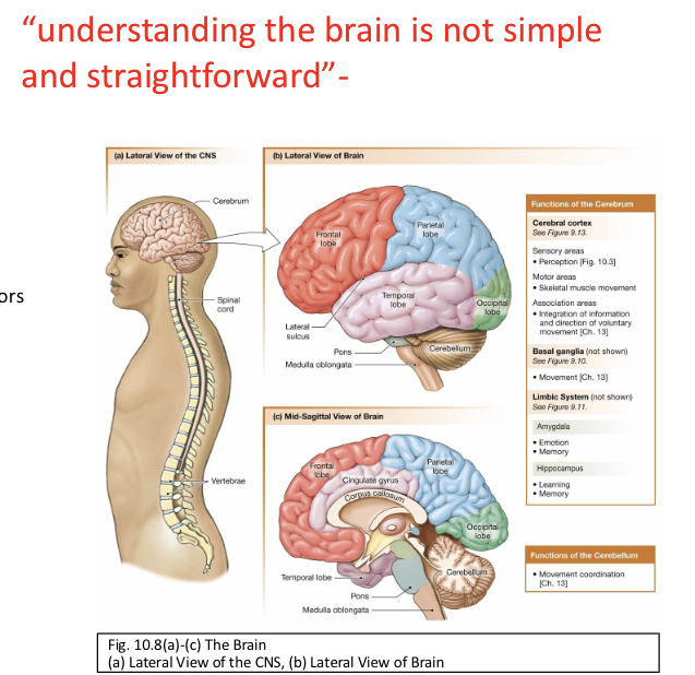
What is the key principle in understanding brain function?
•One function involves multiple brain regions
•One region may support multiple functions
What is plasticity?
the restructuring of the brain networks in
response to sensory input & experience.
What are affective behaviors related to?
Feeling & emotion
What are cognitive behaviors related to?
Thinking
What is the emergent properties of neural networks evolutionary trend?
Increasing complexity from fish to humans
What is the developmental pattern of the CNS?
All vertebrates share a basic CNS structure
• CNS = layers of neural tissue
• Surrounds a fluid-filled central cavity lined w/ epithelium

What is the gray matter of CNS?
• Unmyelinated nerve cell bodies
• Clusters of cell bodies in the CNS
are nuclei
• Dendrites
• Axon terminals
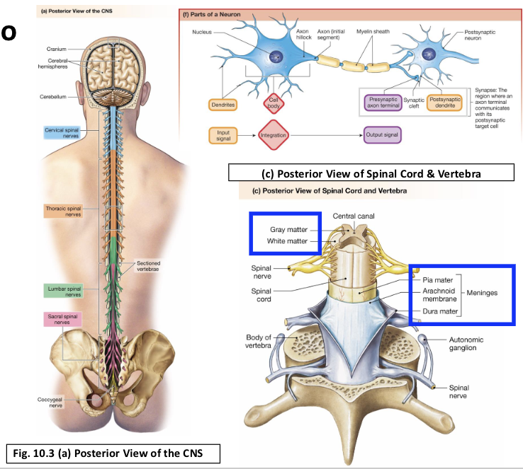
What is the white matter of CNS?
• Myelinated axons
• Axon bundles connecting CNS
regions are tracts
• Contain very few cell bodies

Describe the brain and spinal cord “consistency”.
• soft and jellylike
• individual neurons and glial cells have
highly organized internal cytoskeletons
(maintain cell shape and orientation)
• neural tissue has minimal extracellular
matrix and must rely on external
support for protection from trauma
• support comes from bone, three layers
of connective tissue membrane, and
fluid between the membranes

Bone & Connective Tissue Support the CNS. How?
• Brain is encased in bony skull,
or cranium
• Spinal cord runs through
vertebral column
Meninges lie between bone &
tissues to stabilize neural tissue
and protect from bruising
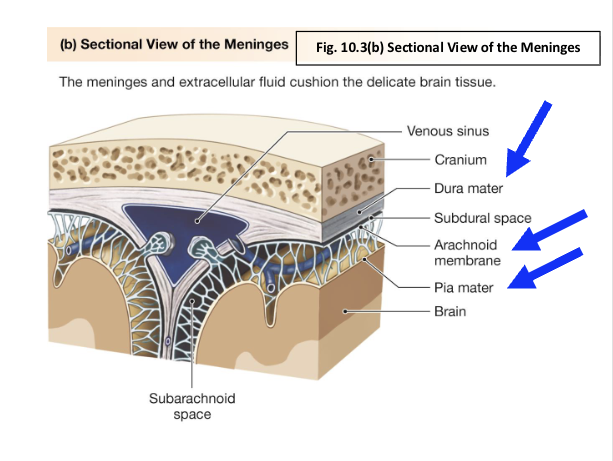
What are meninges? What are the three examples?
Meninges lie between bone &
tissues to stabilize neural tissue
and protect from bruising
• Dura mater
• Arachnoid membrane
• Pia mater

Describe the ventricles of the brain.
• Hollow cavities deep w/in the brain, extensions of the brain’s central cavity
• filled w/ cerebrospinal fluid (CSF) & lined w/ ependymal cells (glial cells)
• continuous w/ each other as well as the central canal of the spinal cord

The Brain Floats in CSF (2 of 3). Describe.
• Salty solution
• Produced by the choroid plexus
in the ventricles
• Materials selectively moved from
plasma to ventricles
• Water follows due to osmotic gradient
• Surrounds entire brain
• Contained w/in subarachnoid space
(between arachnoid membrane & pia
mater)
• Flows from ventricles to subarachnoid
space to return to plasma by villi
• Function in physical & chemical
protection

What produces CSF in the brain?
The choroid plexus

What is the role of villi in the CSF?
CSF is reabsorbed into the blood at fingerlike projections of the arachnoid membrane called villi.
Flows from ventricles to subarachnoid
space to return to plasma by villi
How does CSF help with physical protection?
• Brain and spinal cord float in CSF, reducing effective brain weight.
• Less weight = reduced pressure on blood vessels and nerves.
• Acts as a cushion during impacts—water’s low compressibility helps absorb
shock.
Describe the chemical protection of CSF.
• CSF maintains a stable extracellular environment for neurons.
• Produced by the choroid plexus, which selectively filters substances
• CSF composition differs from plasma
• Lower protein and blood cell content
• Specific ion concentrations regulated

What is the clinical significance of CSF? What spinal procedure is involved with CSF?
Reflects CNS health and chemical
environment
• Normally contains very little
protein and few/no blood cells
• Abnormalities can indicate disease
or injury
• Collected by lumbar puncture
(spinal tap)

A blood brain barrier protects the brain, how? What passes through it?
Highly selective permeability of
brain capillaries
• Not a physical wall, but
a functional barrier
• Protects brain from toxic water
soluble compounds & pathogens
• Small lipid-soluble molecules
cross the blood-brain barrier
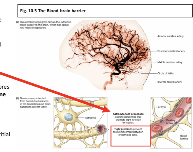
Describe the blood-brain barrier.
Final Layer of Brain Protection
• Isolates brain from harmful substances &
pathogens in the blood
• Maintains a stable internal environment for the
CNS

Describe the structure of the blood-brain barrier. (BBB) What cells are involved? Junctions?
• Formed by tight junctions between endothelial
cells in brain capillaries
• Induced by signals
from pericytes and astrocytes.

Describe the selective membrane permeability of the BBB.
Unlike other capillaries, brain capillaries lack pores
• Only specific substances can cross via membrane
transporters.
Transport Mechanisms:
• Nutrients & essential molecules are actively
transported into the brain.
• Waste products are removed from brain interstitial
fluid to the blood.
• Water-soluble molecules not carried by
transporters cannot cross.

What are the functions of the spinal cord?
• site of attachment for spinal nerves
• two-way conduction pathway between body &
brain
• center for reflexes
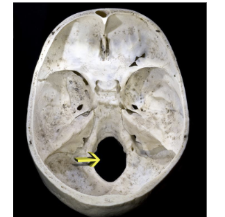
What is the location of the spinal cord? Where does it extend from?
• runs through the vertebral canal
• extends from the foramen magnum to the level
of the vertebra L1 or L2

What are the regions of the spinal cord?
Cervical, thoracic, lumber, and sacral —named after adjacent vertebrae.
Describe the segments of the spinal cord?
Each region has segments that give
rise to bilateral spinal nerves.
What are the two spinal nerve roots?
Dorsal root/ Ventral root
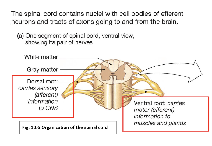
What is the dorsal root?
Carries sensory input to the spinal
cord.
•Contains dorsal root ganglia with sensory
neuron cell bodies

What is the ventral root?
Transmits motor output from
CNS to muscles/glands.

Describe gray matter, dorsal & ventral horns.
Gray Matter: Butterfly/H-shaped core.
• Dorsal Horns: Interneurons for somatic & visceral
sensory info.
• Ventral Horns: Motor neurons for somatic &
autonomic output

Describe white matter in the spinal cords, what are the three tracts it composed of?
surrounds gray matter; composed
of axon tracts.
• Ascending Tracts: Carry sensory info to brain
• Descending Tracts: Carry motor signals from
brain
• Propriospinal Tracts: Stay w/in the spinal cord.

How can the spinal cord function as an integrating center?
• Functions independently for simple spinal
reflexes
• Reflex signals travel:
• From sensory neurons
• Through gray matter
• To efferent (motor) neurons
• Spinal interneurons:
• Route sensory info to the brain via ascending tracts
• Carry motor commands from the brain to motor
neurons
• Modify information during transmission
• Reflexes are essential for coordinating body
movement
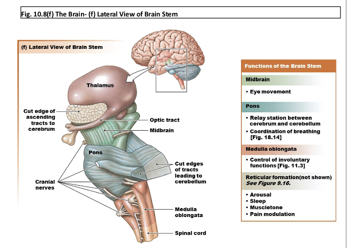
What is the medulla oblongata in the brain?
Controls involuntary
functions: blood pressure,
breathing, swallowing,
vomiting

What are pons in the brain?
Relay station, coordinates
control of breathing (coordination)
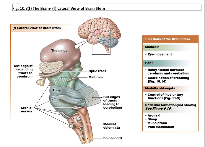
What is midbrain responsible for in the brain?
Eye movement, relay signals
for hearing and seeing
reflexes

What is the reticular formation in the brain? What is it responsible for?
-a diffuse collection of neurons that
extends throughout the brain stem.
-Arousal
-Sleep
-Muscletone
-Pain modulation

The cerebellum coordinates movement, how?
• Second largest structure in
the brain; at base of skull
• “Little brain”
• Process sensory
information and
coordinates the execution
of movement
• Equilibrium and balance
(sensory) from somatic
receptors
• Motor input from cerebrum

The Diencephalon (Group of Structures) Contains the
Centers for Homeostasis What are its three structures?
Thalamus/Hypothalamus/ Endocrine structures
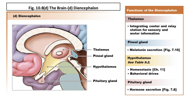
What is the role of the thalamus?
Relay station integrating center (for sensory and motor informatio)

What is the role of the hypothalamus?
• Control of homeostasis
• Center for behavioral drives: Ex: hunger,
thirst,
• Influences autonomic function & endocrine
function

What are the endocrine structures in the diencephalon?
Pituitary gland (anterior & posterior)- hormone secretion
Pineal gland- melatonin secretion

The Cerebrum Is the Site of Higher Brain Functions (1 of 3) Describe. Consists of two hemispheres connected
by_____.
• Largest, most distinctive part of the brain
Corpus callosum

What are the three major regions in the gray matter in the cerebrum?
• Cerebral cortex
• Basal ganglia: control of movement
• Limbic system: link between cognitive
functions & emotions
• Amygdala and cingulate gyrus:
emotion & memory
• Hippocampus: learning & memory
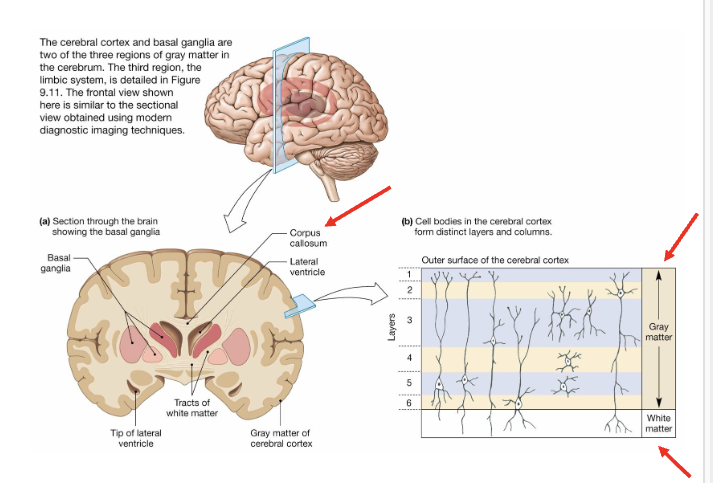
What is the white matter in the cerebrum?
• Found mostly on the interior
• Bundles of fibers connecting the
different regions of the brain
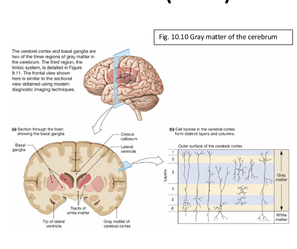
In the gray matter of the cerebrum, what is the cerebral cortex, in depth?
Outer layer of the cerebrum; only a few
millimeters thick
•Organized into vertical columns and
horizontal layers
•Responsible for higher brain functions (e.g.,
reasoning, perception)
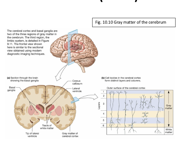
In the gray matter of the cerebrum, what is the basal ganglia (basal nuclei), in depth?
Located deep w/in the cerebrum
•Involved in movement control
•Term “ganglia” used clinically, though
technically refers to structures outside the
CNS

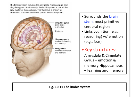
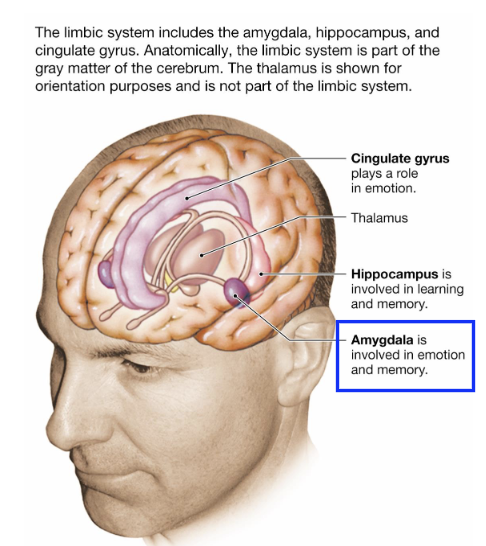
In the gray matter of the cerebrum, what is the limbic system, in depth? Differentiate amygdala & cingulate gyrus from the hippocampus (both involved in memory).
Surrounds the brain stem; most
primitive cerebral region
• Links cognition (e.g., reasoning) w/
emotion (e.g., fear)
• Key structures:
•Amygdala & Cingulate Gyrus –
emotion & memory
•Hippocampus – learning and
memory

Central Nervous System: Simplified Overview—describe.
• CNS functions as an information processor, similar to a computer
• Many processes follow a basic reflex pathway
Sensory Input & Response
• Brain receives sensory information from internal & external
environments
• Integrates and processes incoming signals
• Generates a response when appropriate
Beyond Simple Reflexes
• Brain can generate its own activity w/out external input
• Intrinsic neural activity requires a more complex model than a
simple reflex pathway
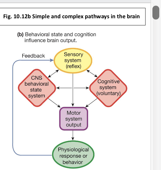
3 Systems Influence Output by
the Motor Systems of the Body: what are they?
1) Sensory system
• Monitors internal & external environments
• Initiates reflex response
2) Cognitive system
• Initiates voluntary responses
3) Behavioral state system
• Governs sleep-wake cycles & other intrinsic
behaviors
Info about the physiological or behavioral
responses created by motor output feeds back to
the sensory system, which in turn communicates w/
the cognitive & behavioral state systems

Reflex Pathways & Homeostatic Control— talk more about it.
• Many physiological systems rely on simple reflex
pathways to maintain homeostasis
• Reflexes are initiated by sensory input & carried
out by motor output

How can the influence of cognitive & behavioral states affect homeostasis?
Voluntary behaviors can override automatic
functions (e.g., breath-holding)
• Emotions can alter normal physiology (e.g.,
stress-induced heart palpitations)
• Circadian rhythms influence physiological
responses (e.g., jet lag, shift-work effects)

Cerebral Cortex is Organized into Functional Areas— what are they?
1) Sensory areas- (Parietal lobe)
• Sensory input translated into
perception (awareness)
2) Motor areas (frontal lobe)
• Direct skeletal muscle
movement
3) Association areas (occipital/ temporal lobe)
• Integrate information from
sensory & motor areas
• Can direct voluntary
behaviors
Information is usually processed in
multiple areas

What are some insights into brain functions from inherited neurological effects or brain injuries?
• Surgical lesions (e.g., for treatment
of severe epilepsy) have revealed
functional roles of specific brain
regions (brainbrow mice)
• Imaging techniques, such as PET
scans, allow noninvasive
observation of the working human
brain
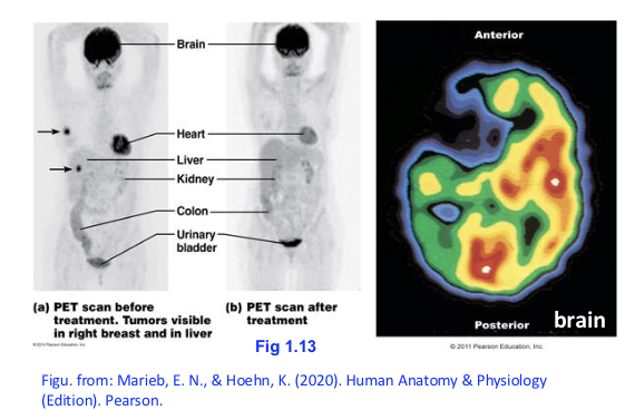
Describe Positron emission tomography (PET).
Radioactive isotopes are injected into
bloodstream (act as a tracer)
• Areas which uptake large quantities
of these isotopes have high cellular
activity
• Can indicate tumor size, location,
response to treatment

The Spinal Cord and Brain Integrate Sensory Information. How?
Sensory System & Reflex Pathways
• The sensory system monitors internal and external environments
• Sends information to neural integrating centers, which initiate
appropriate responses
• The simplest form of this pathway is the classic reflex
Simple Reflexes
• Some reflexes are integrated entirely in the spinal cord, w/out input
from higher brain centers
• Even simple spinal reflexes typically send sensory signals to the brain, creating perception of the stimulus

Describe ascending sensory pathways in the cerebellum and cerebrum.
Sensory information from the body
travels to the brain through
ascending pathways
• signals about muscle & joint
position go to both the
cerebellum & the cerebral cortex
• The cerebellum uses this input for
automatic, subconscious
coordination of movement
• Most sensory information continues
to the cerebral cortex, where five
sensory areas process the incoming
signals
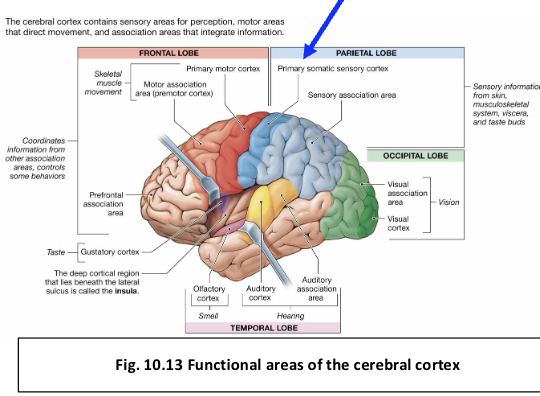
What is the primary somatic sensory cortex?
• Located in the parietal lobe
• Serves as the termination point for sensory pathways
from the skin, musculoskeletal system, & viscera
Somatosensory Pathways
• Sensory fibers cross the midline in the spinal cord
or medulla as they ascend
• this crossing results in opposite-side
representation in the cortex
Processes touch, temperature, pain,
itch, & body position information
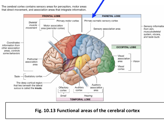
What are the effects of cortical damage in the parietal lobe?
Damage to the somatosensory cortex causes
reduced skin sensitivity on the opposite side of the
body
• Occurs b/c the sensory pathways have already
crossed before reaching the cortex

Special Senses & Their Cortical Processing Areas— describe them.
Different special senses (vision, hearing, taste,
olfaction) have distinct brain regions dedicated to
processing their input

Where is the visual cortex and what is its function?
-The occipital lobe
(receives & processes sensory info from the eyes)

Where is the auditory complex and what is its function?
temporal lobe
receives sensory input from the ear

Where is the olfactory (smell) complex and what is its function?
(small region in the temporal lobe)
• processes signals from chemoreceptors in the
nose

Where is the gustatory (taste) complex and what is its function?
(deep near the edge of the FRONTAL LOBE)
• receives sensory information from the taste buds
Sensory Information Is Processed into Perception. How?
Arrival of sensory information in the appropriate cortical area is only the beginning of processing
• Association areas integrate somatic, visual, auditory, & other sensory inputs
• Integration creates perception — the brain’s interpretation of sensory stimuli
Perception vs. Actual Stimulus
Perception often differs from the actual physical stimulus
• The brain converts light wave frequencies into the perception of color
• Pressure waves reaching the ear are interpreted as sound
• Chemicals binding to chemoreceptors are interpreted as taste or smell
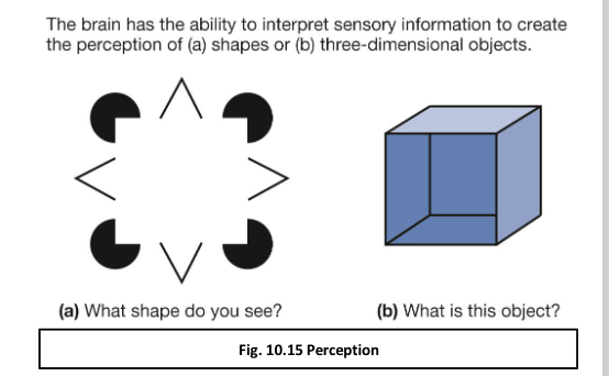
Perception & the Brain’s Interpretive Role— why is that relevant?
• The brain can fill in missing information to create a
complete perceptual picture
• Able to translate 2-D images into 3-D shapes,
based on experience & expectation
• We sometimes perceive what the brain expects
rather than what is actually present
Perception Enables Action
• Perceptual interpretation allows sensory
information to be used effectively
• Supports voluntary motor control
• Essential for complex cognitive functions,
including language

The Motor System Governs Output from the CNS. How? What pathways are involved? Cells?
Originates in the primary motor cortex & motor
association area of the frontal lobes
Inputs to Motor Areas
• Receive information from: sensory areas,
cerebellum, basal ganglia
Descending Motor Pathways
• Pyramidal cells send long axons from motor cortex
→ brain stem → spinal cord
• Additional pathways project from the cortex to the
basal ganglia and lower brain regions
• Motor pathways cross to the opposite side of the
body

What are the effects of motor cortex damage?
b/c pathways cross, damage leads to paralysis or
loss of function on the opposite side of the body
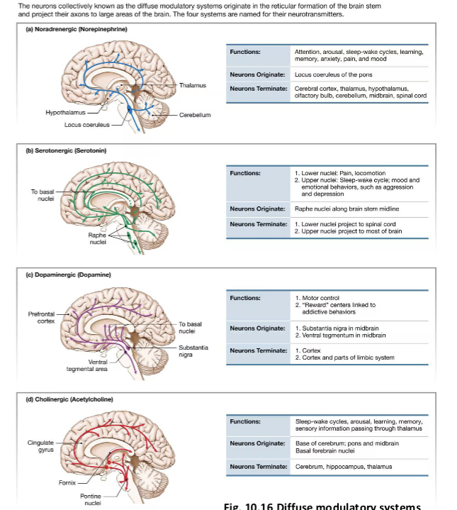
The Behavioral State System Modulates Motor Output— how?
Behavioral State System
• Modulates sensory and cognitive processing
• Involves neurons located outside the cerebral cortex
• includes parts of the reticular formation,
hypothalamus, and limbic system
Diffuse Modulatory Systems
• Neurons originate in the reticular formation of the brain
stem
• Axons project widely throughout the brain
• Four major systems, classified by neurotransmitter:
• Noradrenergic — norepinephrine
• Serotonergic — serotonin
• Dopaminergic — dopamine
• Cholinergic — acetylcholine
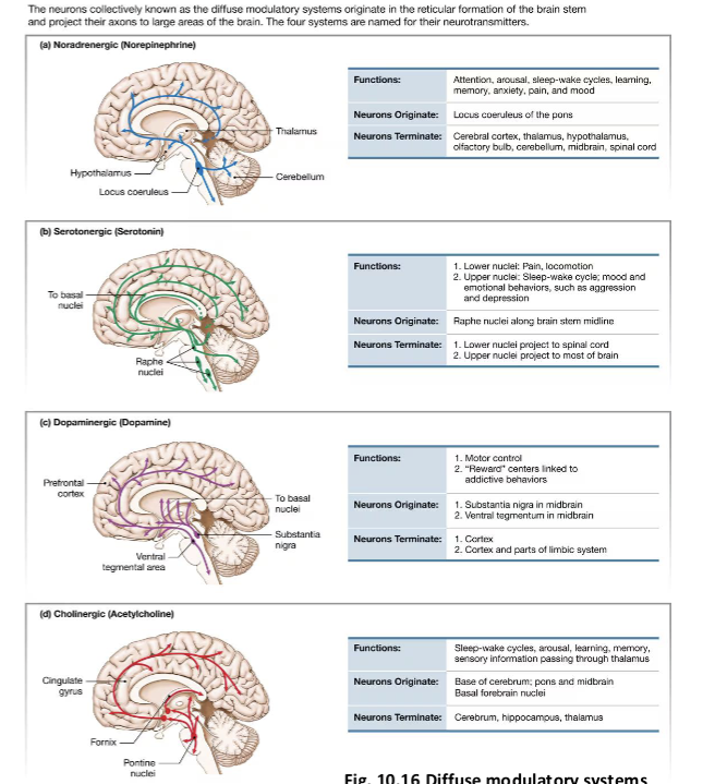
The Behavioral State System Modulates Motor Output— what are the functions of modulatory systems?
Norepinephrine- Influence attention and motivation
Serotonin- Regulate wakefulness and sleep states
Dopamine- Affect memory and motor control
Acetylcholine- Shape mood and contribute to metabolic
homeostasis

What is the dopaminergic system?
• One of the best-studied modulatory
systems due to its role in Parkinson’s
disease
• Dopamine cannot cross the blood–brain
barrier, so treatment uses dopamine
precursors that can be transported (e.g.,
L-DOPA)
• Dopaminergic pathways are involved in
addictive behaviors
• Play a key role in the brain’s reward
centers
Describe the Behavioral State System: Consciousness &
Sleep-Wake Control
What systems are involved?
• Helps regulate levels of consciousness and sleep-wake cycles
• Consciousness = state of arousal and awareness of self and environment
Reticular Activating System (RAS)
• The reticular activating system, located in the reticular formation, is essential for
maintaining wakefulness
• acts as the system that keeps the “conscious brain” awake
What is the evidence for the RAS role?
• Surgical disruption of connections between the reticular formation & cerebral cortex results in coma
• General anesthetics depress synaptic transmission in the reticular formation
• Blocking ascending pathways from the reticular formation to the cortex produces
unconsciousness
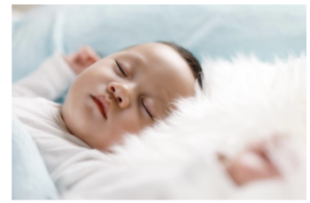
Why Do We Sleep? “outline”
Sleep- easily reversible state of inactivity characterized
by lack of interaction w/ the external environment
• Physiologically, what distinguishes being awake
from various stages of sleep?
• Name the 4 stages of sleep
• What makes us sleep?

Why Do We Sleep?
• Unsolved Mystery in neurophysiology
w/ multiple possible explanations
• Proposed Functions of Sleep:
energy conservation, predator avoidance, physical
repair, memory processing
• Recent Discoveries:
• Sleep helps clear waste from cerebrospinal fluid
• Removes proteins linked to neurodegenerative
diseases like Alzheimer’s

What is an Electroencephalogram (EEG)?
• measures & records brain electrical activity by
detecting neuron impulses through scalp
electrodes
• different wave patterns during wakefulness,
sleep stages, & abnormal brain activity
• helps diagnose neurological conditions like
epilepsy & sleep disorders by analyzing
brain wave patterns
Sleep is active—how?
brain uses as much oxygen as when awake

Describe the different EEG patterns.
Awake (Alert): Rapid, irregular, low-amplitude waves.
•Awake (Resting): More synchronized, low-amplitude, high-
frequency waves (Stage W)

What are the two sleep stages?
REM sleep (stage R) vs. Non-rem sleep
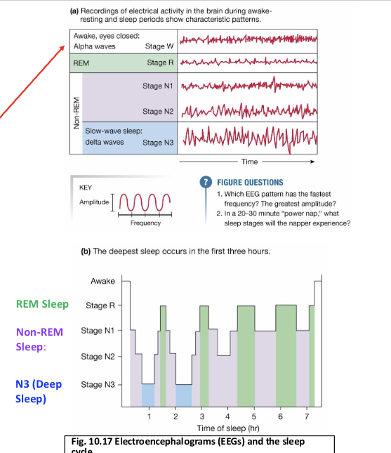
REM Sleep (Stage R):
EEG resembles wakefulness
Muscle paralysis (except eyes & breathing)
Most dreaming occurs
Body temperature regulation is reduced

Non-REM Sleep: what are the two options?
N1 & N2: Light sleep.
•N3 (Deep Sleep): Delta waves—high amplitude, low frequency.

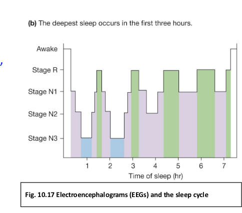
Define the sleep cycle.
Cycles through Non-REM and REM
More REM and light sleep (N1) near morning
What is sleepwalking, who does it affect? When does it occur?
• A sleep behavior disorder involving walking or
performing tasks while asleep
• Not Dreaming: occurs during deep sleep (Stage N3),
not REM (dreaming) sleep
Behavior:
•Eyes open, aware of surroundings
•Can navigate obstacles, stairs, & perform routine tasks
•Little to no memory of the episode upon waking
Who It Affects:
• Most common in children; frequency declines w/ age
• Has a genetic component—runs in families
Emotion & Motivation Involve Complex Neural
Pathways— how?
• The limbic system is the center of emotion in the
human brain
• Motivation is defined as internal signals that
shape voluntary behaviors
• some states known as drives
• Work w/ autonomic and endocrine responses
• Motivated behaviors stop when a person has
reached a certain level of satiety
• Pleasure & addictive behaviors: link to dopamine
(“reward centers”)
What are emotions?
• Hard to define; difficult to control voluntarily.
• Common emotions: anger, fear, pleasure,
happiness, aggression, sexual feelings,
contentment
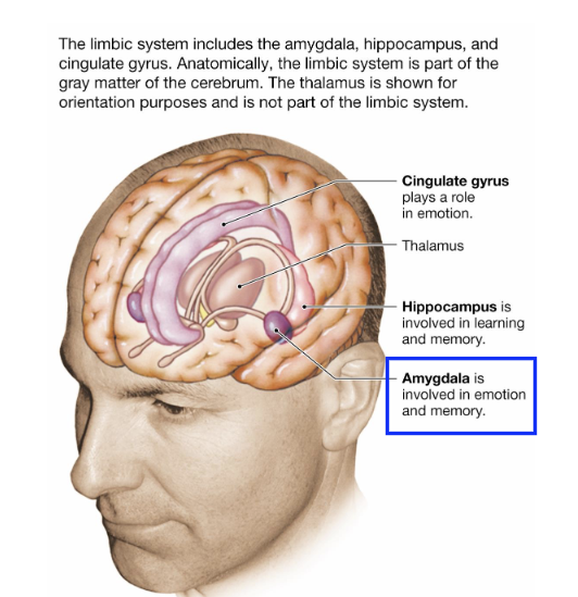
What is the amygdala (emotion center)?
Key part of the limbic system
Stimulation → fear & anxiety
Lesions → tameness & hypersexuality
Linked to basic instincts like fear and aggression
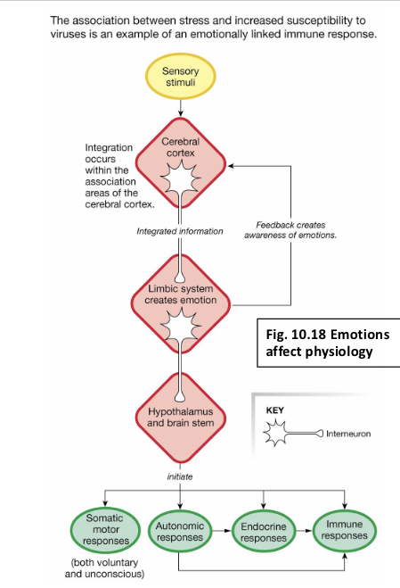
What are the results of emotional Pathways in the Brain?
Emotional pathways are complex, involving multiple
brain regions
• Sensory input → Cerebral Cortex → Limbic System
• Feedback from limbic system creates emotional
awareness
• Descending signals activate:
•Hypothalamus & Brainstem
•autonomic, endocrine, immune, & motor
responses
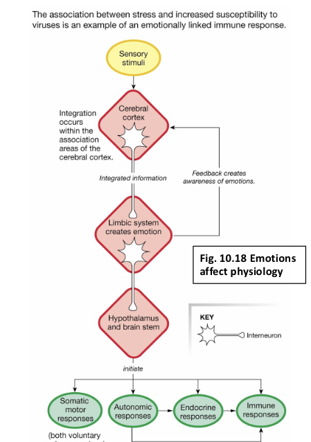
What is the mind-body connection?
• Emotions trigger physical responses:
• Ex: Fight-or-flight → pounding heart
• Long-term stress → irregular heartbeat
• Link between brain and body is not fully understood

What is the broken heart syndrome? (Takotsubo
cardiomyopathy)- linked to the Limbic
System
Emotional Triggers: triggered by intense
emotional stress-→ activates the limbic system
• brain imaging studies have shown that individuals
who develop broken heart syndrome tend to
have heightened activity in the amygdala
Brain-Heart Connection:
• The limbic system communicates w/ the ANS, which
regulates involuntary functions like heart rate
• Disrupted communication between emotional centers &
autonomic control regions can lead to the cardiac symptoms
• Patients with broken heart syndrome show decreased
connectivity between brain regions responsible for
emotional regulation & those controlling heart function

What are moods?
Moods Are Long-Lasting Emotional States
Moods are similar to emotions
• Longer-lasting; related to one’s sense of well being
Mood disorders
• Fourth leading cause of illness worldwide today
• Depression
• Sleep and appetite disturbances
• Alterations of mood and libido
• May affect function at school or work or in personal
relationships
• Antidepressant drugs alter synaptic transmission
• Promote growth of new neurons (long-term
modulation)
• Explains delayed onset of medications

What is associative learning?
involves linking two stimuli or a stimulus with a behavior.
• Ex: Pavlov’s Dogs: Bell + Food → Salivation

What is non-associative learning?
behavior changes after repeated exposure to a single stimulus.
• Habituation: decreased response to harmless
stimuli
=Tuning out the ticking sound of a clock
• Sensitization: increased response to disruptive
stimuli
-Becoming hyper-alert to small noises after a jump scare

Learning and Memory Change Synaptic
Connections in the Brain. How?
How does it disrupt it? (i.e. what is amnesia?)
Memory is the ability to
retain and recall
information
• stored throughout the
cerebral cortex in pathways
known as memory traces
• Anterograde amnesia is the
inability to remember new
information

What is short-term memory?
• Holds 7–12 items briefly
• Requires repetition to transfer to long-term
memory

What is long time memory?
Stores vast amounts of information

How does memory consolidation work?
• Converts short-term to long-term memory
• Takes seconds to minutes
• Involves multiple intermediate stages