Topic 7
1/228
Earn XP
Description and Tags
Name | Mastery | Learn | Test | Matching | Spaced |
|---|
No study sessions yet.
229 Terms
What are the 4 main tissues within the skeletal system? (7.1)
Bones
Tendons
Cartilage
Ligaments
Features & Functions of Bones (7.1)
Features:
Strong & hard
Bone cells embedded in a matrix of collagen and calcium salts
Very strong under compression forces
Is light to reduce the weight moved about
Functions:
support for the body
protection of vital organs
(eg. heart, lungs, brain)
allows movement (with muscles)
What are the 2 different types of Cartilage? (7.1)
Hyaline Cartilage: Smooth, found at ends of bones, the nose, air passages & ear
White Fibrous Cartilage: Has great tensile strength. Found between vertebrae and bones in joints
Features & Functions of Cartilage (7.1)
Features:
Hard but flexible
(Is elastic and able to withstand compressive forces)
Made of cells called chondrocytes (within an organic matrix of collagen fibrils)
Is a very good shock absorber
Frequently found in the joints and between vertebrae
Functions:
Smooth to reduce friction at joints - cartilage lines the joints/covers the ends of the bones preventing damage
Allows smooth movement eg. in hip, knee, ankle
Support eg. in trachea
Shock absorber eg. between vertebrae, to prevent damage during running etc
Features & Functions of Ligaments (7.1)
Features:
Need to be elastic to allow the bones of a joint to move
strong & flexible
Made of yellow elastic tissue which gives both strength and elasticity
Functions:
Join bones to bone
Keep bones in correct alignment
Form around and within the joint
Features & Functions of Tendons (7.1)
Features:
Made of white fibrous tissue
Mainly bundles of collagen fibres
Strong but inelastic
Functions:
Join muscles to bone
enables movement at joints
What are ball and socket joints? (7.1)
allows movement in any direction, round end of long bone fits into hollow of another bone
ex. hips and shoulders

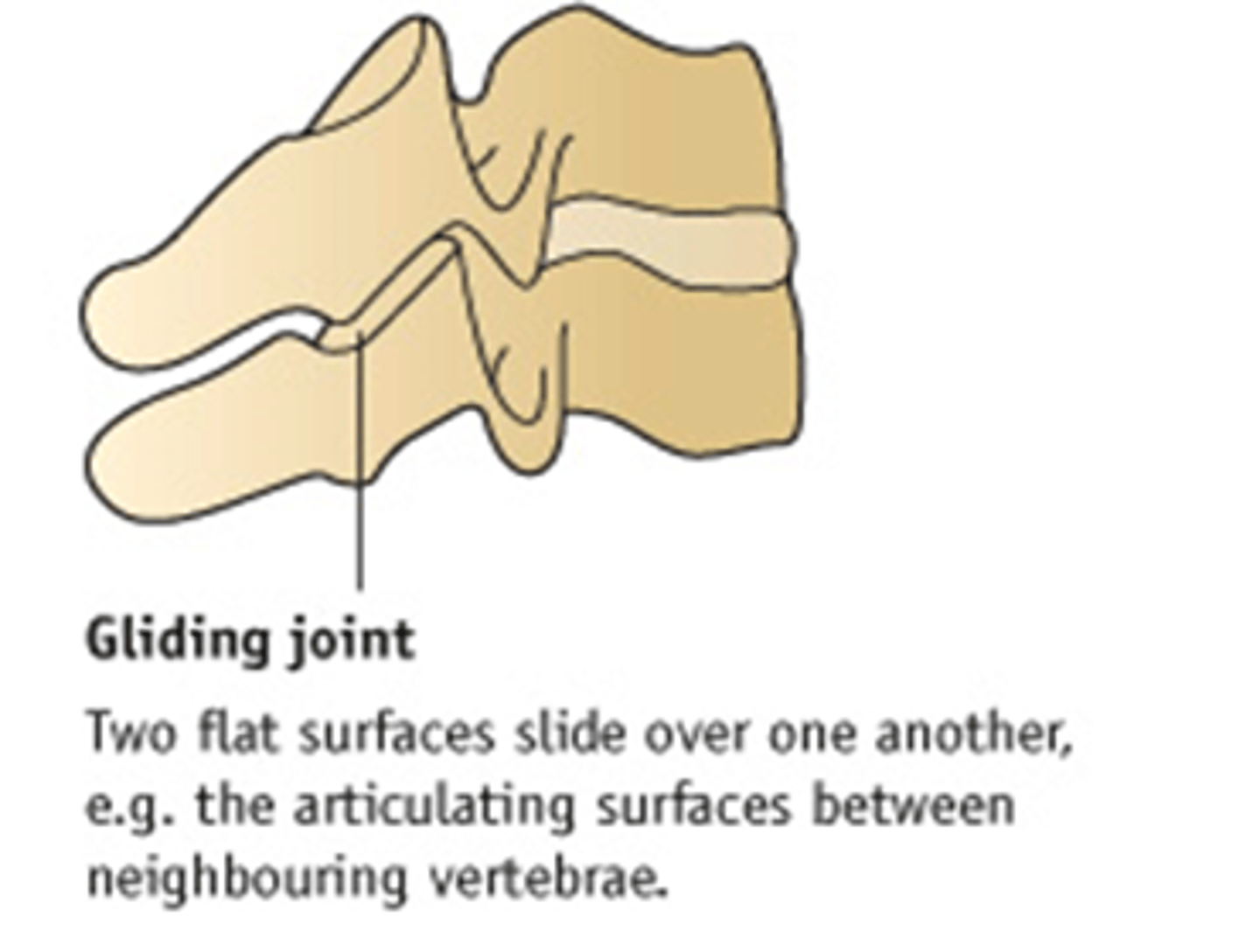
What are gliding joints? (7.1)
Flat surfaces of the bone slide over one another. Ex. Carpal bones of the wrist and Tarsal bones of the ankle
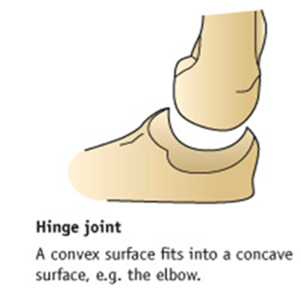
What are hinge joints? (7.1)
movement in one direction only
ex. elbow and knee
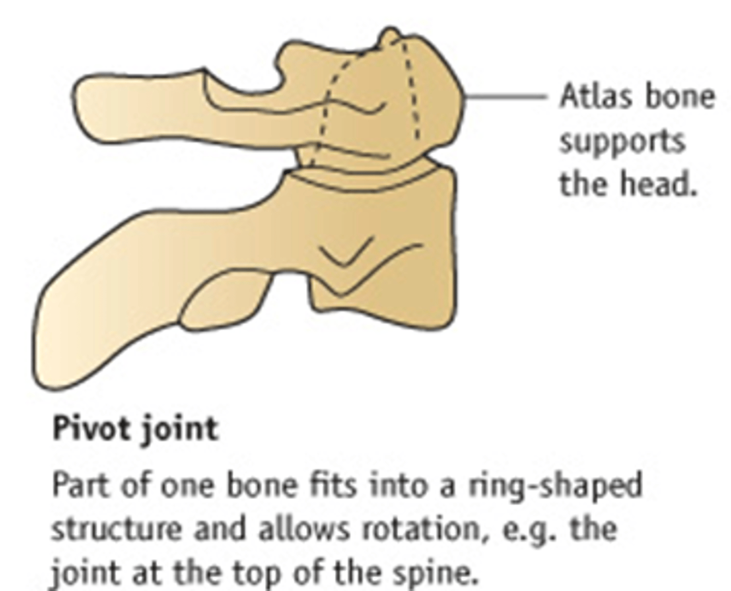
What are pivot joints? (7.1)
Rounded end of one bone conforms to a "sleeve," or ring of another bone
Uniaxial movement only
What is Cartilage? (7.1)
Cartilage - lines joints to prevent bones wearing away
- reduces friction between bones
-Absorbs Synovial Fluid
- acts as a shock absorber
Pad of cartilage provides additional protection
What are fibrous capsules? (7.1)
outer layer of dense irregular connective tissue that encloses joints
These joints also produce a synovial fluid (liquid lubricant) which is held within the fibrous capsule
What is Synovial Fluid? (7.1)
Synovial fluid (liquid lubricant) is produced which is held within the fibrous capsule
What is the synovial membrane? (7.1)
Connective tissue membrane lining joints, produces synovial (oily) fluid
How does the body prevent bones between joints from being eroded? (7.1)
the joint is lined with a replaceable layer of rubbery cartilage
This allows the joint to articulate (move) smoothly
Compare & Contrast: Ligaments vs Tendons (7.1)
Ligaments join Bone & Bone
Tendons Join Bone & Muscle
Ligaments are Elastic
Tendons are Inelastic
Both Ligaments & Tendons are made of Fibrous Connective Tissue
Both Ligaments & Tendons are strong
Ligaments are found between bones
Tendons are found between bones & muscles
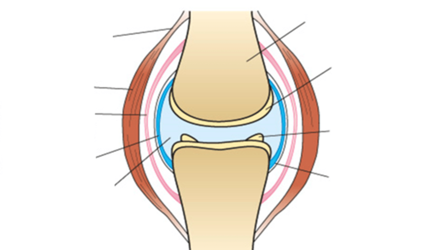
Flip & attempt to label the diagram without looking at the answers (7.1)
going clockwise:
Bone
Cartilage
Pad of Cartilage
Fibrous Capsule
Synovial Fluid
Synovial Membrane
Ligament
Muscle
Tendon
what is the function of a muscle? (7.2)
Muscles bring about the movement of a joint
muscle shortens
pulls on a bone
moves a joint
BUT: PROBLEM
Muscles cannot extend themselves (lengthen)
What are the 3 different types of muscle? (7.2)
Antagonistic muscles = a pair of muscles working together to move a joint
When one relaxes (stretches) the other contracts (shortens)
Extensor muscle: The muscle which contracts to extend the joint
Flexor muscle: The muscle which contracts to reverse the movement
how does Flexion of arm at elbow (bending arm) occur? (7.2)
Biceps contracts
shortens,
thickens
Triceps relaxes
It is pulled and stretches and
becomes thinner
Arm bends
how does Extension of arm at elbow (straightening arm) occur? (7.2)
Triceps contracts
shortens,
thickens
Biceps relaxes
It is pulled and stretches and
becomes thinner
Arm straightens
What are the 3 different types of muscle fiber? (7.2)
Skeletal Muscles
Cardiac Muscles
Smooth Muscles
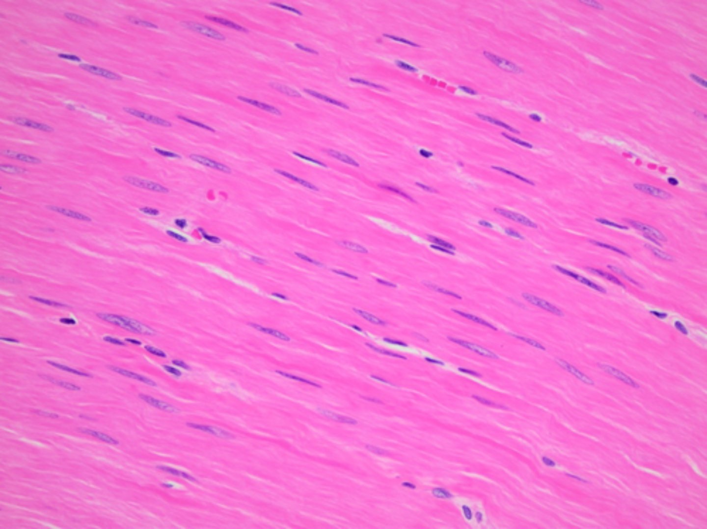
What is smooth muscle? (7.2)
Found in walls of Arteries + Veins
Under control via Involuntary nervous system (Autonomic)
Causes slow contractions of many internal organs (NO FATIGUE)
Long, Spindle-Shaped cells, Each with their own Nucleus
No Striations/Stripes; Parallel Groves
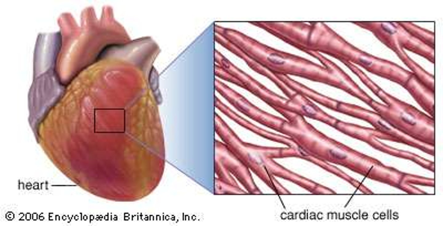
What is Cardiac Muscle? (7.2)
Only found in the heart
Myogenic; Heart can create its own beating (not linked to brain)
Striated/Striped Muscle
Interconnected fibres to ensure coordinated wave of contraction
does not fatigue
capable of short contractions over a long period of time
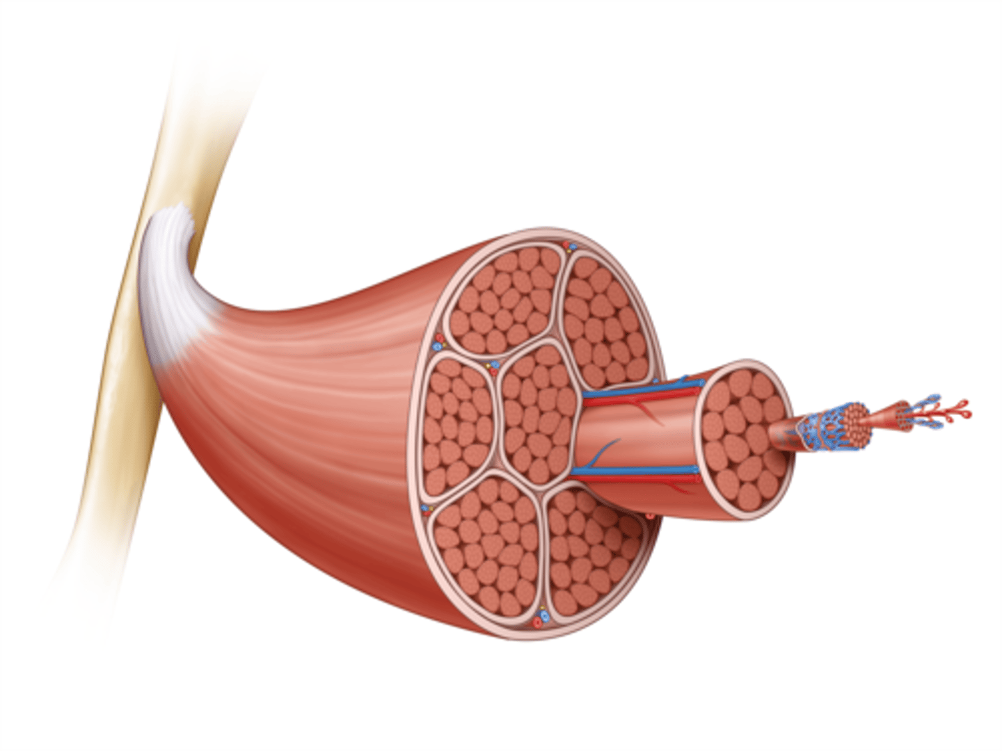
What is Skeletal Muscle? (7.2)
Under control via Voluntary nervous system (Somatic)
Muscle attached to skeleton; involved in locomotion
Contracts rapidly; Fatigues quickly
Capable of strong contractions
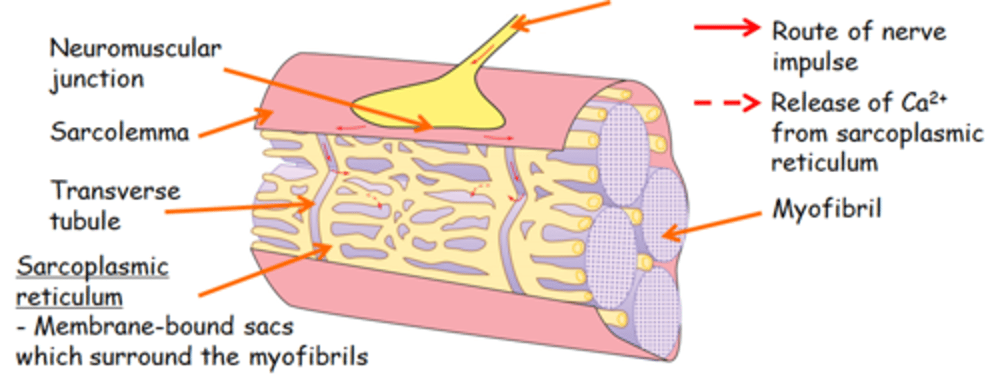
How does the brain cause contraction of skeletal muscles? (7.2)
Muscles contract when nerve impulses from the brain reach the neuromuscular junction at the end of a nerve.
Here there is a synapse (gap) between the nerve and the muscle.
Neurotransmitters are released into the gap and bind to the muscle, causing the release of calcium ions which cause the muscle to contract
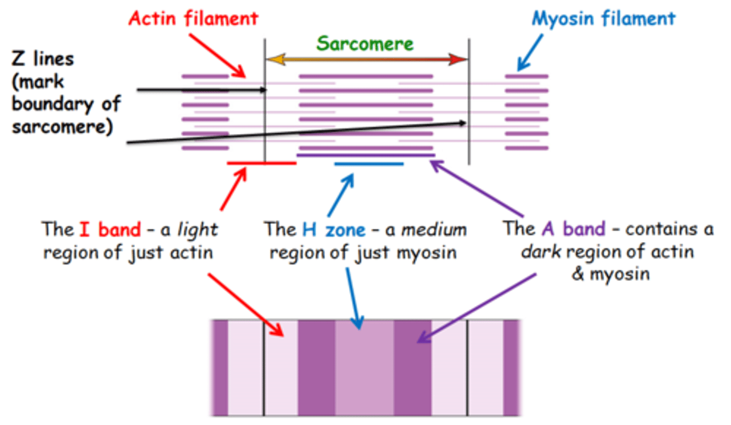
Draw, label & describe the differences between all the components which make the Sarcomere (7.2)
Myosin Filament is Globular
Actin Filament is Fibrous
Draw a simple diagram of the sarcomere pre & post contraction (7.2)
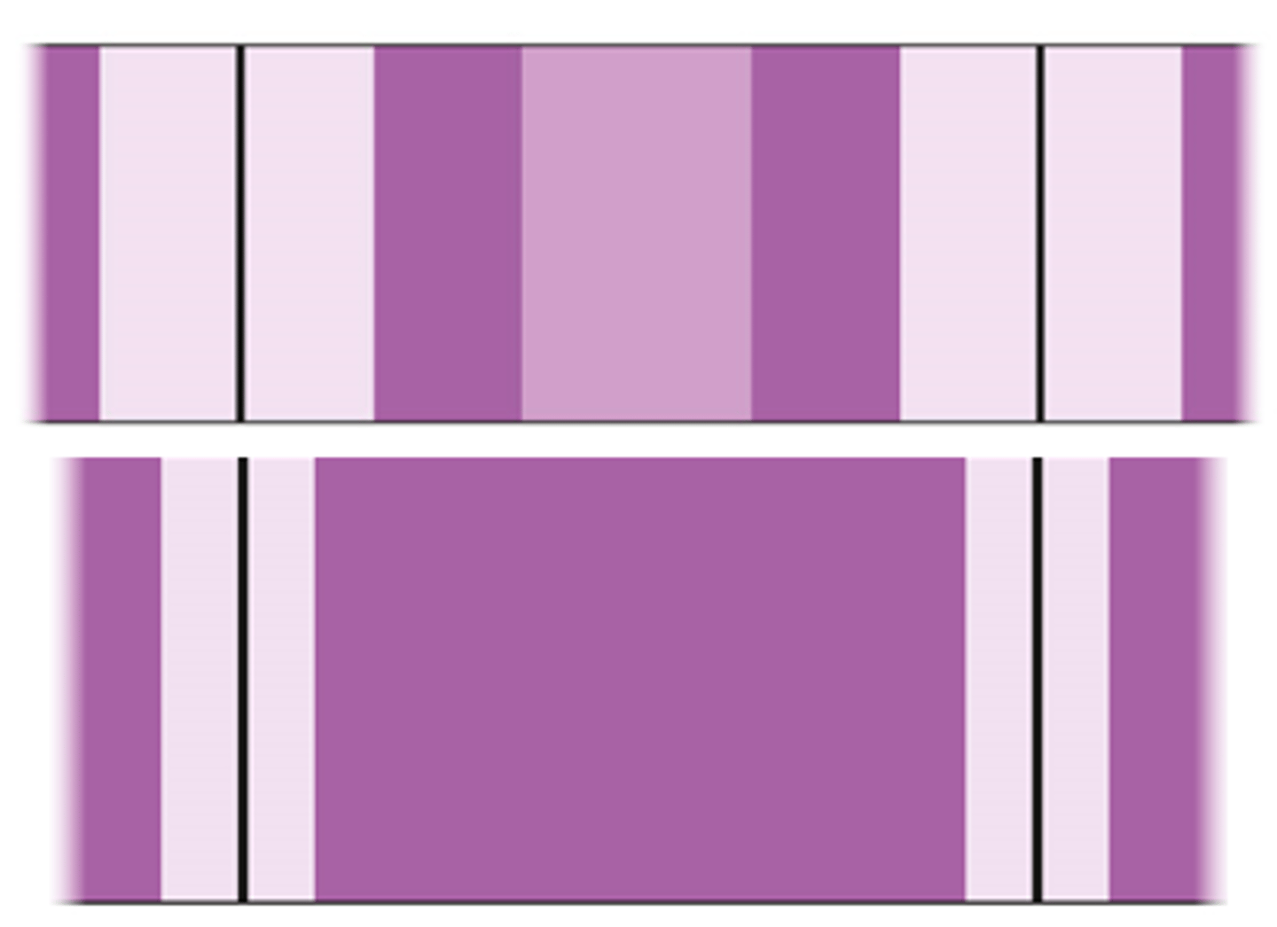
Muscles contract, Actin moves beween myosin towards sarcomere center
This shortens the width of the sarcomere/H-Zone (Z lines get closer together), and hence the length of the muscle
Actin Moves Between Myosin
Sarcomere Length Shortened
H-Zone Disappears
I-Band Narrows
Distance between 2 lines decreases
A-Band stays same width
Describe the sequence of events that occurs at the neuromuscular junction (7.2)
Impulse down motor neurone to neuromuscular junction
Causes calcium ions flooding via Sarcoplasmic Reticulum
Calcium ions bind to troponin via calcium binding site
Troponin moves; pulls on attached Tropomyosin (change of shape)
Tropomyosin moved; reveals myosin (golf) binding site on Actin (main strand)
Myosin (golf) head binds to myosin binding site on Actin (CROSSBRIDGE/ACTOMYOSIN BRIDGE FORMATION)
Myosin Head binds to Actin; ADP + Pi released from myosin head; causes shape change (nod forward)
Nodding forward of myosin head moves/slides the ACTIN (relative movement of Actin filament = 10nm)
Myosin filament stationary; Actin filament moves/slides
Actin moves to centre of sarcomere; shortens overall sarcomere length
ATP binds to myosin head; detaching it from binding sites on the Actin filament
ATPase in myosin head HYDROLYSES ATP upon detachment; ADP + Pi formation
ATPase in myosin head for hydrolysis; requires calcium ions to work; hence calcium flooding
Energy released via hydrolysis of ATP; returns myosin to its original resting position.
Myosin head back in original position; cycle can repeat again.
Sarcoplasmic reticulum sucks all calcium ions back up; calcium pumped back via active transport
Troponin & Tropomyosin returns to original position; contraction is complete.
What is the sarcoplasmic reticulum? (7.2)
Membrane-bound sacs surrounding the myofibrils
Stores/releases Ca2+ ions which flood the sarcoplasm & movement of the protein filaments follows
Why do muscles not contract all the time? (7.2)
Tropomyosin blocks myosin binding sites on actin molecules, preventing muscle contraction when there is no nervous stimulation.
A nerve impulse triggers the release of calcium ions from the sarcoplasmic reticulum (endoplasmic reticulum) into the sarcoplasm of a muscle cell.
This triggers muscle contraction
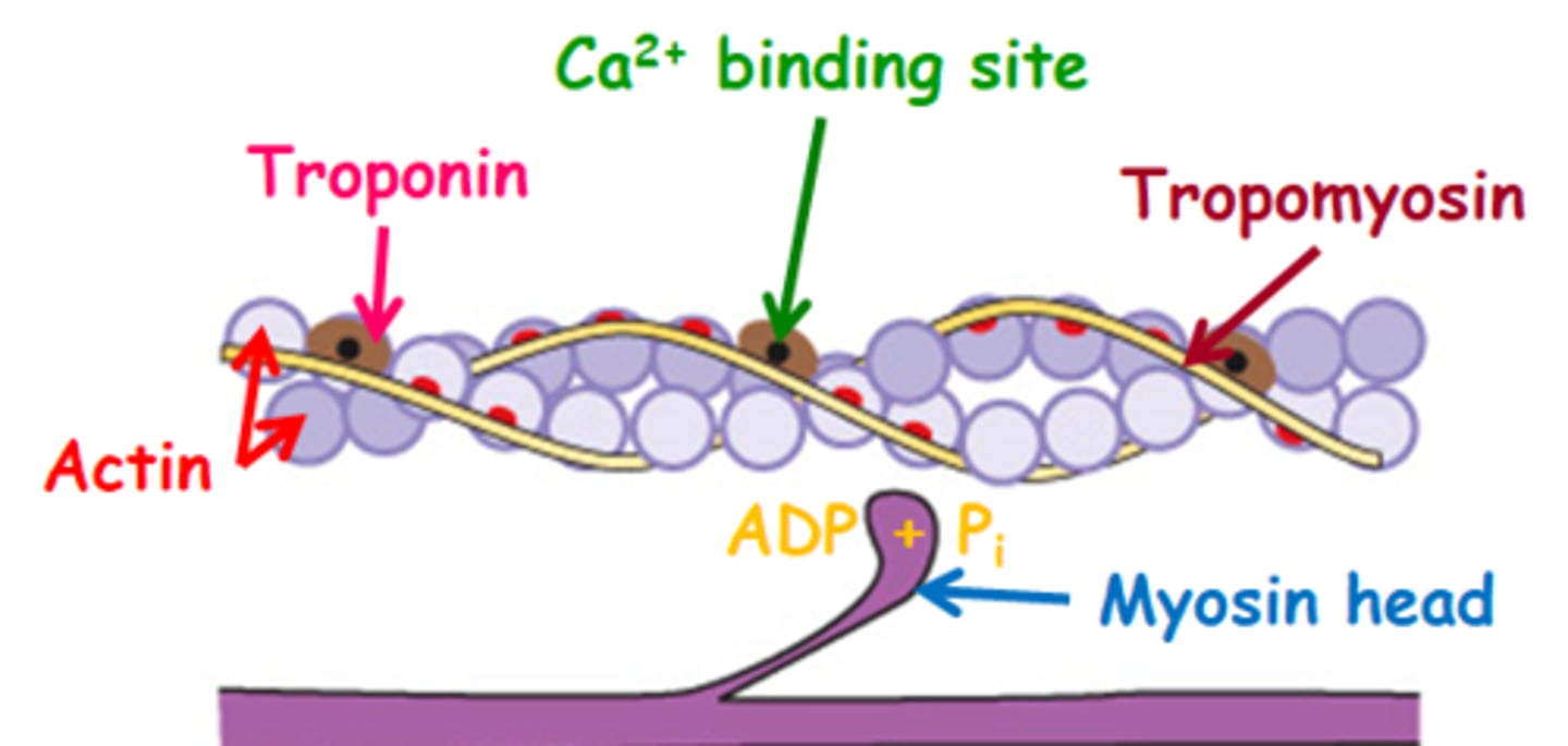
Sliding Filament Theory step 1: Resting State (pre-Contraction) (7.2)
The myosin binding sites
(red dots) on the actin are blocked by tropomyosin
The myosin head cannot bind
ADP + Pi are bound to the myosin head
Sliding Filament Theory step 2: Addition of Calcium ions (7.2)
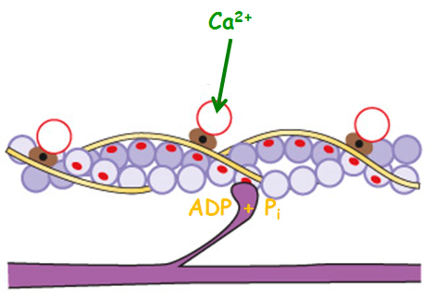
Ca2+ binds to the troponin causing it to move & pull on the tropomyosin
Tropomyosin shifts position exposing the myosin binding
sites on the actin molecule
The myosin head binds with myosin binding sites on the actin molecule
An actomyosin bridge / cross-bridge forms
Sliding Filament Theory step 3: Movement Of Myosin Head (7.2)
When the myosin head binds to the actin, ADP + Pi on
myosin headS are released
The myosin changes shape
The head bends/nods forward
This movement results in the relative movement of the actin filament (10 nm) over the myosin
The actin moves towards the centre of the sarcomere, and shortens the overall sarcomere length
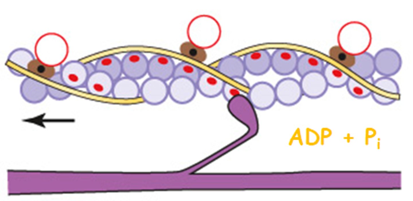
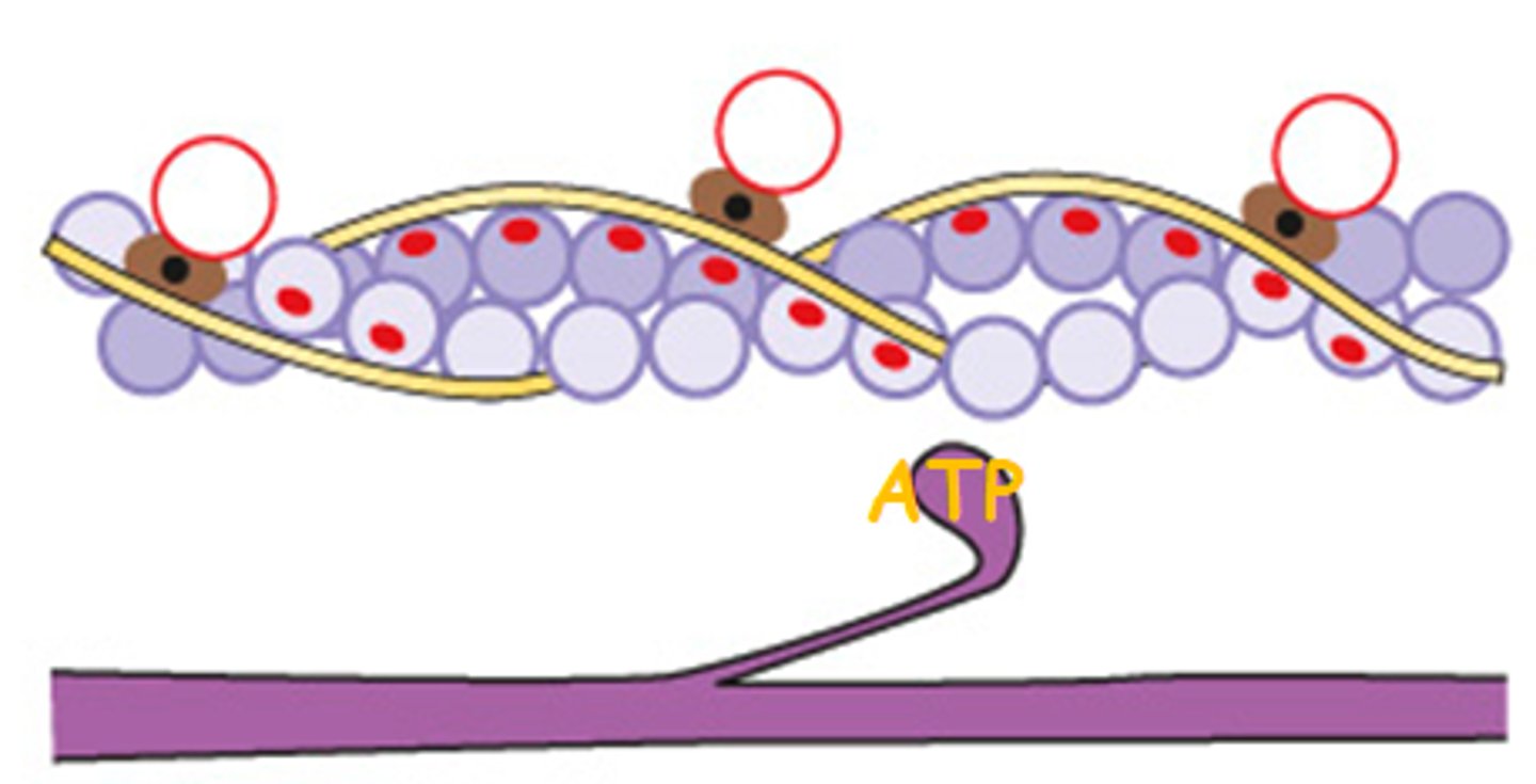
Sliding Filament Theory Step 4: Binding Of ATP (7.2)
An ATP molecule binds to the myosin head causing another shape change
The myosin head detaches from the actin
This activates ATPase in the myosin head, which also needs Ca2+ to work
The ATP is hydrolysed into ADP + Pi are formed and energy is released
The energy returns the myosin head to its original position
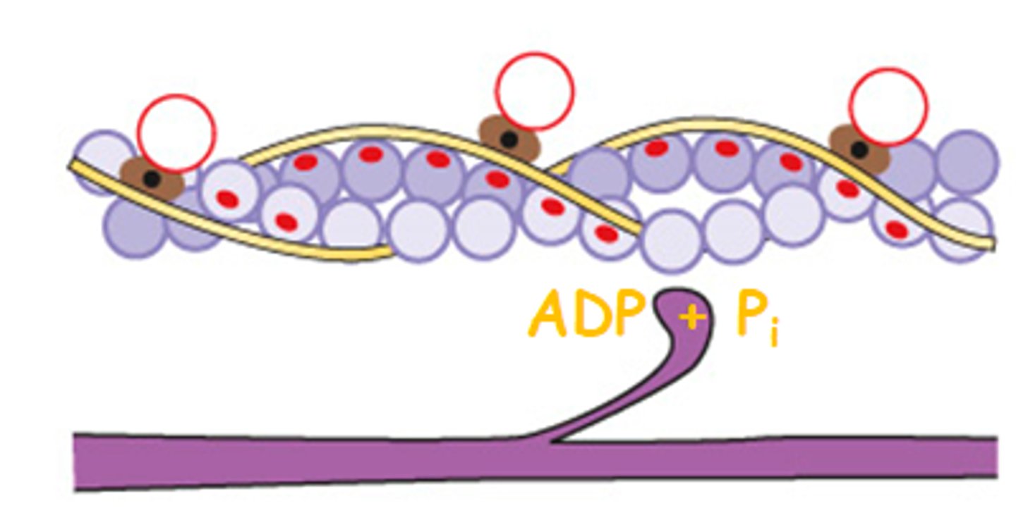
Sliding Filament Theory Step 5: Repeat Cycle (7.2)
With the myosin head in its original position, the cycle then starts again if Ca2+ remains in the sarcoplasm due to further stimulation!
The collective bending of many myosin heads combines to move the actin filaments relative to the myosin filament
This results in muscle contraction
If there is no further stimulation, the Ca2+ is pumped back into the sarcoplasmic reticulum using ATP
The troponin & tropomyosin return to their original positions & the contraction is complete
The Muscle Fibre is Relaxed
why does rigor mortis occur? (7.2)
The energy from ATP is needed to break the bond between actin and myosin
ATP is made by cell respiration in the mitochondria
After death the cells no longer make ATP
So, after death, the actin and myosin bonds are fixed
After death the muscles become stiff with rigor mortis
What is Aerobic Capacity?
Aerobic capacity is the ability to take in, transport and use oxygen
This affects an individual's ability to maintain a continuous supply of ATP for muscle contraction
(Required for long periods of strenuous exercise)
What are the 2 different types of muscle fibre?
Fast twitch muscle fibres
Slow twitch muscle fibres
A muscle can contain both slow and fast twitch muscle
fibres (cells) but the relative amount of each can vary
What is the length of time that a muscle contracts for dependent on?
how long calcium ions remain in the sarcoplasm
What are Slow twitch muscle fibres?
Fibres specialised for slower, sustained contraction
They can cope with long periods of exercise
They therefore, carry out a large amount of aerobic respiration
What are fast twitch muscle fibres?
Fibres specialised to produce rapid, intense contractions
The ATP used in these contractions is produced almost entirely from anaerobic respiration
What is the main disadvantage of fast twitch muscle fibres?
Fast twitch muscle fibres fatigue (get tired) easily.
a poor oxygen supply;
poor oxygen reserves (myoglobin) ;
a poor rate of oxygen breakdown;
very few mitochondria
So muscle cells carry out anaerobic respiration
How does Anaerobic respiration affect a muscle's ability to contract?
causes a rapid build up of lactate, which lowers pH which affects enzymes and prevents muscle contraction
also less ATP is made (as only glycolysis takes place in the cytoplasm/not Krebs cycle and the electron transport chain in the mitochondria
less muscle contraction as ATP is needed for this
How do Fast Twitch Muscle fibres work?
Because fast twitch fibres have much more sarcoplasmic reticulum (therefore more calcium ion pumps) than slow twitch fibres
calcium ions are pumped out of the sarcoplasm back into the sarcoplasmic reticulum more quickly after a nerve impulse
this causes the muscle fibre to relax
so, the effect of nerve stimulation of fast twitch fibres fades away very quickly
How do slow twitch muscle fibres work?
Slow twitch fibres have less sarcoplasmic reticulum, so ...
calcium ions remain in the sarcoplasm of slow twitch fibres for a long time - it cannot be pumped back into the sarcoplasmic reticulum as quickly
this results in a long lasting effect (muscle contraction) from nerve stimulation
What is myoglobin?
Is a protein similar to haemoglobin
Made of 1 chain rather than 4
It has a much higher affinity for oxygen than haemoglobin
Readily accepts oxygen from the blood
Acts as an oxygen store in muscles: Will only release it if
the oxygen concentration in the cell falls very low
Dark red in colour which gives slow twitch muscle fibres their distinctive colour
Fast Vs Slow Twitch comparisons
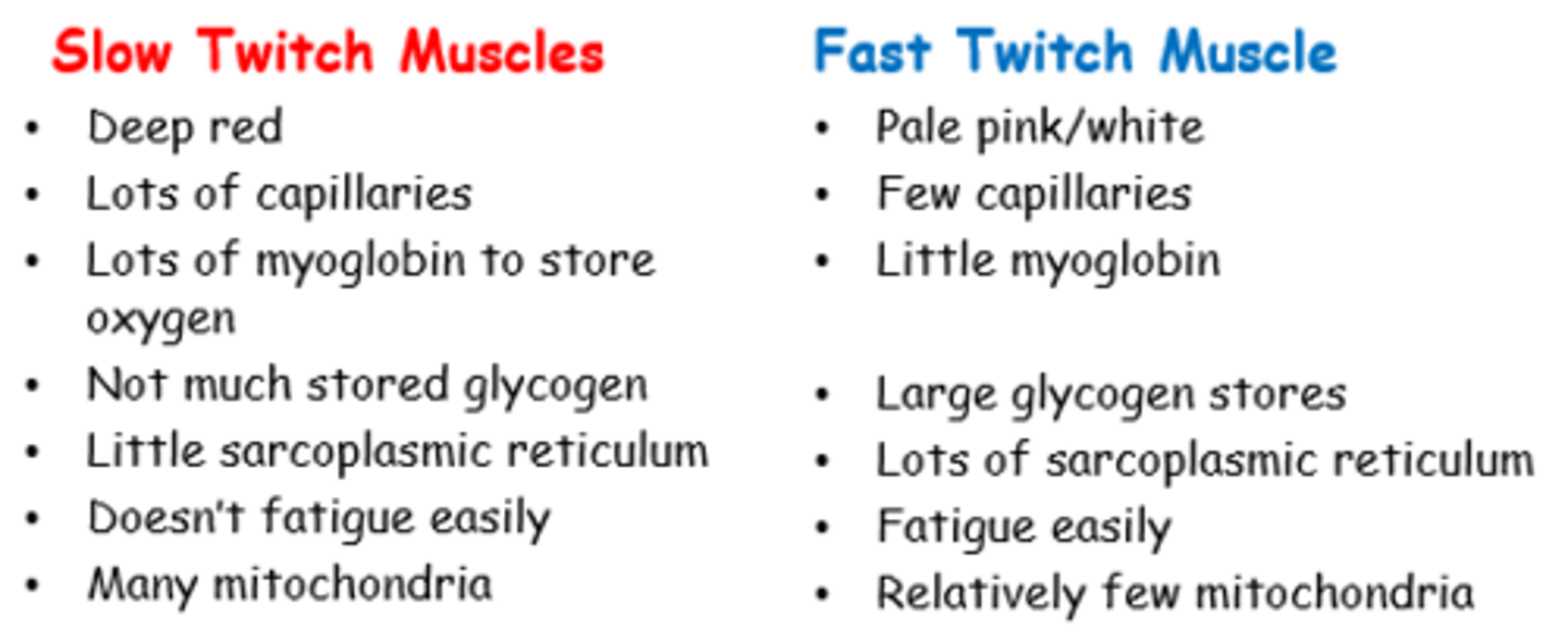
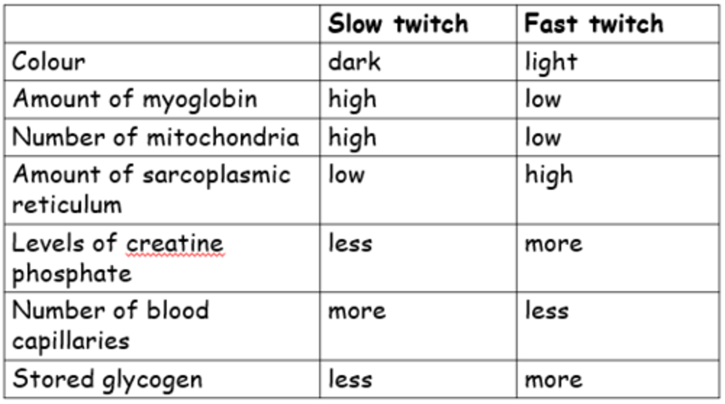
Fast Vs Slow Twitch: structural differences
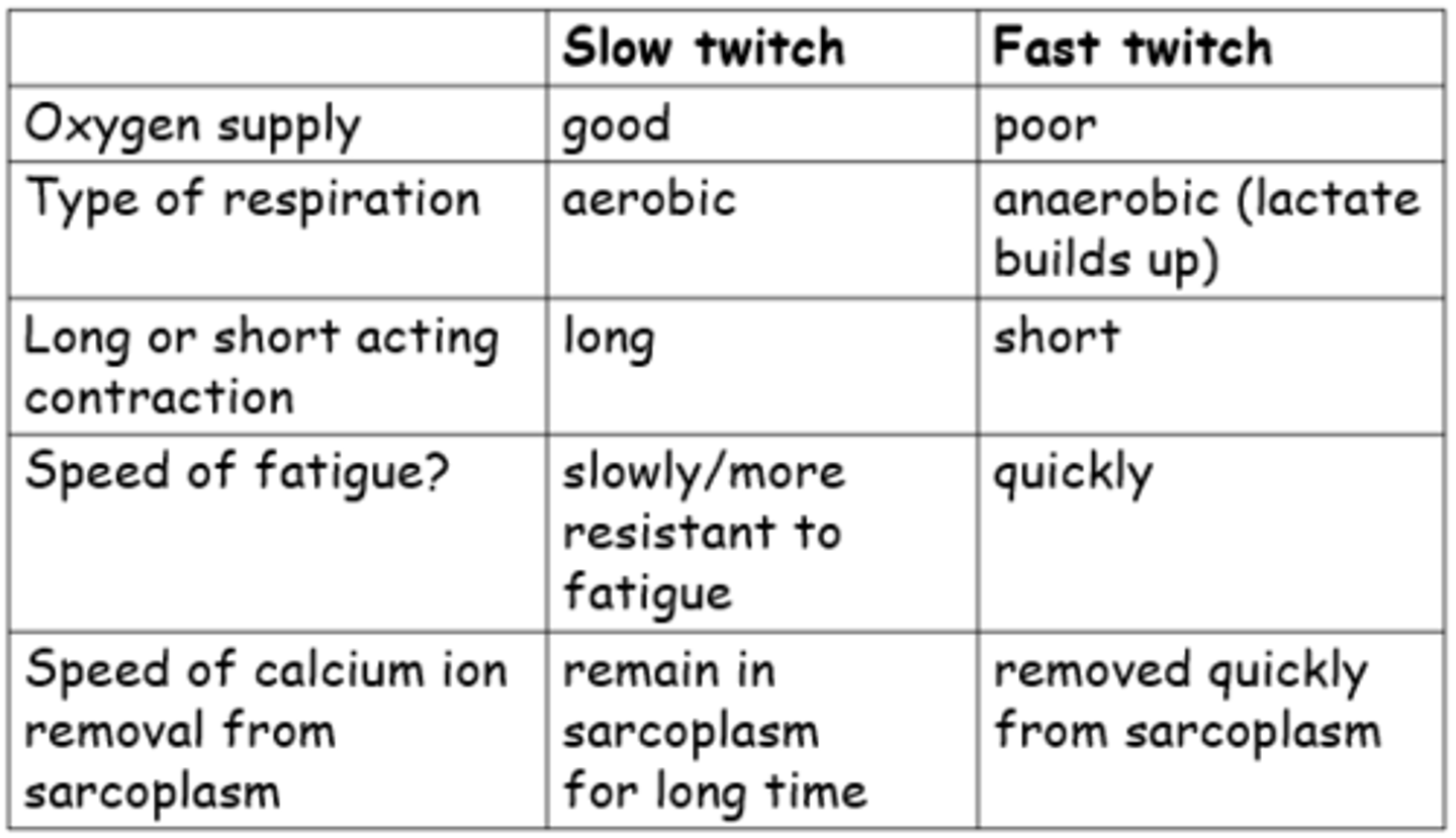
Fast Vs Slow Twitch: Physiological differences
What is cell respiration?
A chemical process that takes place in all living cells to release energy.
It is the chemical breakdown of large molecules, using enzymes, with or without oxygen.
It releases energy which is then stored as ATP
ATP = adenosine triphosphate
The energy is essential for life processes. e.g..
Cell division,
DNA replication,
protein synthesis,
active transport, and
muscle contraction.
Describe what happens during Aerobic Respiration in General
In aerobic respiration, the hydrogen stored in glucose is brought together with oxygen to form water again
The bonds between hydrogen and carbon in glucose are not as strong as the bonds between hydrogen and oxygen atoms in water
So, the input of energy needed to break the bonds in glucose and oxygen is not as great as the energy released when carbon dioxide and water are formed
So the overall energy release can be used to generate ATP

What is Adenosine Triphosphate?
Energy transfer molecule
Able to lose the loosely bonded phosphate groups to become ADP
(Adenoside Diphosphate)
The lost phosphate groups bind to proteins and enzymes activating them so that chemical reactions can take place

What is Phosphoryation?
ATP is created from ADP, from the addition of inorganic
phosphate (Pi) in a reaction called phosphorylation.
In solution, phosphate ions are hydrated, i.e. Water molecules are bound to them
To make ATP, Phosphate must be separated from water, requires energy.
ATP in water is higher in energy than ADP and phosphate ions
in water, so ATP in water is a way of storing chemical potential energy.

What is "substrate level phosphorylation"?
If the phosphate group is transferred to ADP from another molecule, it is called "substrate level phosphorylation"
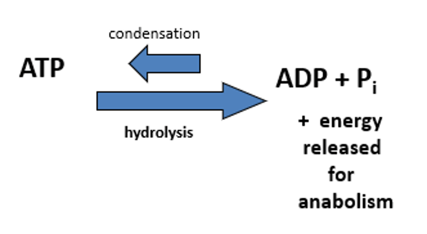
How does Hydrolysis of ATP occur?
ATP keeps the phosphate separated from the water, but the phosphate and water can be brought together in an energy-yielding reaction each time energy is needed for reactions within a cell.
The 3rd phosphate group is losely attached in an ATP molecule. A small amount of energy is required to break the bond holding this end phosphate group to ATP.
ATPase catalyses the breakdown of ATP to ADP.
ATP (Adenosine triphosphate) becomes ADP (Adenosine diphosphate).
The removed phosphate becomes hydrated.
A lot of energy is released as bonds form between water and phosphate. This is the energy used to supply energy-requiring reactions in the cell.
Why are glucose & oxygen not brought together directly?
Glucose & oxygen are not brought together directly
This would result in a large energy release which would damage the cell!!!
How does Aerobic Respiration occur without damaging the cell?
A Biochemical/Metabolic Pathway Exists:
The glucose is dismantled in a series of small steps releasing H+ and CO2 as a waste product
The H+ is eventually reunited with the oxygen to form water and to release large amounts of energy
All these small steps are controlled by specific enzymes at each stage (dont need to know any of them)
Describe the processes and locations involved in the aerobic respiration flowchart
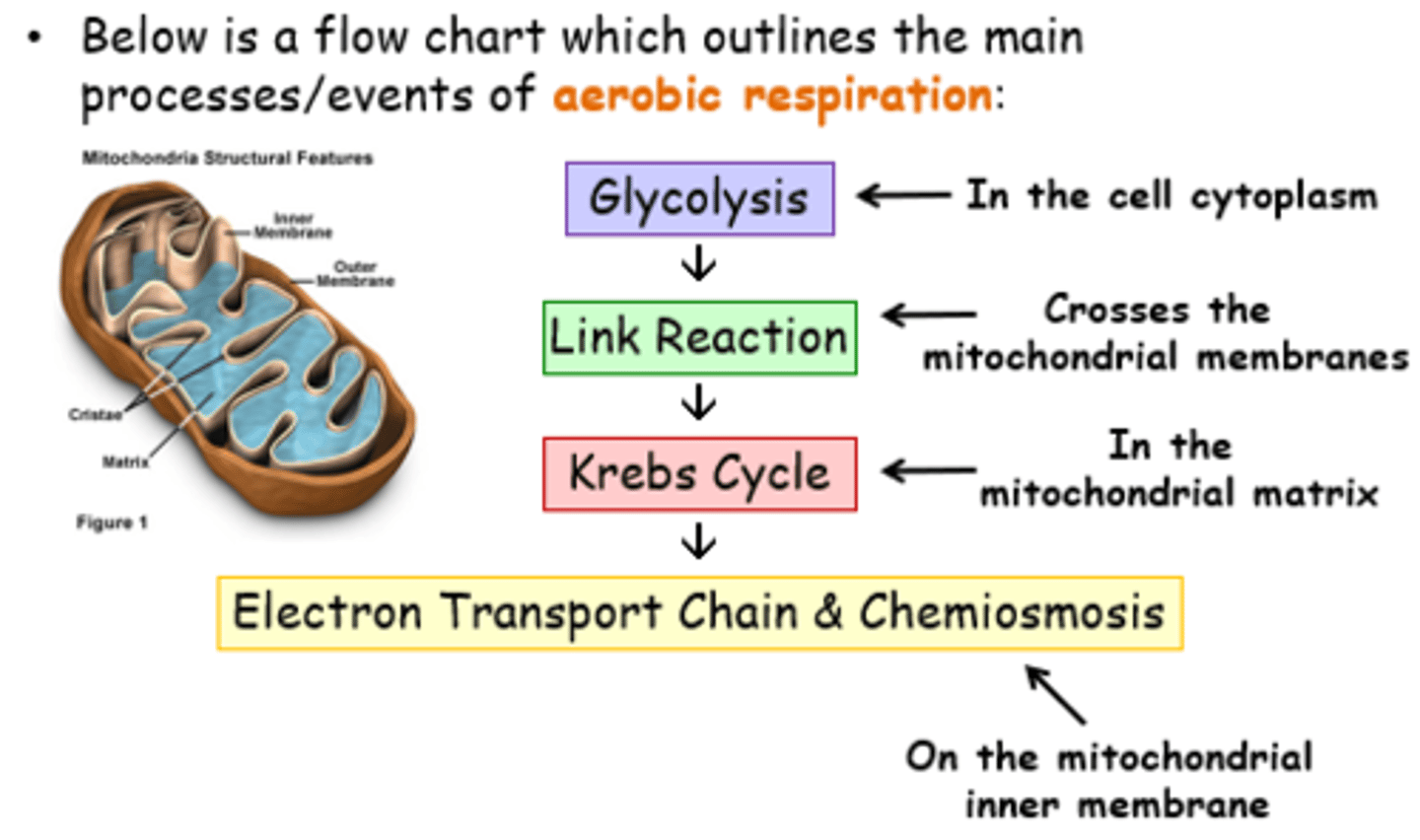
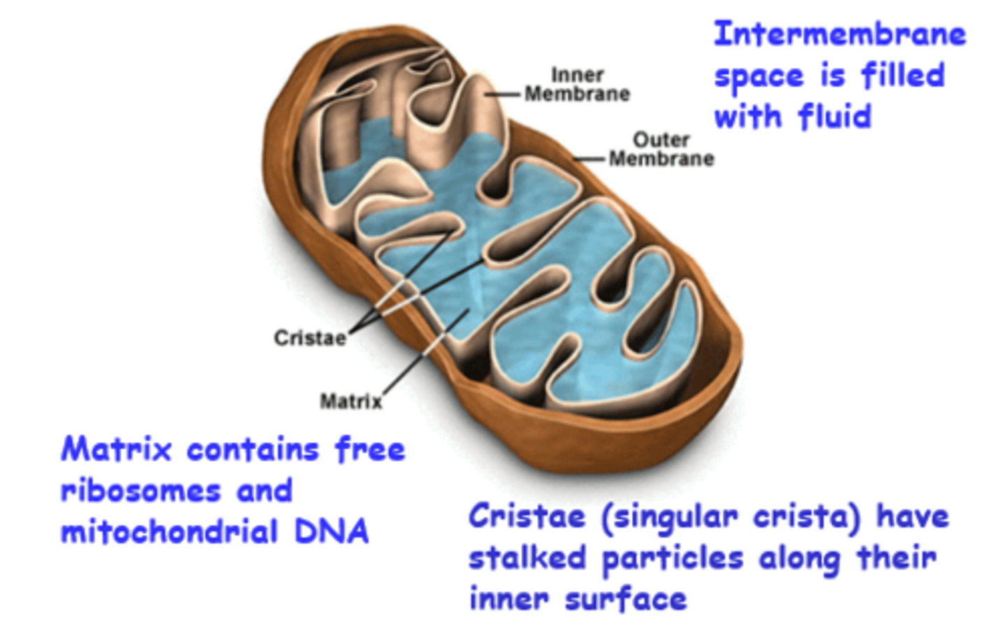
Mitochondria Structure
Matrix is filled with fluid
Stalked particles contain ATP synthase enzymes
Name the enzymes involved in respiration
Removal of carbon is decarboxylation
"decarboxylase enzyme"
Removal of hydrogen is dehydrogenation
"dehydrogenase enzyme"
Addition of phosphate is phosphorylation
"phosphorylase enzyme"
What is Glycolysis?
Literally means 'splitting of glucose'
Occurs In the cytoplasm (or sarcoplasm of muscle)
Needs glucose (hexose monosaccharide) as substrate
(Could come straight from bloodstream or Glycogen in muscle or liver cells is converted to glucose)
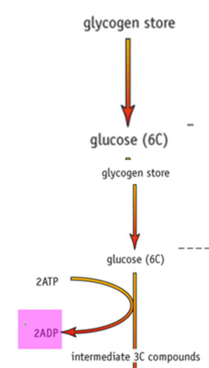
Glycolysis process step 1: GALP Formation
Glucose is quite stable and unreactive so in the first 2 steps ATP must be used to get things going
Stores of glycogen (a polymer of glucose) in the muscle or liver cells are converted to glucose, a hexose (6C) monosaccharide
Energy from ATP is required to split glucose, therefore 2 phosphate groups are added to the glucose, from 2 ATP molecules.
Glucose has been phosphorylated, increasing it's reactivity. Involves a phosphorylase enzyme
The glucose can now be split into 2 intermediate 3C phosphorylated compounds Glyceraldehyde 3-phosphate (GALP)
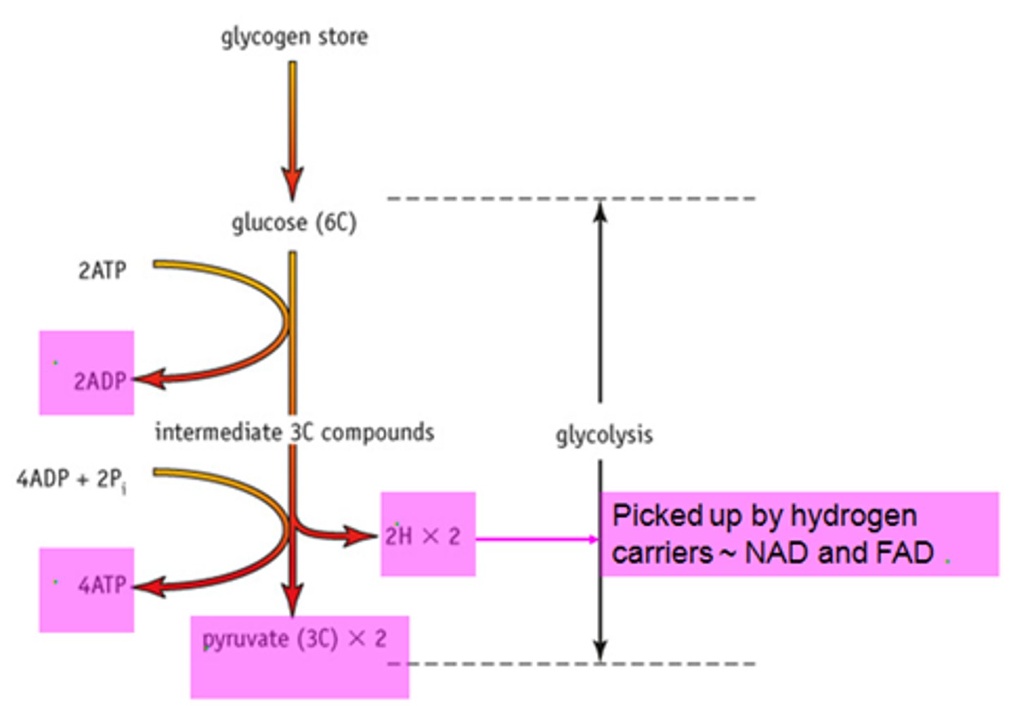
Glycolysis Step 2: GALP oxidation (Pyruvate Formation)
Both GALP compounds are oxidised (lose hydrogen), producing another 2, 3 carbon compounds, pyruvate (pyruvic acid).
In this process, 4 hydrogen atoms are removed during the reaction and taken up by the coenzyme NAD (nicotinamide adenine dinucleotide).
These become 2 molecules of reduced NAD. These hydrogen atoms are used in the last stage of respiration, in oxidative phosphorylation.
Because glucose is at a higher energy level than pyruvate, on its conversion in step 1, energy was released to create ATP. Phosphate from the intermediate 3 carbon compound, GALP, is transferred to ADP, creating ATP.
This is called substrate-level phosphorylation, because
the phosphate and energy for the formation of ATP comes from the substrates, in this case 2 GALP compounds.

What are the overall products produced at the end of Glycolysis?
Glycolysis reactions yield a net gain of:
2 ATPs (used 2, made 2)
2 pairs (total of 4) of hydrogen atoms (reduced NAD): used for oxidative phosphorylation later.
2 molecules of 3-carbon pyruvate: used for the link reaction.
Glycolysis can take place under anaerobic or aerobic
conditions
What are Co-Enzymes?
Co-enzymes are molecules that work closely with enzymes, but they are not proteins .
co-enzymes help transfer electrons. (hydrogen carriers)
Co-enzymes easily accept electrons and become reduced, then give them up and become oxidised.
The coenzymes therefore have two forms, the oxidised form and the reduced.
What is NAD?
NAD and FAD are the most important "co-enzymes" for you to know about. There are others
1. NAD ~ nicotinamide adenine dinucleotide (comes from vitamin B3 ~ nicotinic acid)
Always has a phosphate group attached so is sometimes called NADP
Acts as a hydrogen "carrier" between molecules.
It picks up "H" to become NADPH (NADH)
Also called reduced NADP
What is FAD?
NAD and FAD are the most important "co-enzymes" for you to know about. There are others
2. FAD ~ flavine adenine dinucleotide (comes from vitamin B2 ~ riboflavin)
Simplified Glycolysis process (Marking Points)
series of small enzyme catalysed steps (metabolic pathway);
glucose is phosphorylated (to make it more reactive);
using 2 ATP;
GALP (3C intermediate) is formed;
dehydrogenation/H removed from GALP ;
hydrogen acceptor is NAD ;
Reduced NAD enters electron transport chain;
4 ATP made by substrate level phosphorylation (net 2ATP) ;
final product is pyruvate;
What is the Link Reaction?
The link reaction links glycolysis and the Krebs cycle
Takes place in the mitochondrial matrix
What happens during the Link Reaction?
In the mitchondrial matrix, when oxygen is available:
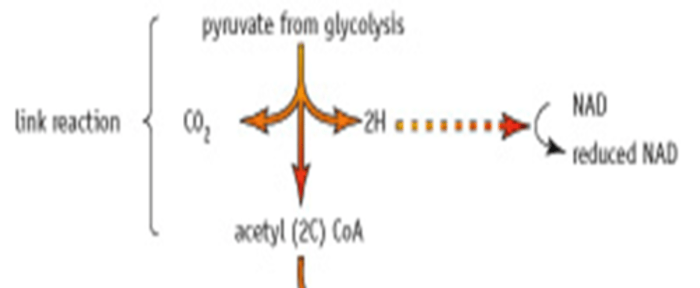
1. Pyruvate (3C) from glycolysis enters the mitochondrion.
2. Pyruvate is decarboxylated (carbon is removed) (1C):
One carbon atom is removed from pyruvate in the form of CO2. This is produced as a waste product, and breathed out of a respiring organism.
3. Pyruvate also loses 2 hydrogen atoms, which are then picked up by the coenzyme NAD. NAD becomes reduced NAD.
4. Because of this loss of carbon and hydrogen, 3C pyruvate molecule becomes a 2C molecule called acetate.
5. Acetate is combined with coenzyme A (CoA) to form acetyl coenzyme A
(acetyl CoA).
The link reaction produces Acetyl Co-enzyme A.
This molecule enters the Krebs cycle
- No ATP is produced in this reaction

Why does the link reaction occur twice for every glucose molecule?
Two pyruvate molecules are made for every glucose molecule that enters Glycolysis. This means the link reaction and the later the kreb cycle, happen twice for every
Glucose molecule.
So for every glucose molecule:
2 molecules of acetyl coenzyme A go into the
Kreb cycle.
2 CO2 molecules are released as waste.
2 molecules of reduced NAD are formed and are used in the last stage - oxidative phosphorylation.
How does the link reaction move to the krebs cycle?
The Acetyl - CoA from the link reaction feeds straight into the Krebs cycle.
Link reaction and Krebs cycle take place in mitochondrial matrix.
What is the Krebs Cycle?
Krebs cycle takes place in the mitochondrial matrix: produces Reduced Coenzyme and ATP
Involves a series of oxidation-reduction reactions controlled by specific intracellular
enzymes.
The cycle happens once for every pyruvate molecule, so it goes round twice for every glucose molecule.
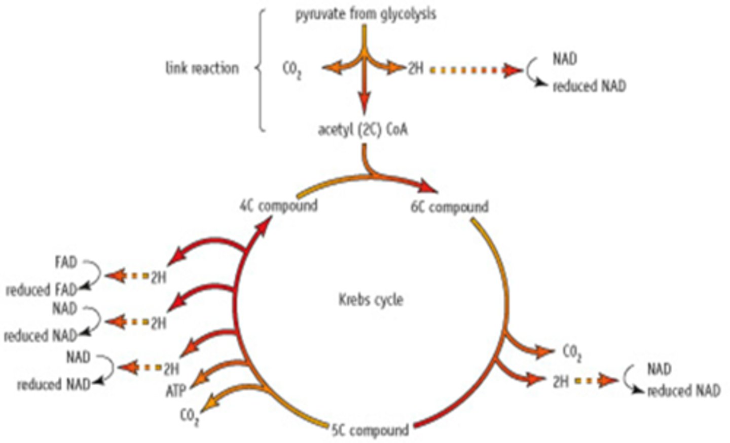
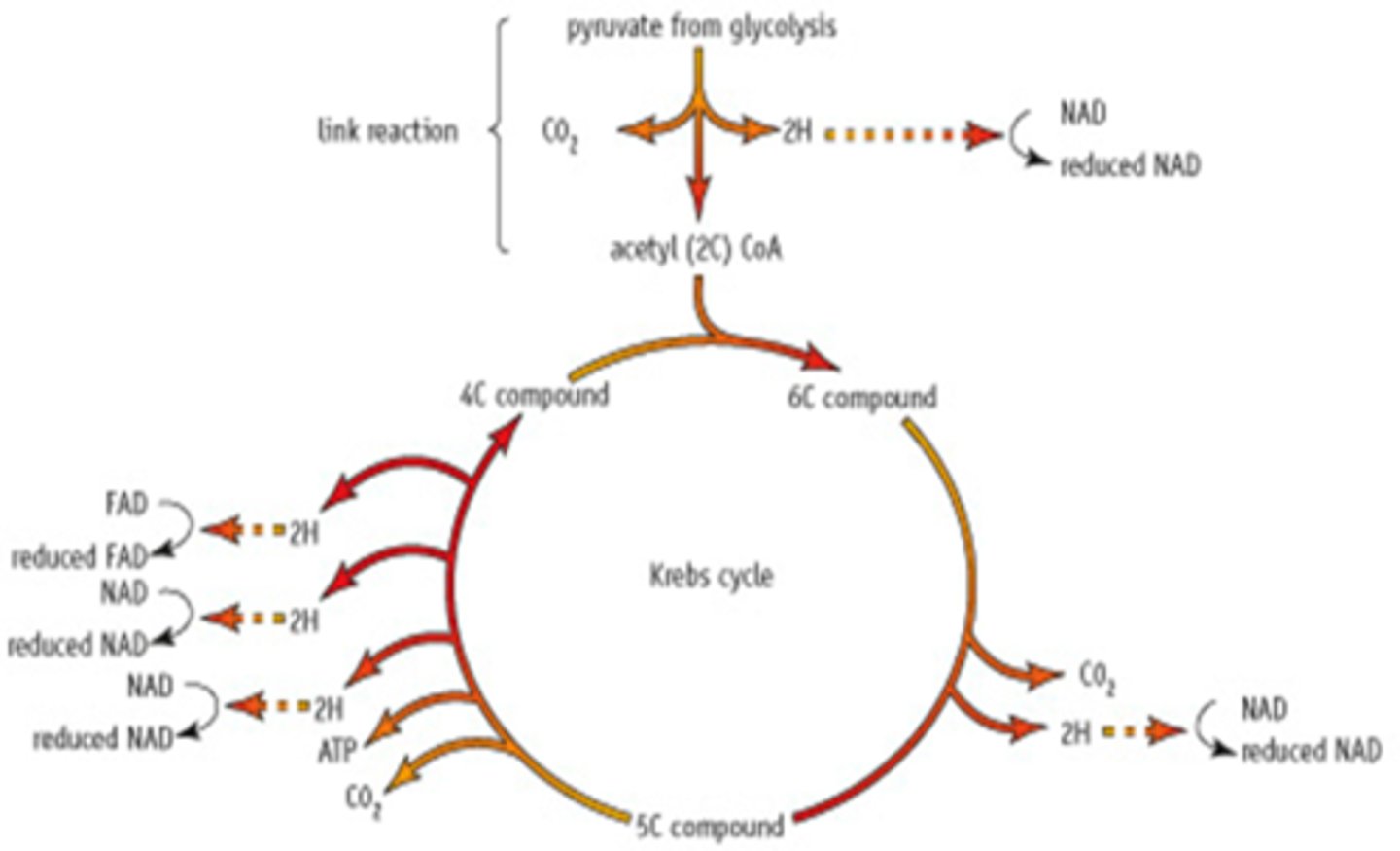
Describe the steps involved in the Krebs Cycle
1. Acetyl CoA from the link reaction combines with oxaloacetate (a 4C compound) to produce citrate (a 6C compound). This is why the Kreb's cycle is sometimes known as the citric acid cycle
The Coenzyme A goes back to the link reaction to be used again.
2. The 6C citrate compound is converted to a 5C molecule (α-ketogluterate).
Decarboxylation occurs where CO2 is removed.
Dehydrogenation also occurs where hydrogen is removed. This produces reduced NAD from NAD (NADH+H+).
3. The 5C compound is then converted to a 4C molecule, oxaloacetate (intermediate compounds are also made here which you don't need to know).
Decarboxylation occurs: removal of another CO2.
Dehydrogenation occurs 3 times:
Total of 2 Reduced NAD (NADH+H+) compounds are produced.
1 compound of Reduced FAD (flavine adenine dinucleotide) (FADH2) is produced.
ATP is produced by the direct transfer of a phosphate group from an intermediate compound to ADP. This is an example of substrate-level phosphorylation.
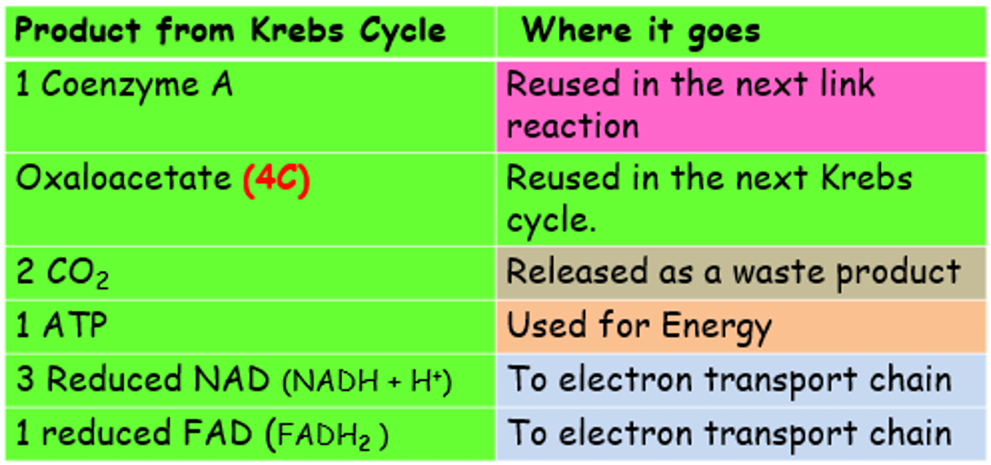
Krebs Cycle Products Summary:
Krebs Cycle Model Answer
series of small enzyme catalysed steps;
4C compound combines with acetyl CoA to form 6C comp;
decarboxylation releases carbon dioxide ;
dehydrogenation releases hydrogen atoms ;
hydrogen acceptors are FAD and NAD ;
reduced NAD and reduced FAD are formed ;
ATP formed by substrate level phosphorylation;
reduced FAD / reduced NAD go to electron transport chain;
4C compound regenerated to keep cycle going ;
What happens during the Electron Transport Chain?
During glycolysis, the link reaction and the Kreb's cycle, many dehydrogenation reactions take place and the hydrogen atoms are passed to the co-enzymes NAD or FAD.
Producing (per glucose molecule);
10 reduced NAD (NADH+H+)
2 reduced FAD (FADH2)
The reduced coenzyme 'shuttles' the hydrogen atoms to the electron transport chain on the mitochondrial inner membrane.
Each hydrogen atom is split into a H+ ion and an electron
The hydrogen atoms are passed to the electron transport chain, where ATP is made by oxidative phosphorylation.
This can only happen in the presence of OXYGEN, therefore the electron transport chain can only function under aerobic conditions.
Describe the processes involved in the electron transport chain
1. The reduced coenzyme
(reduced NAD or FAD) from previous stages pass their hydrogen to an electron carrier fixed in the cristae membrane (inner mitochondrial membrane).
2. The Hydrogen atom is split into a H+ ion and an electron.
The electron is passed to the next carrier in the transport chain (chain is made up of 3 electron carriers)
in a series of redox reactions:
The carrier is reduced when it gains the electrons
The carrier is oxidised when it loses on the electrons
3. Energy is released to pump the H+ ion through the membrane into the inter membrane space (between inner and outer mitochondrial membrane).
As they accumulate here the pH of the intermembrane space falls (it becomes more acidic compared with the inner matrix). This forms an electrochemical gradient (a concentration gradient of ions).
An electrical gradient because.. the H is a charged +ion
and...
a chemical gradient because...
H proton concentration is higher one side of the membrane than the other.
4. At intervals in the cristae membrane, there are protein gates/channels within the stalked particles which allow H+ ions to diffuse back into the matrix. H+ move down an electrochemical gradient.
5. As the H+ diffuses back
into the mitochondrial matrix,
ATP is generated by ATP synthase
enzyme at the gate. This uses
the ADP and inorganic phosphate
from glycolysis.
Hydrogen ions cause a conformational change in the enzyme's active site, so the ADP can bind
This is chemiosmosis.
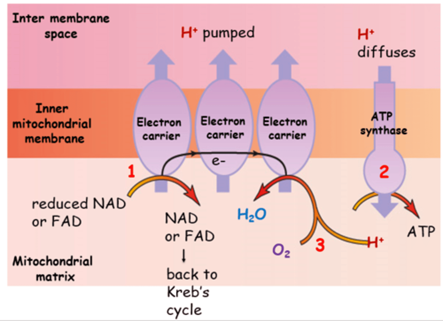
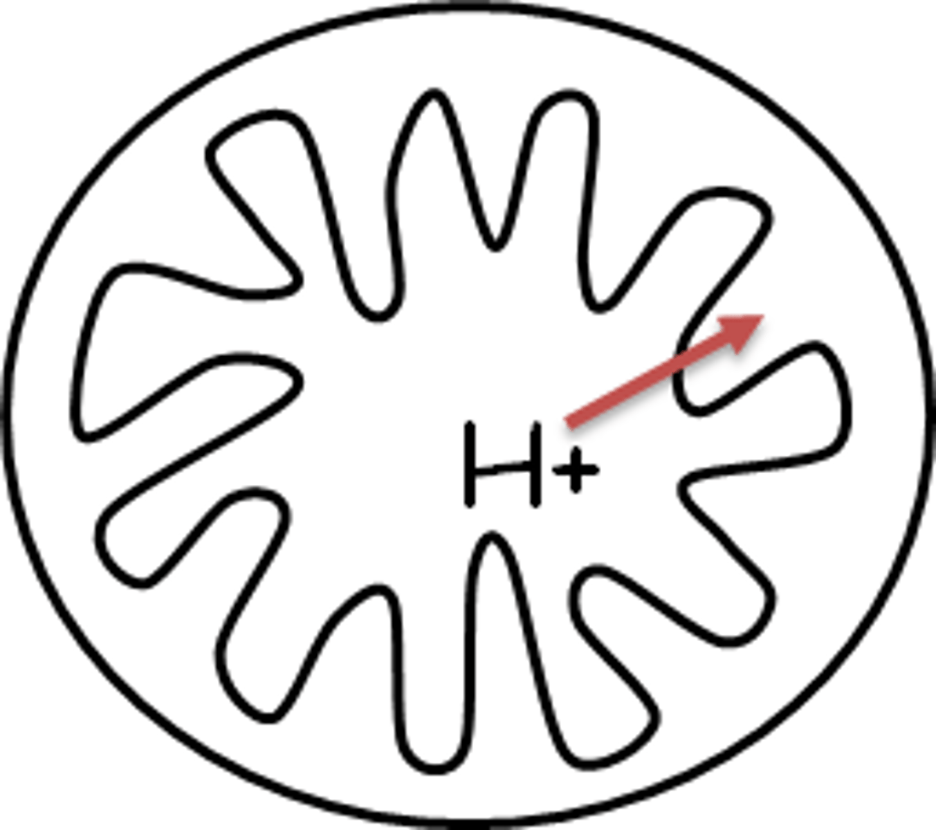
How is the Electrochemical Gradient Created
Electrons lose energy each time they are passed on, so energy is released which is used to pump a H+ ion through the membrane into the intermembrane space
This creates an electrochemical gradient
H+ ions accumulate in the intermembrane space
(pH decreases - more acidic compared with the matrix)
higher concentration of H+ ions in intermembrane space than in matrix
What is Chemiosmosis?
Hydrogen ions diffuse down the electrochemical gradient through hollow protein channels in stalked particles on the membrane.
As hydrogen ions pass through, ATP synthesis is catalysed by ATP synthase in the stalked particle.
Hydrogen ions cause a conformational change in the enzymes active site, so the ADP can bind.
What happens at the end of the Electron Transport Chain?
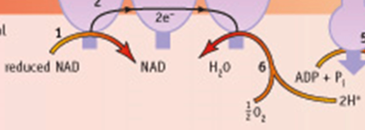
6. Inside the matrix at the end, the H+ ions and the electrons recombine (form hydrogen atom)
The H atoms combine with oxygen to form water
The oxygen, acting as the final carrier in the electron transport chain, is reduced
This method of synthesising ATP is called oxidative phosphorylation
If there is no oxygen, the electron transport chain and ATP synthesis also stop.
Electron transport chain/oxidative phosphorylation model answer
1. Hydrogen atoms split into hydrogen ions + electrons ;
2. Electrons pass along the electron transport chain ;
3. Electrons lose energy each time they are passed on ;
4. Energy is used to phosphorylate ADP to make ATP ;
5. Oxidative phosphorylation
6. Enzyme is ATP synthase ;
7. Process of chemiosmosis ;
8. Oxygen is the final electron (and H+ ion) acceptor ;
9. Water (waste product of respiration) is formed ;
Role of chemiosmosis in Electron transport chain model answer
Chemiosmosis (general) is the movement of ions across a selectively permeable membrane, down their electrochemical gradient.
Diffusion of hydrogen ions ;
Down an electrochemical gradient (high to low conc.) ;
Through ATP synthase (transport protein/enzyme) ;
Shape of active site changes to bind ADP and Pi ;
Phosphorylation of ADP to form ATP ;
ATP is the source of energy for biological processes ;
Role/importance of ATPase Model Answer
Enzyme and transport protein ;
Found on stalked particles on inner mitochondrial membrane ;
Hydrogen ions diffuse through it down an electrochemical gradient ;
Causes active site to change shape and ADP and Pi to bind;
Phosphorylation of ADP produces ATP ;
ATP is the source of energy for biological processes eg. active transport ;
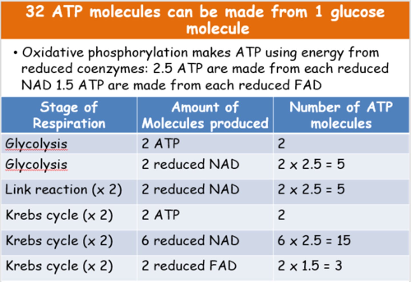
outline how many ATP molecules can be made from one glucose molecule
What variables will affect the rate of respiration?
Respiration is a series of enzyme-catalysed reactions, so the rate will be influenced by any factor affecting the rate of these enzyme controlled reactions:
pH
Concentration of enzyme
Concentration of substrate
Temperature
how does ATP concentration affect rate of respiration?
The concentration of ATP in the cell also has a role in the control of respiration.
In the first stage of glycolysis, the phosphorylation of glucose, ATP needs to be broken down.
The enzyme needed for the phosphorylation of glucose (phosphofructokinase (PFK)) exists in two forms
When ATP is present, enzyme has an inactive; shape and cannot catalyse the reaction, therefore ATP inhibits the enzyme.
When ATP is broken down, the enzyme is converted to its active form and catalyses phosphorylation of glucose.
This enzyme is regulated by levels of;
ATP and citrate
when ATP and citrate levels are high, PFK is inhibited/inactive
How do respirometers work?
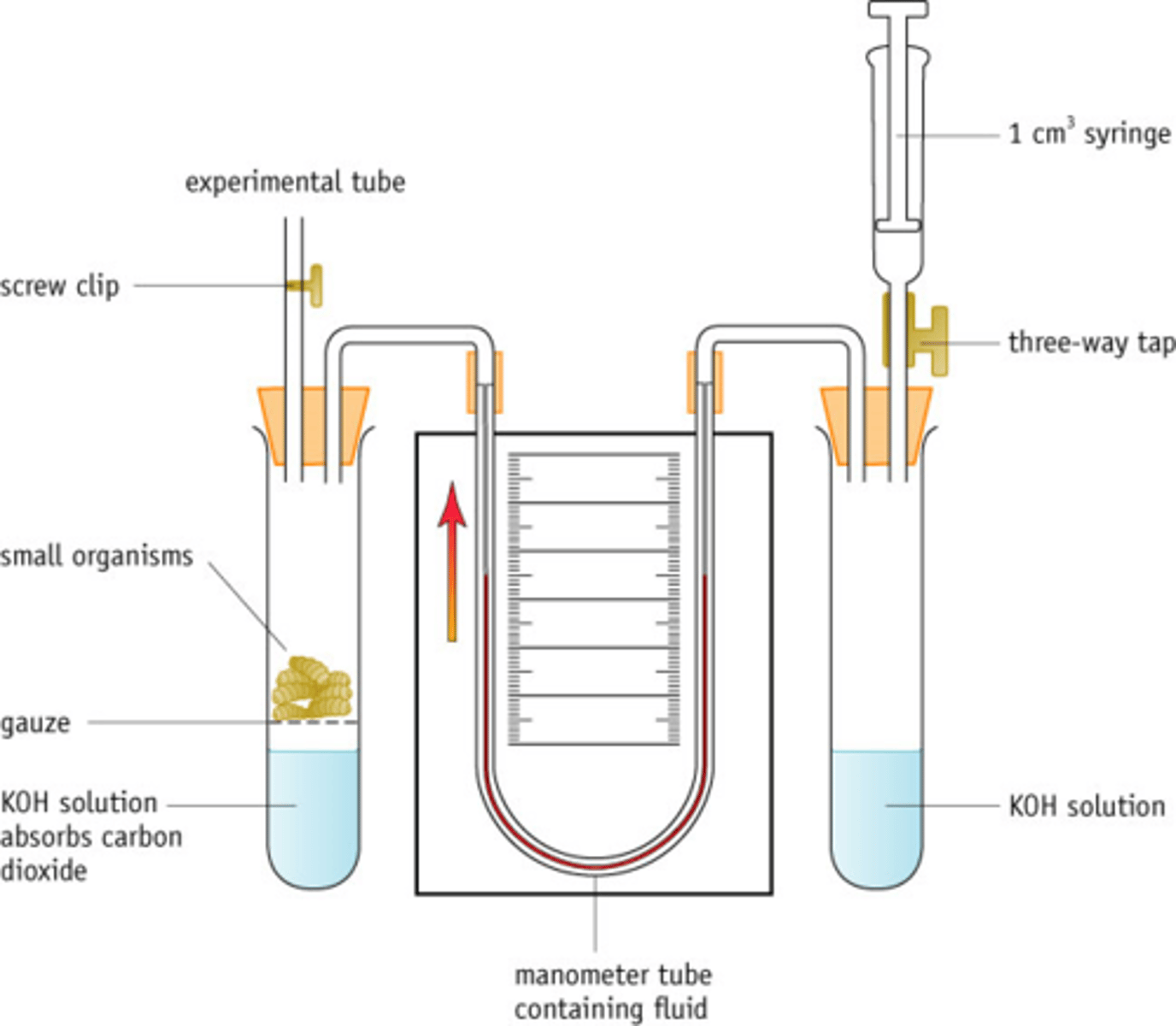
A basic respirometer consists of a sealed chamber containing one or more living organisms, such as germinating seeds or live mice.
The volume of carbon dioxide given off during respiration is the equivalent to the volume of oxygen taken in.
A chemical such as soda lime or potassium hydroxide is used to absorb the carbon dioxide produced during respiration and this loss is measured by observing the movement of fluid in a capillary tube (with a scale).The volume of oxygen used is calculated from this.
By changing the external conditions (e.g. temperature) it is possible to measure their effect on the rate of respiration by recording changes in uptake of oxygen.
The volume (number of molecules) of CO2 given off during respiration is equivalent to the volume (number of molecules) of O2 used for respiration
So if there was no soda lime, the fluid would not move!!
what components are needed to create a respirometer?
Sealed chamber containing living organisms e.g. germinating seeds, leaves, insects, live mice
Soda lime or potassium hydroxide - absorbs carbon dioxide gas produced during respiration, reducing volume / pressure of gas, allowing liquid to move and measurement of oxygen used
A capillary tube with fluid (and scale) - the movement of the fluid allows measurements to be taken
Wire mesh - to separate the organisms from the soda lime
Syringe (sometimes present) - to return the coloured liquid back to zero, for repetition of the experiment
Water bath (sometimes used) - so that respiration rates at different temperatures can be measured or temp controlled
Respirometer exam method
1. ref. to constant temperature ;
2. use of water bath / eq ;
3. reference to suitable time (every min for 5 mins);
4. measure distance fluid moves ;
5. description of how to obtain volume of oxygen (πr2 x
distance moved);
6. calculation of rate described (volume O2 used/time);
7. repeats/replicates ;
8. control (if needed), same but no organism/glass beads ;
9. idea of welfare of organism important ;
10. ref. to {mass / eq} of organism ;
calculating rate of respiraton
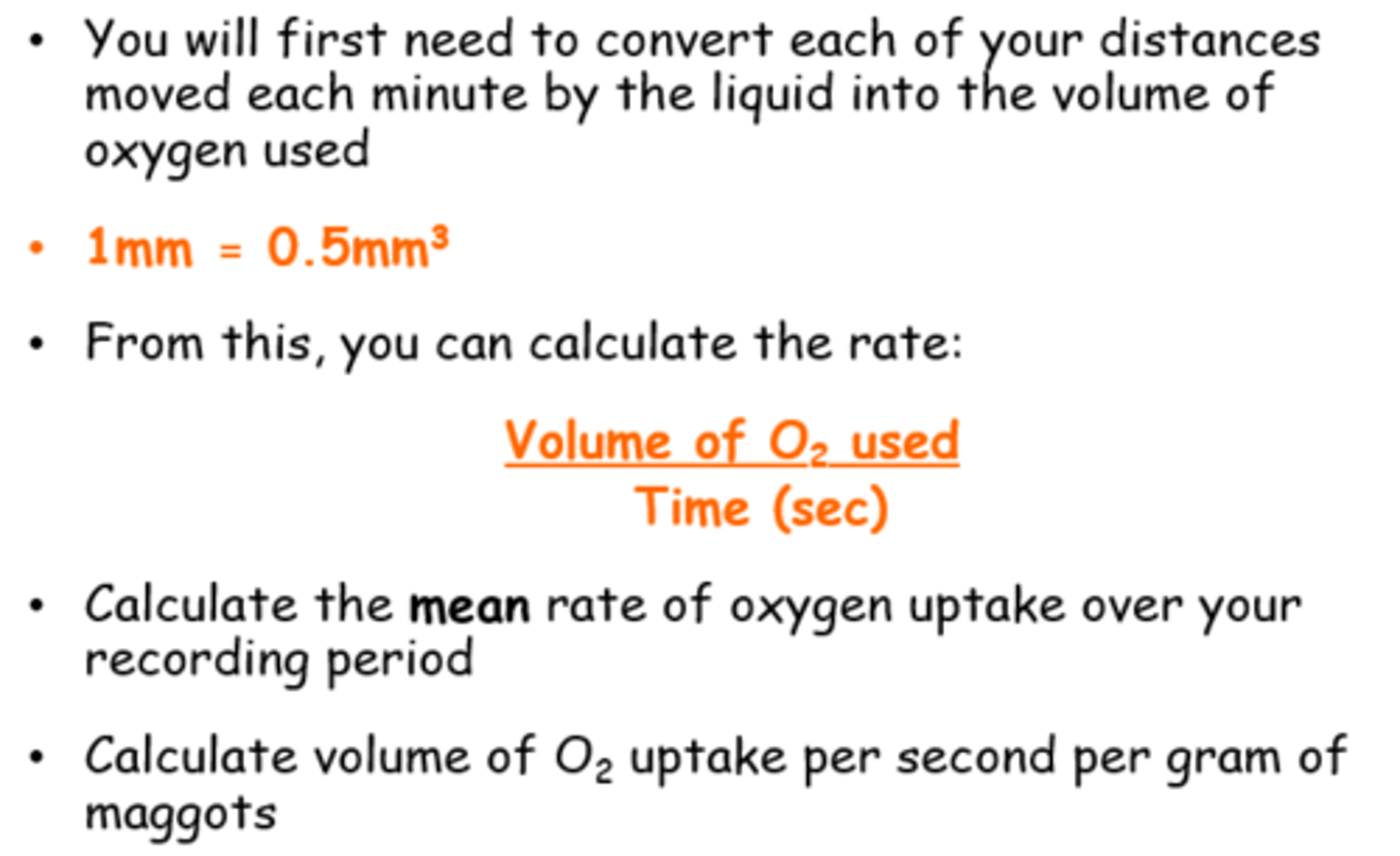
respirometer practical variables
Mass of maggots - use same mass if possible OR express results per unit mass ;
Temperature - control temperature using a water bath ;
Age (size/species) of maggots - hatched at same time ;
Same volume and concentration of KOH solution -measure volume with a pipette ; (mass if soda lime - measure with top-pan balance)
Also
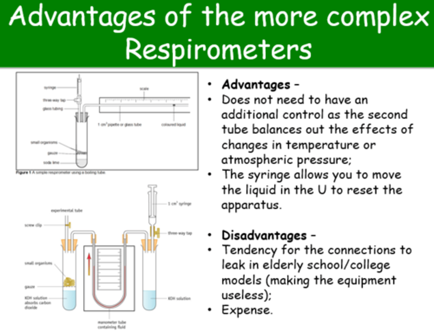
Pressure may affect volume of gas - use control with no organisms, at the same time OR use a complex respirometer with control tube built in
Advantages/disadvantages of the more complex Respirometers
what happens during aerobic respiration when there is no oxygen available?
During intense or lengthy exercise, the oxygen demand in cells exceeds supply (conditions become anaerobic)
Without oxygen to accept the H+ ions and electrons, the electron transport chain stops
The reduced NAD and FAD created in Glycolysis, the Link Reaction and Krebs Cycle cannot be oxidised (release their hydrogen)
Without a supply of oxidised NAD and FAD, aerobic respiration stops (then no ATP would be made!!)
what is the solution to lack of oxygen during exercise?
Another reaction is needed which can take the hydrogen from NAD (oxidise it), to free it up to accept more hydrogens
This will keep glycolysis going
If the cells can keep glycolysis going, a small amount of ATP can be produced!
If there is plenty of oxygen pyruvate will enter the mitochondria and be used in the aerobic reactions of the Krebs cycle.
If there is insufficient oxygen for this, the pyruvate is converted into either ethanol or lactic acid with a little ATP produced.
...This is anaerobic respiration
what is anaerobic respiration?
Anaerobic respiration doesn't use oxygen.
It doesn't involve the link reaction, the krebs cycle or
oxidative phosphorylation to oxidise the NAD or produce
ATP.
There are 2 types, but you only need to know:
Lactate fermentation
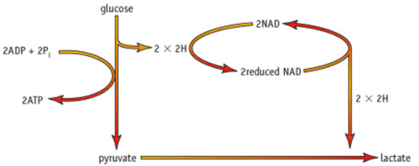
what happens during lactate fermentation?
Occurs in the cell cytoplasm:
Glucose is converted to pyruvate via glycolysis.
Reduced NAD (from glycolysis) transfers H to pyruvate to form lactate (another 3C compound) and NAD (oxidised).
NAD can then be reused in glycolysis.
The production of lactate regenerates NAD.
This means glycolysis can continue even without much oxygen.
For one glucose molecule a net yield of 2 ATP molecules are produced.
Therefore an athlete can continue by partially breaking down glucose to make a small amount of ATP.
anaerobic respiration model answer
1. Anaerobic respiration ;
2. in (cell) cytoplasm ;
3. ATP made from phosphorylation of ADP ;
4. energy required (for phosphorylation) ;
5. glycolysis / glucose converted to pyruvate;
6. pyruvate converted to lactate ;
7. that makes NAD available to accept hydrogen ;
what are the effects of lactate buildup
Lactate builds up in muscle but must be disposed of later
Lactate forms lactic acid in solution
pH of cells falls, inhibiting the enzymes that catalyse the glycolysis reactions
The glycolysis reactions and the physical activity depending on them cannot continue
The animal must slow down or stop so that oxygen supply can meet demand
eventually glycolysis/ ATP production slows down
the physical activity (muscle contractions) depending on the 2 ATP cannot continue, as lactic acid build up causes muscle pain/cramps